Introduction
Entamoeba is a member of the Entamoebidae, a deep lineage within the Archamoebae (Pánek et al., Reference Pánek, Zadrobílková, Walker, Brown, Gentekaki, Hroudová, Kang, Roger, Tice, Vlček and Čepička2016). Entamoeba species use pseudopodia for locomotion and lack flagella, a morphologically identifiable Golgi apparatus, peroxisomes and canonical mitochondria (Loftus et al., Reference Loftus, Anderson, Davies, Alsmark, Samuelson, Amedeo, Roncaglia, Berriman, Hirt, Mann, Nozaki, Suh, Pop, Duchene, Ackers, Tannich, Leippe, Hofer, Bruchhaus, Willhoeft, Bhattacharya, Chillingworth, Churcher, Hance, Harris, Harris, Jagels, Moule, Mungall, Ormond, Squares, Whitehead, Quail, Rabbinowitsch, Norbertczak, Price, Wang, Guillén, Gilchrist, Stroup, Bhattacharya, Lohia, Foster, Sicheritz-Ponten, Weber, Singh, Mukherjee, El-Sayed, Petri, Clark, Embley, Barrell, Fraser and Hall2005; Ptáčková et al., Reference Ptáčková, Kostygov, Chistyakova, Falteisek, Frolov, Patterson, Walker and Cepicka2013). Entamoeba species have trophozoite and cyst stages. The latter may have one nucleus or as many as eight, each with peripheral chromatin prominently visible. Historically, cyst size and nuclear number and appearance, along with host range information, were considered taxonomically important features and used to identify and group species of Entamoeba. However, in recent years it has become obvious that morphological features alone are not sufficient to adequately discriminate species known to be genetically distinct (Clark et al., Reference Clark, Kaffashian, Tawari, Windsor, Twigg-Flesner, Davies-Morel, Blessmann, Ebert, Peschel, Le Van, Jackson, Macfarlane and Tannich2006; Stensvold et al., Reference Stensvold, Lebbad, Victory, Verweij, Tannich, Alfellani, Legarraga and Clark2011). For example, morphology does not distinguish the morphologically identical E. histolytica and E. dispar, yet only the former is a human pathogen (Gonin and Trudel, Reference Gonin and Trudel2003; Fotedar et al., Reference Fotedar, Stark, Beebe, Marriott, Ellis and Harkness2007a; Hooshyar et al., Reference Hooshyar, Rostamkhani and Rezaeian2015). The advent of molecular tools has shed light on the taxonomic landscape of Entamoeba and clarified several issues associated not only with taxonomy but also epidemiology and host range (Verweij et al., Reference Verweij, Laeijendecker, Brienen, van Lieshout and Polderman2003; Fotedar et al., Reference Fotedar, Stark, Beebe, Marriott, Ellis and Harkness2007b; García et al., Reference García, Ramos, Pérez, Yañez, Estrada, Mendoza, Martinez-Hernandez and Gaytán2014). Screening of fecal samples from a broad range of hosts using SSU rRNA gene primers has uncovered several new and distinct lineages of Entamoeba, indicating a richly diverse genus (Santos et al., Reference Santos, Bandea, Martins, de Macedo, Peralta, Peralta, Ndubuisi and da Silva2010; Stensvold et al., Reference Stensvold, Lebbad, Victory, Verweij, Tannich, Alfellani, Legarraga and Clark2011; Jacob et al., Reference Jacob, Busby, Levy, Komm and Clark2015). Much of this diversity had not been previously recognized.
Members of the genus Entamoeba generally inhabit the gastrointestinal tract of vertebrates and invertebrates, but they have also been observed within other protist cells (Ghosh, Reference Ghosh1973; Stensvold et al., Reference Stensvold, Lebbad, Victory, Verweij, Tannich, Alfellani, Legarraga and Clark2011; García et al., Reference García, Ramos, Pérez, Yañez, Estrada, Mendoza, Martinez-Hernandez and Gaytán2014; Shilton et al., Reference Shilton, Šlapeta, Shine and Brown2018). Several Entamoeba species are parasitic, but commensals are more common (Hooshyar et al., Reference Hooshyar, Rostamkhani and Rezaeian2015). Uniquely among members of the genus, E. gingivalis inhabits the human oral cavity (Ghabanchi et al., Reference Ghabanchi, Zibaei, Afkar and Sarbazie2010; Luszczak et al., Reference Luszczak, Bartosik, Rzymowska, Sochaczewska-Dolecka, Tomaszek, Wysokinska-Miszczuk and Bogucka-Kocka2016; Maybodi et al., Reference Maybodi, Ardakani, Bafghi, Ardakani and Zafarbakhsh2016). In addition, a few members of the genus have also been isolated from the environment (Clark and Diamond, Reference Clark and Diamond1997; Shiratori and Ishida, Reference Shiratori and Ishida2015).
Most Entamoeba gene sequences in public databases originate from species living in endothermic hosts, while relatively few derive from species living in ectotherms. To date, the latter hosts include amphibians, reptiles and insects (Silberman et al., Reference Silberman, Clark, Diamond and Sogin1999; Garcia et al., Reference García, Ramos, Pérez, Yañez, Estrada, Mendoza, Martinez-Hernandez and Gaytán2014; Clark and Stensvold, Reference Clark, Stensvold, Nozaki and Bhattacharya2015; Jacob et al., Reference Jacob, Busby, Levy, Komm and Clark2015; Kawano et al., Reference Kawano, Imada, Chamavit, Kobayashi, Hashimoto and Nozaki2017). Herein, we report a new species of Entamoeba, isolated from the gastrointestinal tract of the fish Monopterus albus (the Asian swamp eel) in Chiang Rai, Thailand. We examine its morphological features using light microscopy of living and stained specimens and provide the first SSU rRNA gene sequence of an Entamoeba isolated from fish.
Methods
Sample collection and establishment of culture
Two Asian swamp eels were purchased at a local market at Sanpong village, Phan district, Chiang Rai Province, northern Thailand. The eels were obtained at two separate times, in May and July 2018. Colonic contents were placed in modified (no mucin was added) LYSGM medium (Diamond, Reference Diamond1982, http://entamoeba.lshtm.ac.uk/xenic.htm) and incubated at room temperature (25–27 °C). After 24 h, sediment was transferred to fresh medium and cells were subcultured every 2 weeks. The culture has been maintained since July 2018.
Light microscopy and staining
A wet mount of live amoebae was prepared and cells were observed using Nikon inverted light microscope. Trophozoites (n = 10) and cysts (n = 125 live; n = 125 stained with iodine) were measured using the same microscope. For a more detailed view of the cells, iron hematoxylin staining was performed by the Diagnostic Parasitology Laboratory, London School of Hygiene and Tropical Medicine. Stained cells were observed with a Leica DMRB microscope fitted with a DFC 420 camera.
DNA extraction, amplification, purification and sequencing
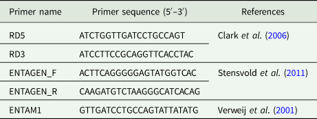
Total genomic DNA was extracted from the culture using an AccuPrep® Genomic DNA Extraction Kit (Bioneer, South Korea, catalog No: K-3032) according to the manufacturer's specifications. Polymerase chain reaction (PCR) using the broad specificity primers RD5 and RD3 was used to amplify almost the entire SSU rRNA gene (Table 1). Emerald Amp® GT PCR Master Mix for PCR reactions was obtained from TaKaRa Bio USA, Inc. Cycling conditions were as follows: initial denaturation at 94 °C for 3 min, followed by 40 cycles of: denaturation at 94 °C for 1.3 min, annealing at 60 °C for 1 min and extension at 72 °C for 2 min, ending with a final extension of 10 min at 72 °C.
Primers used to amplify and sequence Entamoeba chiangraiensis

The resulting PCR products were purified from gels with the GeneJET Gel Extraction Kit (Thermo Scientific; Wardmedic, Thailand) according to manufacturer's specifications. Samples were sequenced with RD5 and RD3 primers, along with ENTAM1, ENTAGENF and ENTAGENR (Table 1).
Phylogenetic analysis
The chromatogram quality of raw reads was checked individually with Sequencher software and ambiguous bases from the ends were removed. Sequences were combined into contigs and checked against the NCBI nr database, where they were identified as Entamoeba. A dataset was assembled including the newly derived sequences along with sequences spanning the breadth of molecular diversity of Entamoeba. In total, 90 sequences were used. Sequence alignment was performed on the EBI online platform (https://www.ebi.ac.uk/Tools/msa/mafft/) using MAFFT v.7.394 (Katoh and Toh, Reference Katoh and Toh2010). Ambiguously aligned positions were removed using Trimal v.1.3 (Capella-Gutierrez et al., Reference Capella-Gutierrez, Silla-Martinez and Gabaldon2009) available on the online platform Phylemon 2.0 (http://phylemon.bioinfo.cipf.es). After trimming 1434 sites remained. Maximum likelihood analysis was conducted using RAxML v.8 (Stamatakis, Reference Stamatakis2006) on the online platform CIPRES Science Gateway (http://www.phylo.org/index.php/). For ML analysis, the general time reversible + Γ model of nucleotide substitution was employed as dictated by jModelTest v.2.1.10 using the Akaike criterion. Bootstrap support was computed from 1000 bootstrap replicates.
Results
Culture, light microscopy and phylogenetic analysis
Colonic gut contents were inoculated into modified LYSGM, a medium widely used for xenic cultivation of Entamoeba species, and incubated at room temperature overnight. No live amoebae or cysts were observed in any tubes incubated at 37 °C, indicating that this species does not survive at that temperature.
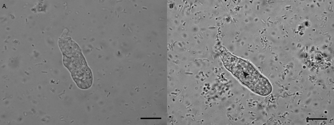
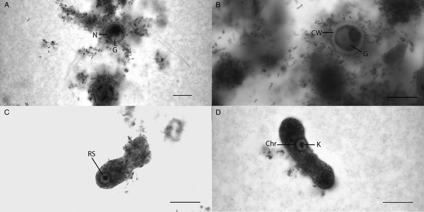
The trophozoite of the amoeba is longer than it is wide (Fig. 1, Fig. 2C and D). Length is 40–50 µm (mean 44.31 µm) when the amoeba swims, but when it glides on the slide it ranges from 30 to 40 µm, while width ranges from 9 to 13 µm (mean 11.18 µm). The cell changes shape slowly while in motion and has a single prominent pseudopodium, while the posterior end is smooth with no obvious uroid (Fig. 1, Fig. 2C and D). The granuloplasm has multiple vesicles while the hyaloplasm is narrow (Fig. 1A). Unstained spherical cysts range from 10.0 to 17.50 µm in diameter (mean 14.15 µm; ±1.42 standard deviation; ±0.13 standard error). Stained cysts range from 10.0 to 17.50 µm in diameter (mean 13.75 µm; ±1.54 standard deviation; ±0.14 standard error). All observed cysts in both live and stained samples were uninucleate (Fig. 2A and B), with the exception of a single stained example where it looked like there were two nuclei. Large, prominent glycogen vacuoles were present in both live and stained cysts, indicating that all observed cysts were immature (Fig. 2A and B). Therefore, we cannot state the number of nuclei per cyst definitively, as we were not able to observe mature cysts. Cysts have no distinctive appearance (Fig. 2A and B).
Light micrographs of living trophozoites of Entamoeba chiangraiensis n. sp. Arrowhead indicates the nucleus. Scale bar = 25 µm.

Light micrographs of trophozoites and cysts stained with iron hematoxylin. (A and B) Stained cysts. N, nucleus; G, glycogen vacuole; CW, cyst wall. (C and D) Stained trophozoites. RS, radial spokes connecting karyosome to peripheral chromatin; Chr, peripheral chromatin forming an even fine lining around nuclear membrane; K, karyosome consisting of granules. Scale bar = 10 µm.

The size of the nucleus in both cysts and trophozoites ranges in diameter from 2.5 to 7.5 µm (mean 3.97 µm; ±1.46 standard deviation; ±0.13 standard error) and is generally found in the anterior half of the trophozoite. The trophozoite nucleus has a karyosome that has the appearance of a cluster of granules (Fig. 2C and D). Karyosome size is variable depending on how tightly the granules cluster. Chromatin forms a delicate, even lining along the inner membrane of the nucleus (Fig. 2D). Unlike many other Entamoeba species, there are no clearly visible clumps of peripheral chromatin. Radial spokes are present in the nucleus joining the karyosome to peripheral chromatin (Fig. 2C).
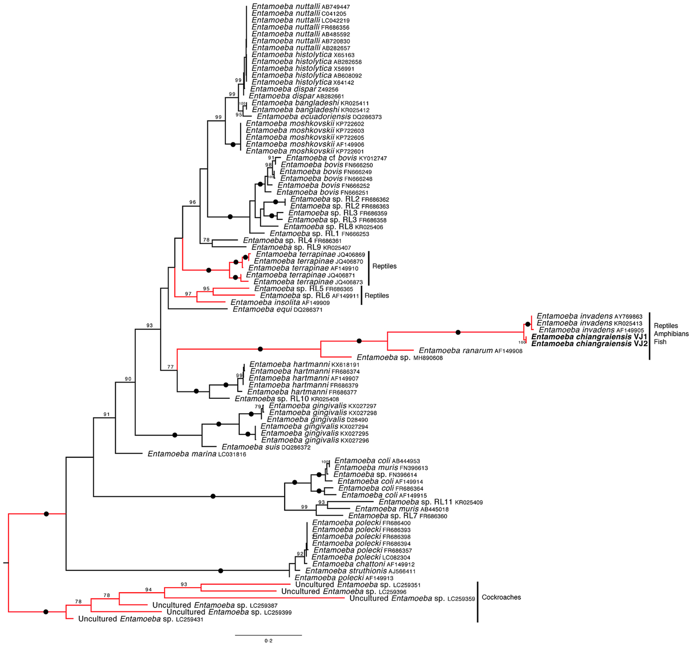
The SSU rRNA gene sequences of the two isolates are nearly complete (1849 and 1856 bp). Both sequences have been deposited in GenBank under accession numbers MK652887 and MK652888. The overall topology of the phylogenetic tree is similar to previous studies (Jacob et al., Reference Jacob, Busby, Levy, Komm and Clark2015). The tree is artificially rooted to the clade containing the cockroach sequences. These were the earliest diverging Entamoeba sequences in the eukaryotic supergroup tree of Kawano et al., Reference Kawano, Imada, Chamavit, Kobayashi, Hashimoto and Nozaki2017. The new SSU rRNA gene sequences are sister to those from E. invadens and this relationship has maximum bootstrap support (Fig. 3). The genetic distance between the new sequences and E. invadens sequences ranges from 3.4 to 3.8% (Table S1). All observed nucleotide differences (including insertion and deletion events) are taxon-specific. Intraspecific genetic divergence for the new amoeba and E. invadens is 0 and 0.4%, respectively. These sister species are in a clade that also includes E. ranarum and an unnamed Entamoeba sp., both from amphibian hosts. All members of this clade have been isolated from ectothermic hosts. This clade also has maximum bootstrap support.
Maximum likelihood phylogenetic tree inferred from 90 SSUrRNA sequences and 1434 sites. The tree is artificially rooted to cockroach-derived Entamoeba sequences. Newly generated sequences are depicted in bold lettering. Numerical values indicate bootstrap support. Only values above 70 are shown. Full circles represent maximum bootstrap support. Clades in red consist of sequences exclusively from ectothermic hosts.

Taxonomic Summary
Amoebozoa Lühe 1913, emend. Cavalier-Smith 1998
Archamoebae Cavalier-Smith 1983
Entamoebidae Chatton 1925, emend. Cavalier-Smith 1993
Entamoeba Casagrandi & Barbagallo Reference Casagrandi and Barbagallo1895
Entamoeba chiangraiensis n. sp. Jinatham, Clark & Gentekaki 2019
Diagnosis: Amoeba inhabiting the gut of Monopterus albus (Asian swamp eel). Trophozoite is much longer than it is wide; length in motion is 30–50 µm, width 9–13 µm. A trailing end is smooth and devoid of visible uroid processes. Cysts are spherical, appearing smooth and thick-walled. Immature cysts have a single nucleus and a prominent glycogen vacuole, which often obscures the nucleus. Cyst diameter is 10.0–17.5 µm (mean 14.15 µm; ±1.42 standard deviation; ±0.13 standard error), nucleus 2.5–7.5 µm (mean 3.97 µm; ±1.46 standard deviation; ±0.13 standard error). There is a karyosome composed of granules. Chromatin is evenly distributed around the inner nuclear membrane, forming a thin, uniform lining. Radial spokes connect the karyosome to the peripheral chromatin.
Etymology: the epithet chiangraiensis refers to Chiang Rai province, Thailand, in which the organism was isolated
Host: Monopterus albus
Type location: isolated from the gut of Asian swamp eel, Sanpong, Phan, Chiang Rai, Thailand
Type material: permanent slide stained with iron-hematoxylin was deposited in the Smithsonian Museum under accession number USNM 1484171.
Type sequence: GenBank accession number MK652887
Discussion
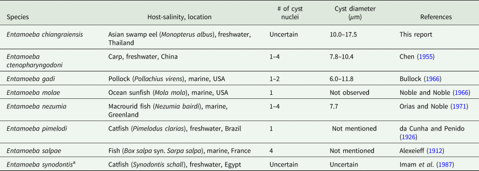
Like all members of the genus Entamoeba, the new species has a nucleus with the characteristic ‘ring and dot’ appearance corresponding to peripheral chromatin and central karyosome (Clark and Stensvold, Reference Clark, Stensvold, Nozaki and Bhattacharya2015). Entamoeba chiangraiensis n.sp. was isolated twice from the Asian swamp eel, Monopterus albus, which inhabits rivers across Southeast Asia. Only a few species of Entamoeba from fish have been documented: four from marine hosts and three from freshwater (Table 2 and references therein). Molecular data for any of these species is absent.
Species of Entamoeba isolated from fish

a Description is incomplete in the original text
Pathogenicity of the new species is unknown. Only a few species of Entamoeba are definitively pathogenic based on histology evidence. These are E. histolytica, a human pathogen, E. nuttalli, a pathogen of non-human primates, E. invadens, a reptile pathogen and Entamoeba sp., a toad pathogen (Clark and Stensvold, Reference Clark, Stensvold, Nozaki and Bhattacharya2015; Shilton et al., Reference Shilton, Šlapeta, Shine and Brown2018). Microscopic examination of E. chiangraiensis cells immediately after sample collection did not reveal ingestion of red blood cells, suggesting that the species is commensal rather than invasive. Nonetheless, to definitively determine pathogenicity further studies will be needed, including histology of infected fish to detect whether E. chiangraiensis invades host tissue.
We observed a single nucleus in cysts of the new species. However, the number of nuclei in mature cysts remains undetermined as cysts degenerated before reaching maturity. In the literature, the number of nuclei in cysts of Entamoebae from fish varies from one to four (Table 2 and references within). Species of Entamoeba from other ectothermic hosts commonly have four nucleated cysts, although octo-nucleated cysts have been observed in some reptiles, including E. barreti from a snapping turtle (Taliaferro and Holmes, Reference Taliaferro and Holmes1924).
The host range of our and other species of Entamoeba from fish is unknown. We screened a number of fish inhabiting the same environment as the Asian swamp eel (Synbranchiformes) including: Anabas sp. (Anabatiformes, n = 3), Tilapia sp. (Cichliformes, n-5), Trichogaster sp. (Anabatiformes, n = 3), Trachinocephalus (Aulopiformes, n = 2) and Siluriformes (Siluriformes, n = 4). Our examination included both microscopy and a molecular survey using combinations of the primers described in the methods section. Intestinal contents from all fish were placed in the same culture medium in an attempt to grow amoebae. We were unable to find Entamoeba in any of the other hosts using any of the methods described. Although we tried to be as inclusive as possible in our screening, we cannot exclude the possibility that E. changraiensis might also inhabit the gut of fish that we have not examined. Host ranges of many Entamoeba species remain incompletely known, but they keep expanding. For instance, E. coli has traditionally been reported from humans and non-human primates but is now known in rodents (Clark and Stensvold, Reference Clark, Stensvold, Nozaki and Bhattacharya2015). Nonetheless, it seems likely that body temperature will pose a constraint on host range, as Entamoebae from ectotherms have not been found in endotherms and vice versa. Entamoeba moshkovskii is a notable exception, having been found in both reptiles and mammals (Garcia et al., Reference García, Ramos, Pérez, Yañez, Estrada, Mendoza, Martinez-Hernandez and Gaytán2014); it seems to be the only species of Entamoeba that has crossed the ectotherm/endotherm barrier. Within ectotherms, Entamoeba species show host specificity at the higher level of classification. Thus, reptilian isolates have never been isolated from amphibians and vice versa.
Entamoeba SSU rRNA gene sequences that have been detected exclusively in ectothermic hosts are diverse and dispersed across the phylogenetic tree, forming four distinct clades. The first clade comprises E. chiangraiensis, E. invadens, E. ranarum and an unnamed Entamoeba sp. (MH890608) from a toad. The latter represents only the second amphibian-derived Entamoeba sequence. The SSU rRNA gene sequences from two eels sampled at two separate time points were identical, indicating low intra-specific diversity of this gene in E. chiangraiensis. This is similar to E. invadens, whose SSU rRNA gene sequences also display a high degree of genetic similarity, even when isolated from different hosts and from different countries (Jacob et al., Reference Jacob, Busby, Levy, Komm and Clark2015). The new species groups together with E. invadens. When comparing their SSU rRNA sequences, the genetic distance is a little below 4%, almost 4-fold than that between E. histolytica and E. dispar. The second clade contains several variants of E. terrapinae derived from aquatic turtles (Garcia et al., Reference García, Ramos, Pérez, Yañez, Estrada, Mendoza, Martinez-Hernandez and Gaytán2014). The third clade contains Entamoeba insolita, along with Entamoeba RL5 from tortoise and Entamoeba RL6 from iguana. These organisms are each represented by a single sequence (Silberman et al., Reference Silberman, Clark, Diamond and Sogin1999; Stensvold et al., Reference Stensvold, Lebbad, Victory, Verweij, Tannich, Alfellani, Legarraga and Clark2011). Finally, the fourth clade consists of numerous sequences of Entamoeba from cockroaches (Kawano et al., Reference Kawano, Imada, Chamavit, Kobayashi, Hashimoto and Nozaki2017). In their study, Kawano et al. (Reference Kawano, Imada, Chamavit, Kobayashi, Hashimoto and Nozaki2017) examined 186 cockroaches and found Entamoebae in 134. In their phylogenetic analyses, cockroach-derived sequences formed a distinct clade with nine separate groups within. This strongly hints at the presence of a vast diversity of Entamoeba that has yet to be uncovered. It seems likely that screening of additional hosts, especially ectotherms, will reveal an ever greater number of novel Entamoeba species.
Supplementary material
The supplementary material for this article can be found at https://doi.org/10.1017/S0031182019000775.
Author ORCIDs
Eleni Gentekaki, 0000-0002-3306-6714.
Acknowledgements
The authors thank Mrs. Noppadon Jinatham for her assistance in collecting the samples. We are grateful to the Diagnostic Parasitology Laboratory, London School of Hygiene and Tropical Medicine for undertaking the staining.
Financial support
This work was supported by the Thailand Research Fund (grant number RSA6080048) awarded to E.G.
Conflict of interest
None.
Ethical standards
No animals were sacrificed specifically for this work. Asian swamp eel is a popular food in Thailand and can be purchased at local markets. Intestinal contents were obtained from eels that had been purchased for food consumption. Permission and approval for obtaining such contents were obtained from the Mae Fah Luang University Animal Care and Use Committee (protocol no. AR01/62).