INTRODUCTION
The infraorder Thalassinidea has been split in two infraorders recently, Axiidea de Saint Laurent, 1979 and Gebiidea de Saint Laurent, 1979. Upogebia is one of the 13 genera of the family Upogebiidae, infraorder Gebiidea (De Grave et al., Reference De Grave, Pentcheff, Ahyong, Chan, Crandall, Dworschak, Felder, Feldmann, Fransen, Goulding, Lemaitre, Low, Martin, Ng, Schweitzer, Tan, Tshudy and Wetzer2009). Species from this family are commonly found in shallow waters, with 44.6% of species occurring in depths of less than 2 m (Dworschak, Reference Dworschak2000). Upogebia vasquezi Ngoc-Ho, 1989 inhabits the western Atlantic in the regions of southern Florida, the Bahamas, Central America and Brazil (from the States of Maranhão to São Paulo (Melo, Reference Melo1999)) and its occurrence has been extended to the coast of the State of Pará (northern Brazil) in present study. This species occurs in the intertidal zone in Y-shaped holes in the sand, the geological significance of which is quite important, especially with regard to the recognition of sea levels in the past (Curran & Martin, Reference Curran and Martin2003).
The description of larval morphology is one of the keys to the elucidation of the lifecycle, taxonomy and systematics of diverse species of decapods and is fundamental to phylogenetic analyses (Mantelatto & Cuesta, Reference Mantelatto and Cuesta2010). There is little knowledge on the larval biology of thalassinidean species (Ngoc-Ho, Reference Ngoc-Ho1981; Thessalou-Legaki, Reference Thessalou-Legaki1990; Abrunhosa et al., Reference Abrunhosa, Arruda, Smith and Palmeira2008a) and only about one-third of larval accounts comprise complete larval descriptions of species (Pohle et al., Reference Pohle, Santana, Jansen and Greenlaw2011). The Gebiidea represent approximately 45% of the larval descriptions and Upogebia represent the genus with most known larvae (Pohle et al., Reference Pohle, Santana, Jansen and Greenlaw2011).
Recent studies involving species of this genus offer an analysis of larval emission patterns and the effect of temperature and salinity on larval stages of U. africana (Paula, Reference Paula1989; Wooldridge & Loubser, Reference Wooldridge and Loubser1996; Paula et al., Reference Paula, Mendes, Paci, McLaughlin, Gherardi and Emmerson2001; Teske et al., Reference Teske, Papadopoulos, Zardi, McQuaid, Edkins, Griffiths and Barker2007, Reference Teske, Papadopoulos, Newman, Dworschak, McQuaid and Barker2008), the characterization of the lifecycle of U. major (Kinoshita et al., Reference Kinoshita, Nakayama and Furota2003) and an analysis of the best food sources in the early larval stages of U. pusilla (Faleiro & Narciso, Reference Faleiro and Narciso2009). Some studies are available on the morphological characteristics of Upogebia larvae and post-larvae, with descriptions of the larval stages from plankton-collected larvae (Sandifer, Reference Sandifer1973; Ngoc-Ho, Reference Ngoc-Ho1981; Andryszak, Reference Andryszak1986) and from hatching (Ngoc-Ho, Reference Ngoc-Ho1977; Konishi, Reference Konishi1989; Siddiqui & Tirmizi, Reference Siddiqui and Tirmizi1995; Shy & Chan, Reference Shy and Chan1996; Melo & Brossi-Garcia, Reference Melo and Brossi-Garcia2000; Santos & Paula, Reference Santos and Paula2003). However, the number of these descriptions remains quite small considering the estimated 116 known species of this genus (De Grave et al., Reference De Grave, Pentcheff, Ahyong, Chan, Crandall, Dworschak, Felder, Feldmann, Fransen, Goulding, Lemaitre, Low, Martin, Ng, Schweitzer, Tan, Tshudy and Wetzer2009).
In Brazil, have been registered the occurrence of ten species of Upogebia: U. acanthura Coelho, 1973, U. brasiliensis Holthuis, 1956, U. paraffinis Williams, 1993, U. omissa Gomes Corrêa, 1968, U. omissago Williams, 1993, U. marina Coelho, 1973, U. careospina Williams, 1993, U. noronhensis Fausto-Filho, 1969, U. vasquezi Ngoc-Ho, 1989 and U. inomissa Williams, 1993 (Melo, Reference Melo1999; Nucci & Melo, Reference Nucci and Melo2001). Among these species, Coelho et al. (Reference Coelho, Cooper and Rodrigues2000) and Candisani et al. (Reference Candisani, Sumida and Pires-Vanin2001) studied the burrow morphology and mating behaviour of U. noronhensis and U. omissa. Regarding the larval development of these species, U. brasiliensis, U. omissa and U. vasquezi have been studied, but on unpublished data (Melo, Reference Melo2001). A description of complete larval development is only available on published data for U. paraffinis (Melo & Brossi-Garcia, Reference Melo and Brossi-Garcia2000).
For the coast of the State of Pará (northern Brazil) only four species of Upogebia have previously been recorded: U. acanthura, U. brasiliensis, U. marina and U. paraffinis (Melo, Reference Melo1999; Barros & Pimentel, Reference Barros and Pimentel2001). However, the distribution of U. vasquezi has been extended to this region. Regarding the coast of the State of Pará, few studies have addressed the larval development of thalassinidean species, like Lepidophthalmus siriboia Felder & Rodrigues, 1993 and Callichirus major (Say, 1818) (Abrunhosa et al., Reference Abrunhosa, Pires, Lima and Coelho-Filho2005, Reference Abrunhosa, Melo, Lima and Abrunhosa2006, Reference Abrunhosa, Arruda, Smith and Palmeira2008a, b) of the family Callianassidae. The aim of the present study was to describe the early two larval stages in U. vasquezi and compare them to larvae of other Upogebia species occurring in Brazil, contributing for the expansion on thalassinidean larval literature and identification of these larval forms from zooplankton samples.
MATERIALS AND METHODS
In October 2009, six (6) ovigerous female specimens of Upogebia vasquezi were collected two days prior to the new moon from rocky outcrops in the lower mediolittoral zone of the estuary of the Marapanim River, which is located on the north-eastern coast of the State of Pará, Brazil (0°38′12″S 47°38′74″W). The collection was carried out manually, using small iron spades to aid in the removal of rocks in the search for thalassinidean burrows. The specimens were placed in small plastic containers containing seawater and substrate from the collection site and transported to the Laboratory of Fish Biology and Management of Aquatic Resources of the Universidade Federal do Pará (Brazil), where the specimens were kept individually in plastic containers with seawater from the field (salinity: 26; temperature: 24°C) under constant aeration and changed every two days until the hatching of the eggs.
After hatching, the larvae were individually transferred to small transparent acrylic 5-ml containers with previously filtered seawater changed daily. Recently hatched nauplii of Artemia sp. were used to feed the U. vasquezi larvae. Salinity, temperature and pH were monitored daily using an optical refractometer (Atago), thermometer and pH strips. The larvae and exuviae were preserved in a solution of 70% alcohol and glycerin (1:1).
Carapace length was measured from the tip of the rostrum to the posterior edge of the carapace. Total length was measured from the tip of the rostrum to the posterior edge of the telson. The illustrations and measurements of the larvae were made under a binocular microscope (ZEISS) equipped with an ocular micrometer. The nomenclature used in the descriptions and length measurements followed Sandifer (Reference Sandifer1973), Ngoc-Ho (Reference Ngoc-Ho1981) and Melo & Brossi-Garcia (Reference Melo and Brossi-Garcia2000).
Upogebia vasquezi females and larvae in zoea I and zoea II will be deposited in the invertebrate collection of the Museu Paraense Emílio Goeldi (Pará, Brazil).
RESULTS
Only four of the six ovigerous U. vasquezi collected from the Marapanim estuary successfully hatched their eggs and released their larvae. The first spawning occurred on the third day of captivity, but the larvae died soon after hatching. The larvae of the other females survived seven to eight days under laboratory conditions. Approximately 20% of them reached the second stage of larval development (zoea II). During the study, the water temperature remained virtually constant (23°C to 24°C), salinity was 27.5 ± 2.8 (mean ± standard deviation) and pH was 8.5 ± 0.2 (mean ± standard deviation).
The two early larval stages of U. vasquezi (zoeas I and II) are described below:
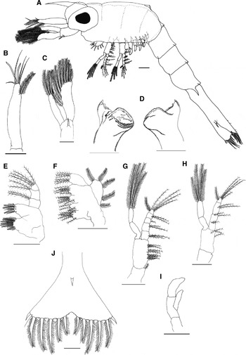
Carapace length: (Figure 1A): 1.06 mm (0.99 to 1.20 mm).
Upogebia vasquezi, zoea I: (A) side view; (B) antennule; (C) antenna; (D) mandibles; (E) maxillule; (F) maxilla; (G) maxilliped 1; (H) maxilliped 2; (I) maxilliped 3; (J) telson, dorsal view; scale bar 0.1 mm.

Total length: (Figure 1A): 2.15 mm (2.10 to 2.19).
Carapace (Figure 1A): longer than broad, long rostral spine with tip curved slightly downward; sessile eyes.
Antennule (Figure 1B): uniramous; peduncle unsegmented, with 2 aesthetascs and 4 simple setae distally and 1 subterminal plumose seta.
Antenna (Figure 1C): biramous; protopod with 1 denticulate spiniform seta; exopod with small distal spine and 9 marginal plumose setae; endopod unsegmented, with 3 terminal plumose setae.
Mandibles (Figure 1D): symmetrical, but different in dentition on each side; incisor process denticulated bearing 1 projection with 2 teeth; molar process semicircular with several small teeth arranged in rows and 4 teeth laterally; palp absent.
Maxillule (Figure 1E): endopod 3-segmented with 4 plumose setae distally, 2 plumose setae medially and 2 plumose setae proximally; basial endite with 3 plumodenticulate setae + 3 simple setae; coxal endite, with 7 plumodenticulate setae.
Maxilla (Figure 1F): scaphognathite with 5 marginal plumose setae; endopod unsegmented, with 3 + 3 distal plumose setae; basial endite, 2-lobed, proximal lobe with 6 plumose setae, distal lobe with 6 plumose setae; coxal endite 2-lobed, proximal lobe with 7 plumose setae, distal lobe with 3 plumose setae.
Maxilliped 1 (Figure 1G): basipodite with 11 plumose setae; exopod 2-segmented with 4 apical plumose setae; endopod 5-segmented with 3 + 2 + 1 + 2 + 5 plumose setae.
Maxilliped 2 (Figure 1H): basipodite with 3 plumose setae; exopod 2-segmented with 4 apical plumose setae; endopod 4-segmented with 2 + 2 + 2 + 5 plumose setae.
Maxilliped 3 (Figure 1I): biramous; exopod and endopod unsegmented and without setae.
Pereiopods 1–3: biramous; underdeveloped and without setae.
Pereiopods 4–5: small underdeveloped buds.
Abdomen (Figure 1A): 5-segmented; 4th segment with 1 pair of posterolateral spines; 5th segment fused to telson.
Telson (Figure 1J): triangular, with central concavity forming 2 symmetrical parts with 7 plumose setae each (7 + 7), 2nd ‘anomuran hair’; anal spine present.
Pleopods: absent.
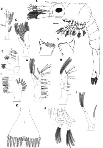
Carapace length (Figure 2A): 0.99 mm (0.98 to 1.00 mm).
Upogebia vasquezi, zoea II: (A) side view; (B) antennule; (C) antenna; (D) mandibles; (E) maxillule; (F) maxilla; (G) maxilliped 1; (H) maxilliped 2; (I) maxilliped 3; (J) pereiopods; (K) telson, dorsal view; scale bar 0.1 mm.

Total length (Figure 2A): 2.33 mm (2.30 to 2.35 mm).
Carapace (Figure 2A): longer than wide; eyes stalked.
Antennule (Figure 2B): biramous; protopod with 2 plumose setae on the inner margin and 2 simple setae on the outer margin; exopod with 3 aesthetascs, 3 medium-sized setae and 1 small seta; endopod with 1 medium-sized plumose seta.
Antenna (Figure 2C): biramous; protopod with denticulate spiniform setae and 1 small distal spine on the outer margin; exopod with 1 small distal spine and 10–11 marginal plumose setae; endopod unsegmented, with 3 terminal plumose setae.
Mandibles (Figure 2D): symmetrical, but different in dentition on each side; incisor process denticulated bearing 1 projection with 3 teeth; molar process semicircular with several small teeth arranged in rows; palp absent.
Maxillule (Figure 2E): endopod 3-segmented, with 2 + 2 plumose setae distally, 2 plumose setae medially and 1 large + 1 small setae, both plumose, proximally; basial endite with 4 plumodenticulate setae + 3 simple setae; coxal endite with 8 plumodenticulate setae.
Maxilla (Figure 2F): scaphognathite with 8 marginal plumose setae; endopod unsegmented, with 2 + 3 distal plumose setae; basial endite, 2-lobed, proximal lobe with 5 plumose setae, distal lobe with 7 plumose setae; coxal endite 2-lobed, proximal lobe with 6 + 2 plumose setae, distal lobe with 4 + 1 plumose setae.
Maxilliped 1 (Figure 2G): basipodite with 2 + 3 + 3 + 3 plumose setae; exopod 2-segmented with six apical plumose setae; endopod 5-segmented with 3 + 2 + 1 + 2 + 1 + 4 plumose setae.
Maxilliped 2 (Figure 2H): basipodite with 3 plumose setae; exopod 2-segmented with five apical plumose setae; endopod 4-segmented with 2 + 2 + 2 + 4 + 1 plumose setae.
Maxilliped 3 (Figure 2I): exopod 2-segmented, with 5 apical plumose setae; endopod small and unsegmented, with 1 apical plumose seta.
Pereiopod 1 (Figure 2J): exopod 2-segmented, with 6 apical plumose setae; endopod unsegmented and without setae.
Pereiopod 2 (Figure 2J): exopod 2-segmented, with 5 apical plumose setae; endopod unsegmented and without setae.
Pereiopod 3 (Figure 2J): exopod and endopod unsegmented and without setae.
Pereiopod 4 (Figure 2J): exopod absent; endopod larger than in previous stage, unsegmented and without setae.
Pereiopod 5 (Figure 2J): exopod absent; endopod larger than in previous stage, unsegmented and without setae.
Abdomen (Figure 2A): 5-segmented; 4th segment with 1 pair of posterolateral spines; 5th segment fused to telson.
Telson (Figure 2K): triangular, posterior margin with 1 central spine followed by 6 plumose setae and 1 cuspidate spine at each of the extremities; ‘anomuran hair’; anal spine present.
Pleopods: apparent as small ventral protuberances on abdominal somites 2 to 5.
DISCUSSION
Although most decapods have long initial stages of larval development, the incidence of abbreviated development is frequent in some groups (Rabalais & Gore, Reference Rabalais, Gore, Schram and Wenner1985). Some species of Upogebia have a short developmental period, such as U. edulis (Shy & Chan, Reference Shy and Chan1996), now considered in a new genus of Upogebiidae, Austinogebia (Ngoc-Ho, Reference Ngoc-Ho2001) and U. quddusiae (Siddiqui & Tirmizi, Reference Siddiqui and Tirmizi1995). However, U. vasquezi apparently does not have a short larval development period, as it needed seven days to reach zoea II during laboratory rearing, whereas U. edulis is reported to have reached the post-larval stage on the third day of laboratory rearing (Shy & Chan, Reference Shy and Chan1996).
Feeding is a critical factor to the successful rearing of decapod larvae (Abrunhosa & Kittaka, Reference Abrunhosa and Kittaka1997). However, a number of groups exhibit lecithotrophic behaviour, such as some species of Lepidophthalmus, including L. siriboia (Abrunhosa et al., Reference Abrunhosa, Smith, Palmeira and Arruda2008b). However, for other thalassinideans, such as Callichirus major (Abrunhosa et al., Reference Abrunhosa, Arruda, Smith and Palmeira2008a) and Upogebia paraffinis (Melo & Brossi-Garcia, Reference Melo and Brossi-Garcia2000), feeding is essential during the stages of larval development. Therefore, a previous assessment is needed to determine the best food for a given species. Factors such as prey size, digestibility and nutritional quality should be considered in this evaluation (Faleiro & Narciso, Reference Faleiro and Narciso2009). In the present study, the U. vasquezi larvae were only fed on Artemia sp. nauplii and reached the second larval stage (zoea II). This may not be the most adequate food source for the species and may have impeded the larvae from completing their development. For example, rotifers are essential to the survival and development of the early larval stages of U. pursilla, but may be replaced with Artemia sp. nauplii once the larvae have reached zoea IV (Faleiro & Narciso, Reference Faleiro and Narciso2009).
Studies that provide a description of the first larval stage of decapods are essential to the identification of these larvae in meroplankton samples. A number of studies of this type are currently available in the literature (Rodrigues, Reference Rodrigues1994; Sankarankutty et al., Reference Sankarankutty, Hong and Kim1999; Lopes et al., Reference Lopes, Abrunhosa and Reis2000; Guerao et al., Reference Guerao, Schubart and Cuesta2001; Hong et al., Reference Hong, Sankarankutty and Cunha2001; Fransozo et al., Reference Fransozo, Mantelatto and Bertini2002; Martins & Calazans, Reference Martins and Calazans2003; Negreiros-Fransozo et al., Reference Negreiros-Fransozo, Hirose, Fransozo and Bolla2009; Mantelatto & Cuesta, Reference Mantelatto and Cuesta2010). However, there is much left to describe in order to understand the complete development of the majority of decapod species. Future studies on U. vasquezi testing different types of food sources and rearing conditions are needed in order to achieve the complete larval development of this species.
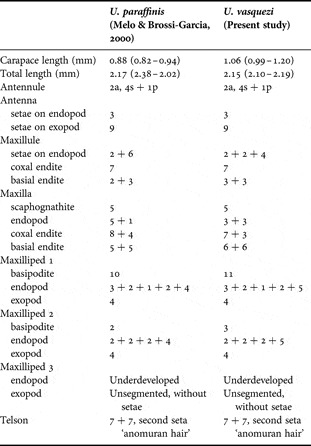
Regarding the main morphological characteristics of the zoea I stage of U. paraffinis and U. vasquezi, the differences were in the number of setae in the basial endite of the maxillule, in the coxal and basial endites of the maxilla and in the basipodite and endopod of maxilliped 1 and maxilliped 2 (Table 1). The setal pattern of the maxillary scaphognathite in zoea I is generally constant among Upogebia species, as described for U. affinis (Sandifer, Reference Sandifer1973), but differences have been noted in this pattern for some species, such as U. major, which have seven setae on the scaphognathite (Konishi, Reference Konishi1989) and U. pusilla, with ten marginal plumose setae on the scaphognathite in zoea I larvae (Santos & Paula, Reference Santos and Paula2003).
Comparison of main morphological characteristics among species of Upogebia occurring on the Brazilian coast in zoea I; a, aesthetasc; s, simple setae; p, plumose setae.

The two species occurring on the Brazilian coast compared in the present study have the same morphological characteristics in relation to the antennules, with two aesthetascs and five setae, with one subterminal setae (Table 1). Other species of Upogebia present different patterns, such as U. affinis, with four to five aesthetascs and one plumose setae (Sandifer, Reference Sandifer1973); and U. major, with four aesthetascs and two plumose setae, with one subterminal setae (Konishi, Reference Konishi1989). It is possible that these differences occurred in descriptions written prior to the proposition of present terminology for the types of antennular setae and are not actual morphological differences among the species in question.
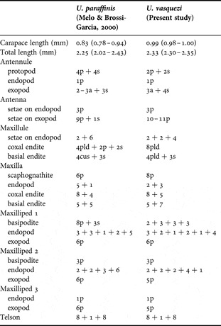
Regarding the zoea II morphology of U. vasquezi and U. paraffinis, there are differences in the number of setae in the antennule, in the basial endite of the maxillule, coxal and basial endites of maxilla, and in the basipodite and endopod of maxilliped 1 and maxilliped 2 (Table 2). About the other characters, they are quite conservative between these species.
Comparison of main morphological characteristics among species of Upogebia occurring on the Brazilian coast in zoea II; a, aesthetasc; s, simple setae; p, plumose setae; pld, plumodenticulate setae; sp, spine; cs, cuspidate setae.

Upogebia larvae do not constitute a homogeneous zoogeographical group with regard to the presence of abdominal spines, as larvae collected from different regions exhibit differences in a given character, with either the presence or absence of a pair of lateral spines on the fifth segment of the abdomen (Ngoc-Ho, Reference Ngoc-Ho1981). The larvae of U. affinis, U. paraffinis and U. vasquezi have these abdominal spines (Sandifer, Reference Sandifer1973; Melo & Brossi-Garcia, Reference Melo and Brossi-Garcia2000), whereas U. major larvae do not (Konishi, Reference Konishi1989).
There is considerable variation in the larval development between thalassinideans (Abrunhosa et al., Reference Abrunhosa, Arruda, Smith and Palmeira2008a). Regarding species of the genus Upogebia, however, the morphology of zoea I is quite conservative, with virtually no differences in the appendages among some species (Ngoc-Ho, Reference Ngoc-Ho1981). In general, the endopod of the maxillule in zoea I of Upogebia is 3-segmented, which is a characteristic shared by Axiidae, Callianassidae and Upogebiidae (Melo & Brossi-Garcia, Reference Melo and Brossi-Garcia2000). Upogebia vasquezi follows this pattern of a 3-segmented maxillular endopod, which is a characteristic also found in U. affinis (Sandifer, Reference Sandifer1973) and U. major (Konishi, Reference Konishi1989), whereas U. paraffinis has a 2-segmented endopod (Melo & Brossi-Garcia, Reference Melo and Brossi-Garcia2000) and U. pusilla has an unsegmented endopod in zoea I larvae (Santos & Paula, Reference Santos and Paula2003).
The morphology of the telson is quite conservative in Upogebia and is therefore not a distinctive characteristic among species of this genus (Melo & Brossi-Garcia, Reference Melo and Brossi-Garcia2000). However, the telson is one of the main characteristics for distinguishing the different zoea stages in species of Upogebia (Melo & Brossi-Garcia, Reference Melo and Brossi-Garcia2000), as for many other decapods. For U. vasquezi in zoea II, the main morphological differences found on the telson in comparison to zoea I are the addition of two plumose setae and one small central spine.
ACKNOWLEDGEMENTS
The authors are grateful to the colleagues who helped in the field collections, to Adelson Silva de Souza for the illustrations of the mandibles and to Richard Boike for the translation into English. All experiments conducted in this study complied with current applicable state and federal laws of Brazil (Proc. DIFAP/IBAMA No 02018.008516/2005 − 51; number 94-2005). This work was supported by the Conselho Nacional de Desenvolvimento Científico e Tecnológico (CNPq) through research project CT-AMAZÔNIA (grant 553106/2005-8) and by the Pró-Reitoria de Pesquisa/Universidade Federal do Pará (PROPESP/UFPA) and Fundação de Amparo e Desenvolvimento da Pesquisa (FADESP). The first author was supported by a postgraduate fellowship from CNPq (grant 132847/2008-6). The authors thank a referee for critical review of the manuscript and constructive comments.