Introduction
Diplozoidae have been reported from Africa on four instances, namely Paradiplozoon aegyptense (Fischtal & Kuntz, 1963) in Egypt, Kenya and Uganda from Labeo forskali, L. coubie, L. victorianus, L. cylindricus, Barbus loati and B. paludinosis (Fischtal & Kuntz, 1963; Yamaguti, Reference Yamaguti1963; Paperna, Reference Paperna1979, Reference Paperna1996; Petr & Paperna, Reference Petr and Paperna1979; Khotenovskii, Reference Khotenovskii1985; Puchachev et al., 2010) and Paradiplozoon ghanense (Thomas, Reference Thomas1957) in Ghana from Alestus macrolepidotus and Alestus baremoze (Thomas, Reference Thomas1957; Yamaguti, Reference Yamaguti1963; Paperna, Reference Paperna1973). An underscribed diplozoid was collected from Alestus in Uganda (Thurston, Reference Thurston1970). The most recent description was made by Mashego (Reference Mashego2000) concerning Neodiplozoon polycotyleus from Barbus marequensis, B. trimaculatus and B. neefi in the Limpopo Province in South Africa (Khotenovskii (1985) refers to this species as Afrodiplozoon polycotyleus). During the course of surveys in the Vaal Dam and Vaal Barrage, in the Vaal River in South Africa diplozoid specimens were collected from the gills of Labeobarbus aeneus (Burchell, 1822) and are described as a new taxon. This species was previously the topic of a paper discussing a fluorescence technique (Milne & Avenant-Oldewage, Reference Milne and Avenant-Oldewage2006).
Materials and methods
Collection and examination of fishes
Bimonthly surveys were carried out in the Vaal Dam and Vaal River Barrage between December 1998 and February 2000. Labeobarbus aeneus, L. kimberleyensis, Cyprinus carpio, Micropterus salmoides, Labeo capensis and Labeo umbratus were collected with the aid of gill nets. The fish were kept in a holding tank supplied with circulating dam water. They were killed by a single cut through the spinal cord and dissected. Gills were removed and parasites collected and fixed in a prostrate position in warmed aceto-formaldehyde alcohol and preserved in 70% ethanol.
Of the 117 adult pairs and nine diporpa larvae collected, five adult pairs were studied using a JEOL JSM 5600 scanning electron microscope (JEOL Ltd, Tokyo, Japan) at acceleration voltages of between 10 and 15 kV. Specimens were prepared either by freeze drying or critical point drying. Twenty adult pairs were stained with borax carmine, Mayer's haematoxylin or Horen's trichrome. Drawings of the parasite were made from whole mounts with the aid of a Zeiss standard 16 light microscope and drawing tube attachment (Carl Zeiss, Jena, Switzerland). Seven adult specimen pairs were dehydrated and embedded in a low-viscosity aliphatic resin. Transverse and sagittal sections were made with glass knives fixed to a rotary microtome at a thickness of 5 and 7 μm, respectively. Sections were stained with AZAN trichrome stain and graphic reconstructions were made according to the method of Pusey (Reference Pusey1939). The haptor of some specimens of the pair was excised and fixed in a mixture of glycerine-ammonium-picrate (Malmberg, Reference Malmberg1957). A light microscope and drawing attachment were used for measurements of the attachment clamps and central hooks and for drawing the hard parts of the haptor.
Molecular analysis
DNA was extracted from the anterior section of three parasites using the DNeasy® tissue kit (QIAGEN Inc., Crawley, West Sussex, United Kingdom) following standard protocols suggested by the manufacturer. The anterior section of the parasites was chosen to allow preservation of the posterior section which is important for morphological assessments. The internal transcribed spacer of the ribosomal gene (ITS2) was targeted as this fragment has been used previously in DNA assessments of Diplozoidae and readily distinguishes between taxa (Matejusová et al., Reference Matejusová, Koubkova, D'Amelio and Cunningham2001; Gao et al., Reference Gao, Chen, Yao, Gao, Song and Wang2007). Amplification was done using the universal primers D and B1 (Bachellerie & Qu, Reference Bachellerie, Qu and Hyde1993). All the polymerase chain reactions (PCRs) were performed using 10 ng of DNA under the following cycling parameters: 95°C for 10 min, (95°C for 30 s, 55°C for 30 s and 72°C for 75 s) for 30 cycles, with a final extension step at 72°C for 5 min. Amplification was verified on a 1% ethidium bromide stained agarose gel. Sequencing reactions were performed with quarter-reactions (2 μl) of BigDye® Terminator 3.1 mix (Applied Biosystems, Warrington, Cheshire, UK). Purified sequencing products were run on an ABI 3137 automated sequencer (Applied Biosystems). The sequences generated in this study were deposited in the European Molecular Biology Laboratory (EMBL) (accession number HF566124).
DNA sequences generated from the current study were aligned and edited in GENEIOUS Pro™ 5.0 software (Biomatters Ltd, New Zealand). To determine the distinctness of the South African species, sequences were aligned to previously published sequences of various Diplozoidae. Specifically, the following sequences were retrieved from GenBank for comparisons: Paradiplozoon homoion (accession numbers AF36,9760, AJ30,0715), P. sapae (accession number AJ30,0713), P. bliccae (accession numbers AF36,9761, AJ30,0712), P. pavlovskii (accession number AJ30,0714), P. megan (accession number AJ30,0711), Sindiplozoon ctenopharyngodoni (accession numbers DQ098,898), Paradiplozoon hemiculteri (accession numbers DQ098,884, DQ098,887), P. opsariichthydis (accession numbers DQ098,890, DQ098,891), P. parabramisi (accession numbers DQ098,883, DQ098,889), P. parapeleci (accession number DQ098,882), P. diplophyllorchidis (accession number DQ098,891) and P. jiangxiensis (accession numbers DQ098,885). Eudiplozoon nipponicum (accession numbers AJ30,0710, AF36,9758) was included to root resultant phylogenies. Genealogical relationships were constructed using both parsimony (equally weighted characters) and neighbour joining (using uncorrected p-distances) methods as implemented in PAUP 4* (Swofford, 2002). The robustness of all the resultant topologies was assessed by 1000 bootstrap replicates.
Results
Paradiplozoon ichthyoxanthon Avenant-Oldewage, 2013
Type host. Labeobarbus aeneus (Burchell, 1822).
Type locality. Vaal Dam, Gauteng, South Africa (26°52.249′S, 28°10.249′E). Other locality: Vaal River.
Site. Gills.
Prevalence. 44.7% at Vaal Dam; 26.6% in Vaal Barrage.
Material studied. RAU 11 A–H.
Type material. Type material in Iziko South African Museum (Cape Town); Holotype SAMC A61795; Paratypes SAMC A61796 -SAMC A61801; additional specimens RAU 11A-11D stored in the collection of the University of Johannesburg under the auspices of the Albany Museum, Grahamstown.
Etymology. The species name refers to yellowfish; the host of this parasite.
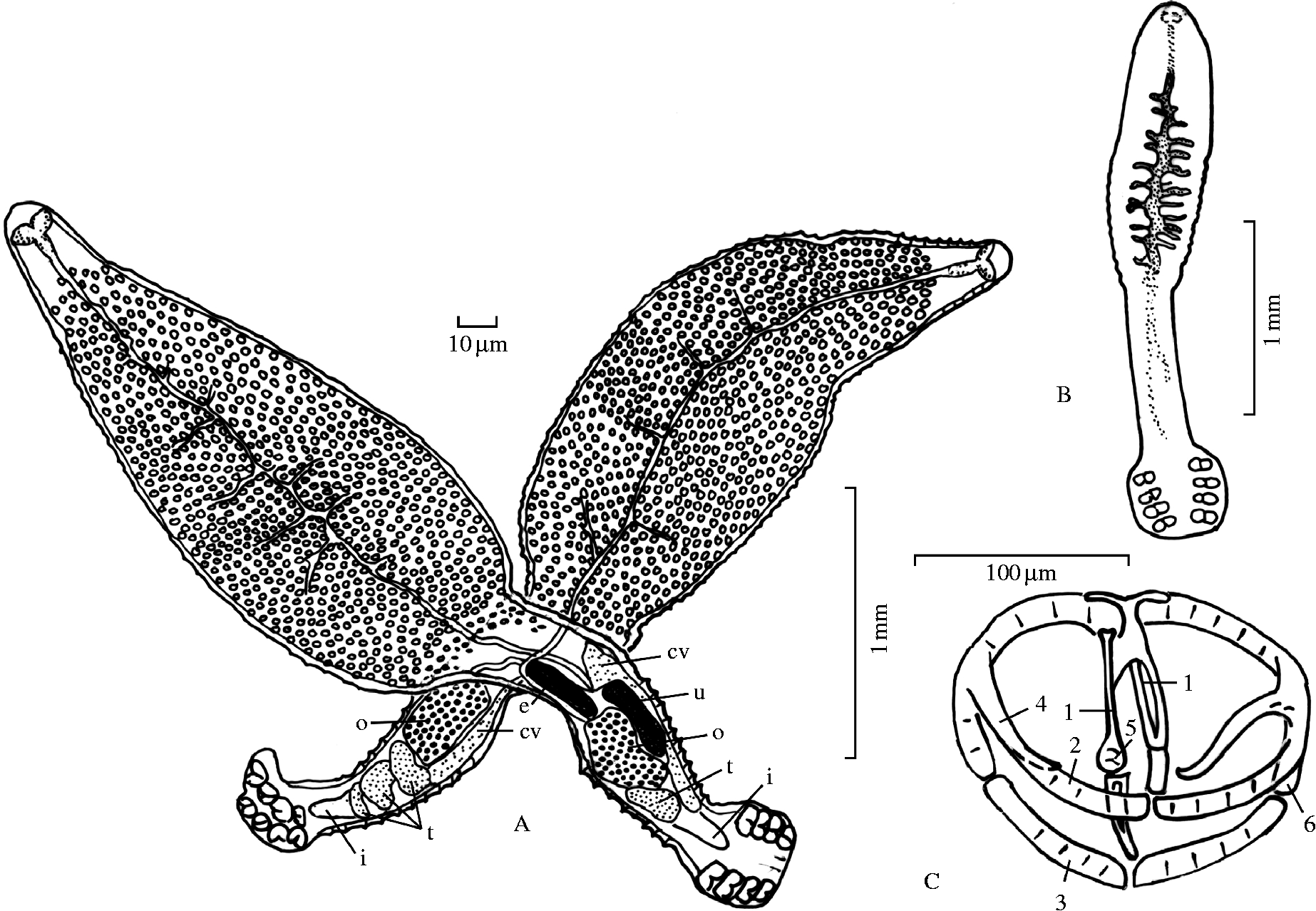
Description. Illustrated by figs 1–3. Two adult specimens fused together in permanent copula resemble the letter ‘X’ (fig. 1A). Mean total body length 3.88 mm (n= 219, 1.30–6.00 mm). Body transparent.
Paradiplozoon ichthyoxanthon n. sp. (A) Adult pair, ventral view, (B) diporpa larva and (C) clamp. e, egg; cv, common vitelline duct; i, end of intestine; o, ovary; t, testis; u, uterus; 1, median plate; 2, anterior jaw; 3, median sclerite of posterior jaw; 4, spiky spur; 5, joining sclerite of posterior jaw; 6, lateral sclerite of posterior jaw.

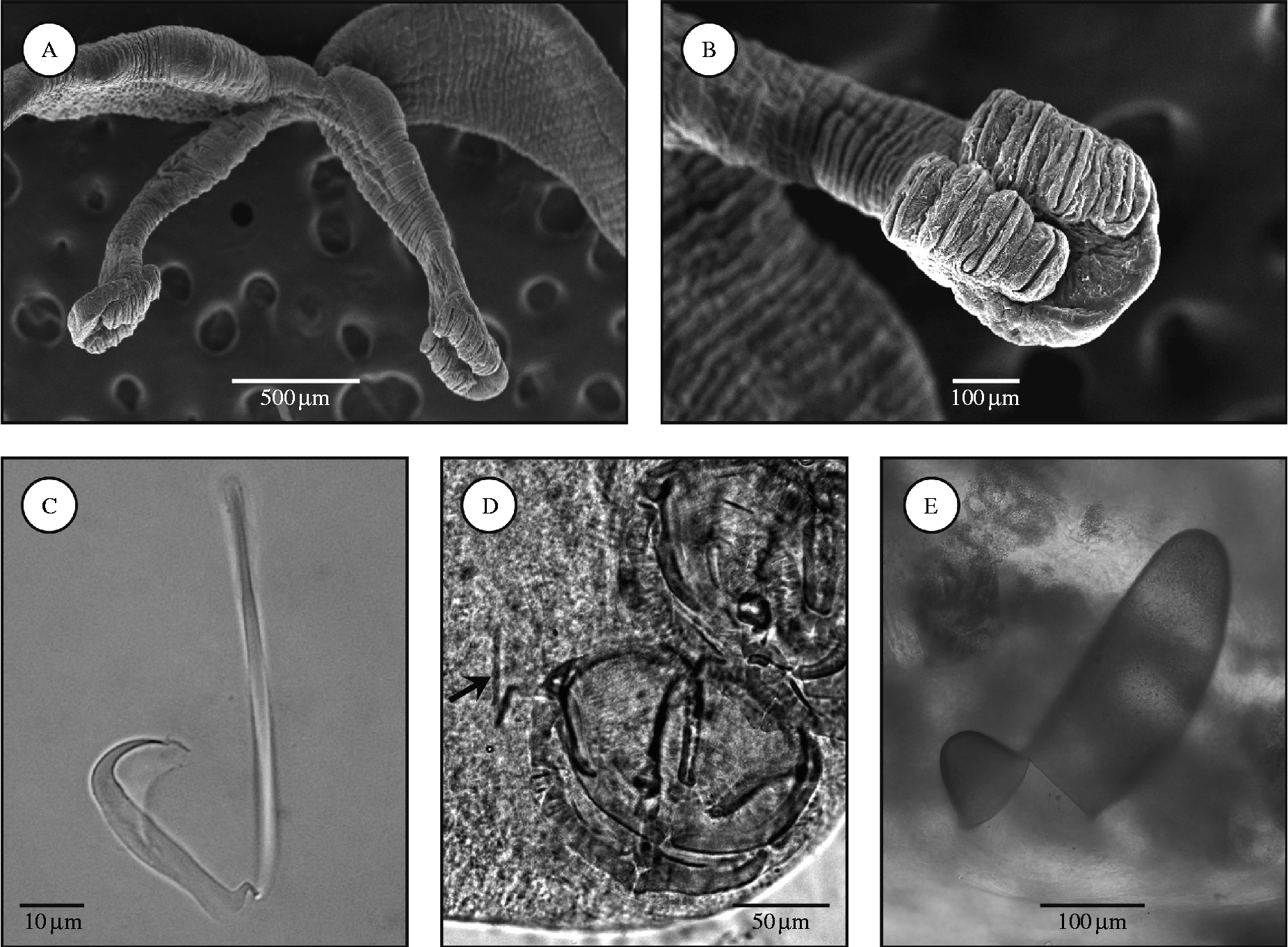
Paradiplozoon ichthyoxanthon n. sp. Scanning electron micrographs of (A) adult pairs and (B) the haptor. Light micrographs of (C) the central hook, to show the handle and sickle; (D) clamps (arrow indicates central hook); and (E) egg with operculum.
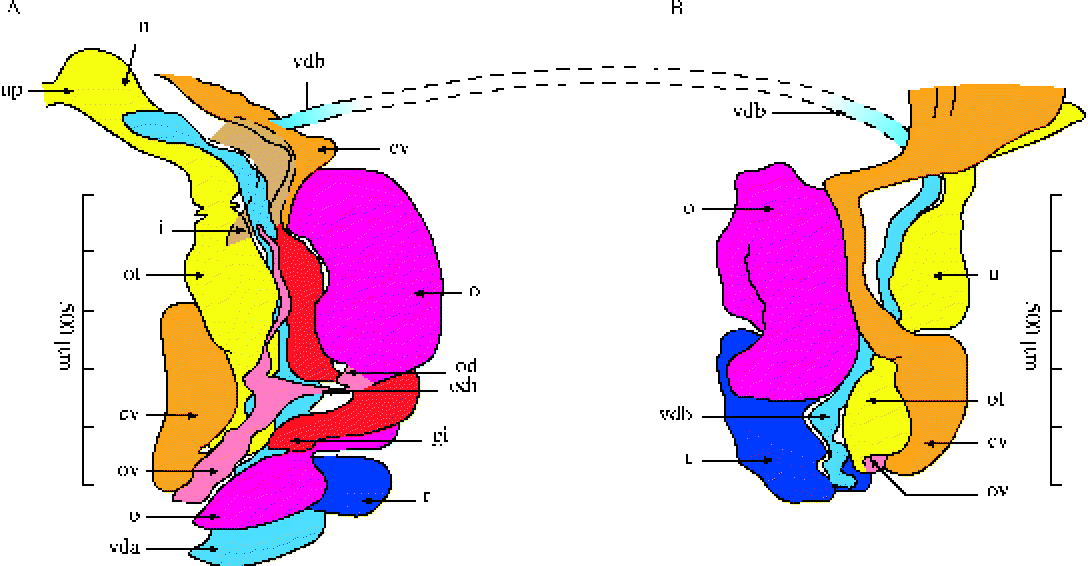
(colour online) Paradiplozoon ichthyoxanthon n. sp. Graphic reconstruction of the reproductive system. cv, Common vitelline duct; gi, genito-intestinal canal; i, intestine; o, ovary; od, oviduct; odi, oviducto-intestinal canal; ot, ootype; ov, ovovitelline duct; u, uterus; up, uterine pore; t, testis; vda, the vas deferens of individual A; vdb, the vas deferens of individual B.
Tegument relatively smooth anteriorly, but with small inconspicuous annular transverse folds or plicae. Posteriorly, in region anterior to haptor these plicae become more conspicuous, forming transverse striations (fig. 2A and B). Mean length of anterior region of adult specimen 2.35 mm (n= 229, 0.65–3.8 mm). Relatively large, crescent-shaped mouth sub-terminal on antero-ventral surface. Mouth opens into buccal cavity containing two large, muscular buccal suckers, joined proximally. The suckers are 0.08 mm long (n= 192, 0.01–0.12 mm) and 0.08 mm wide (n= 192, 0.05–0.11 mm). Cavity leads to oval, muscular, glandular pharynx that opens into highly branched intestine (fig. 1A). Throughout this region, from just posterior to the pharynx to start of the fusion region, the vitellarium occurs, displacing parenchyma between branches of intestine (fig. 1A). Vitelline cells migrate posteriorly in vitelline ducts, and drain into the common vitelline duct. In the posterior part of the fusion area, the common vitelline duct widens to form the vitelline reservoir which contains mature vitellaria and spermatozoa from another individual. Intestine and vitellarium occur throughout the fusion region. Reproductive organs are situated in this region: the most anterior part of the ovary, uterine pore, proximal part of the common vitelline duct and vasa deferentia of both individuals (fig. 1A).
Mean length of posterior region in adult specimen 1.15 mm (n= 222, 0.9–1.85 mm). Haptor disk-like, 0.44 mm long (n= 138, 0.4–0.43 mm) and 0.46 mm wide (n= 138, 0.45–0.46 mm). Ventrally it bears four pairs of clamps arranged laterally in parallel rows (2A). Each clamp contains a typical framework. In P. ichthyoxanthon it consists of a U-shaped median plate, connected to the lateral sclerites of the posterior jaw. The median sclerite of the posterior jaw extends the full width of the clamp. The joining sclerite of the posterior jaw is round. A well-developed muscular system is attached to the clamps. Mean measurements of 110 clamps: clamp 1 is 0.07 mm long (0.05–0.11 mm) by 0.11 mm wide (0.06–0.19 mm); clamp 2 is 0.07 mm long (0.04–0.10 mm) by 0.13 mm wide (0.06–0.2 mm); clamp 3 is 0.0.07 mm long (0.05–0.15 mm) by 0.13 mm wide (0.05–0.18 mm); clamp 4 is 0.07 mm long (0.06–0.15 mm) by 0.12 mm wide (0.07–0.12 mm). Note: all clamps are wider than long, with the first pair, situated posteriorly on the haptor, the smallest. Central hooks are present, internally, in the median region between the most posterior pair of clamps. Central hooks consist of a handle (53 μm) and a half-moon-shaped sickle (25 μm) with a small barb and a wing (fig. 2C and D).
Male reproductive organs (figs 1A and 3) consist of a single, lobed testis sub-terminally in the posterior lobe of the body, adjacent to anterior part of the haptor. The lumen carries mature spermatozoa to the common vas deferens. Centrally, the distal lobe extends and connects to the single vas deferens, which is convoluted mid-ventrally, parallel to the common vitelline duct. It runs parallel to the digestive tract towards the fusion area where it bends 90° before transversing towards other individual. It carries mature aflagellate sperm and facilitates cross-fertilization by entering laterally into the other individual's common vitellarium duct. The vas deferens wall consists of simple cuboidal epithelial cells with central nuclei and a relatively thick basal membrane.
The female reproductive organs (figs 1A and 3) consist of a single large ovary folded over to form a U-shaped structure, with a dorsal and a ventral component. Posteriorly the ovary overlaps part of the anterior testis lobe distally. The remainder of the ovary is situated anterior to the testis. A single oviduct arises posteriorly from the ventral part of the ovary. It bends dorso-laterally and receives the genito-intestinal canal. It widens, continues postero-laterally towards the dorsally situated common vitellarium duct. These ducts fuse postero-laterally, forming the ovovitellarium duct. The vas deferens of the other individual drains into the common vitellarium duct, where fertilization probably takes place in the ovovitellarium duct via the ootype. Zygote and vitelline cells will thereafter progress in the ovovitellarium duct to the uterus. The ovovitelline duct curves 90° dorsally, then 90° laterally and continues dorsally towards the anterior region, to Mehlis' gland, thereafter to the ootype situated dorsally, median between the common vitelline duct and vas deferens. The two individuals' uteri continue in opposite directions, so that the uterine pores finally break through the tegument laterally on opposite sides of worm pair. Oval eggs (320 × 130 μm), with a thick, smooth, yellow-brown operculated shell present without filaments or protrusions (figs 1A and 2E).
Diporpa larva: elongated body with ventral haptor; mean total length 1.78 mm (min. 1.10 mm; max. 2.63 mm; n= 9). Four pairs of clamps on haptor, arranged in two rows. First clamp is smaller than the other three. Mouth opens into buccal cavity containing two large, muscular suckers, adjoined proximally. Digestive system ramified in anterior part of body (fig. 1B). Central hooks similar shape to that in adult.
Molecular analysis
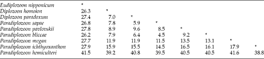
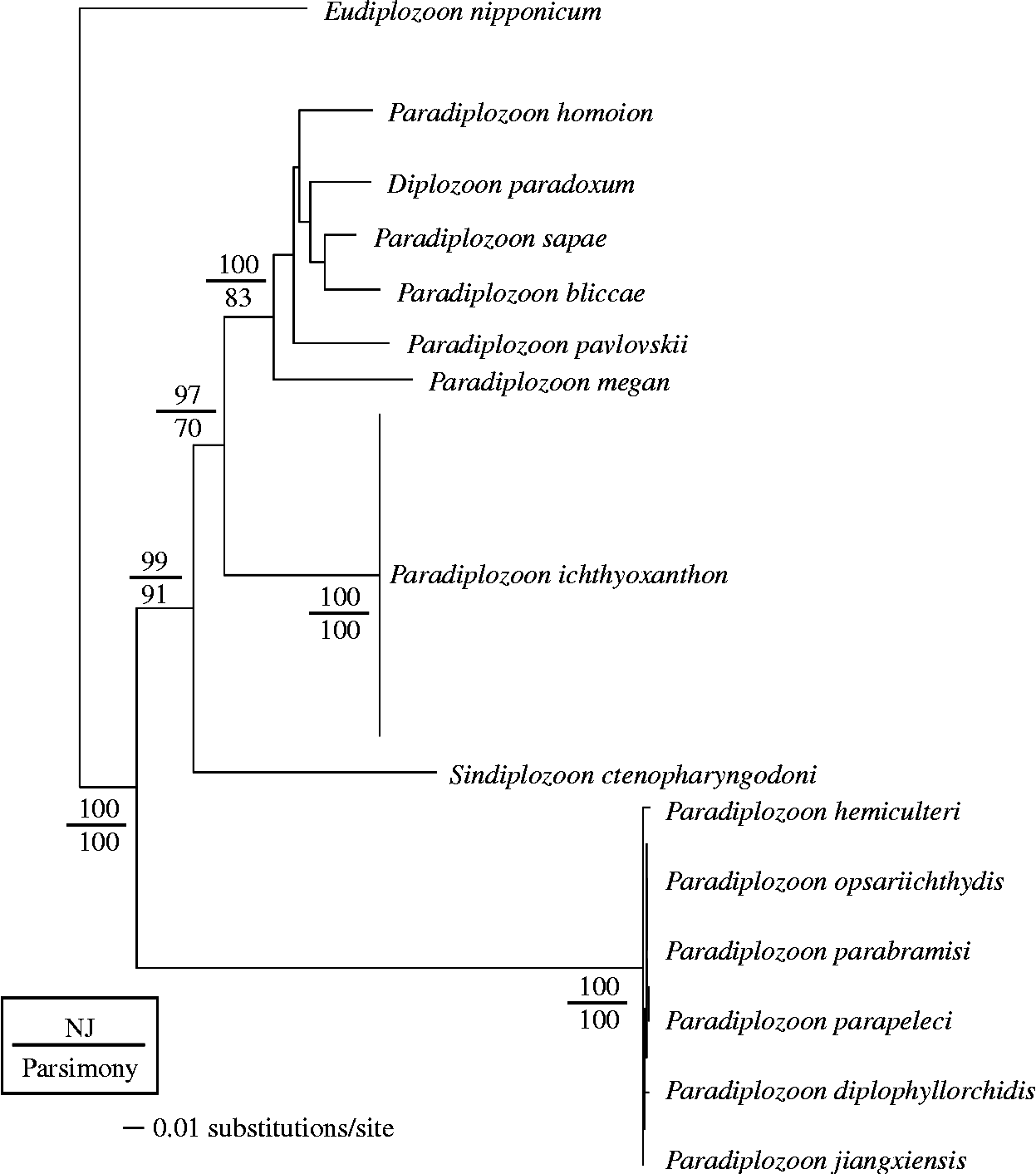
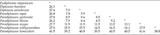
The three South African parasite specimens included here had identical sequences across the 732 bp of the ITS2 region. This is not unexpected as sequence divergences within species are generally low (less than 0.5% uncorrected p-distances). Uncorrected sequence divergences separating distinct taxa are given in Table 1. The South African specimens are separated from other taxa included here by 14.5–38.8% uncorrected p-distance. Similar topologies were retrieved irrespective the method of analyses (parsimony or distance). For ease of representation, the neighbour-joining tree is shown in fig. 4. The South African individuals formed a distinct clade, receiving 100% statistical support.
Average uncorrected (p-distance) sequence divergence separating the different diplozoid taxa. Values are given as a percentage, * indicates no distance.
Distance-based phylogenetic tree indicating the evolutionary relationships among different diplozoid taxa. The tree is rooted with Eudiplozoon nipponicum. Bootstrap support for nodes is indicated on the tree.
Species diagnostics
The Paradiplozoon species collected during the present study differs from other Paradiplozoon species on the basis of the combined morphological characteristics, namely: disk-like haptor, eggs without filaments, the internal structure of the clamps and shape of the central hook. Furthermore, the DNA sequence across the 732 bp of the ITS2 region is unique and distinct.
Discussion
Paradiplozoon ichthyoxanthon is compared to diplozoids previously found in Africa; P. ghanense, P. aegyptense and Neodiplozoon polycotyleus. Unfortunately neither the sclerite morphology nor the DNA sequences of these species has been published.
Paradiplozoon ghanense is characterized by the fact that the surface of the posterior part of the body is not plicated (Khotenovskii, Reference Khotenovskii1985), while that of P. ichthyoxanthon is. The posterior parts of the body of both parasites are shorter than the anterior part. The mean diameter of the third clamp of P. ghanense is from 3 to 8 times less than the length of the posterior part of the body (Khotenovskii, Reference Khotenovskii1985), while that of P. ichthyoxanthon is 8.8 times less. The median hooks of P. ghanense are more than 25 μm in length (Khotenovskii, Reference Khotenovskii1985), while those of P. ichthyoxanthon are approximately 54 μm. The width of the attachment clamps of P. ghanense is between 120 and 160 μm (Khotenovskii, Reference Khotenovskii1985), while that of P. ichthyoxanthon is approximately 130 μm. The eggs of P. ghanense have filaments (Thomas, Reference Thomas1957), while those of P. ichthyoxanthon lack filaments. Paradiplozoon ghanense has previously been found in African countries, but only as far south as Ghana on Alestus macrolepidotus, while P. ichthyoxanthon is found on Labeobarbus aeneus in the Vaal Dam and Vaal Barrage, South Africa. The Vaal River forms part of the Orange River system and is not linked to any of the rivers where Paradiplozoon have previously been found in Africa, and is separated by a number of large river systems such as the Congo and Zambezi.
In P. aegyptense the mean diameter of the third clamp is from 10 to 20 times less than the length of the posterior part of the body (Khotenovskii, Reference Khotenovskii1985), while that of P. ichthyoxanthon is 8.8 times less. The width of the clamps of P. aegyptense is from 80 to 117 μm (Khotenovskii, Reference Khotenovskii1985), while the width of the clamps of P. ichthyoxanthon is on average 130 μm (minimum 50 and maximum 200 μm). The eggs of P. aegyptense have filaments, while those of P. ichthyoxanthon do not; the lengths of the buccal suckers of P. aegyptense are from 95 to 125 μm (Khotenovskii, Reference Khotenovskii1985), while those of P. ichthyoxanthon are on average 80 μm long (minimum 10 and maximum 120 μm); and P. aegyptense has an average total body length of 4.529 mm (Fischthal & Kuntz, Reference Fischthal and Kuntz1963), while that of P. ichthyoxanthon is smaller, at an average of 3.88 mm.
According to Khotenovskii (Reference Khotenovskii1985) Afrodiplozoon polycotyleus (now Neodiplozoon polycotyleus) falls under the subfamily Neodiplozoinae. Although in a different subfamily than P. ichthyoxanthon, it is mentioned here because it occurs in South Africa on various Barbus species. Neodiplozoon polycotyleus has 8–10 pairs of clamps per individual (Paperna, Reference Paperna1973), while P. ichthyoxanthon has only four pairs of clamps per individual. The eggs of N. polycotyleus have filaments, while those of P. ichthyoxanthon do not.
Various morphological differences between P. ichthyoxanthon and other members of the family are present. After comparisons with P. ichthyoxanthon, it was concluded that this is in fact a new species, in a new locality, on a new host fish species. The characteristics specific to this species are a disk-like haptor, intestine without lateral branches, eggs without filaments, the internal structure of the clamps, as well as the morphological measurements and a unique and distinct DNA profile for the ITS2 gene region.
Acknowledgements
The authors wish to thank the National Research Foundation for funding to A.A.-O., Professor Hank Swanepoel (UJ) for translation from Russian. Professor Bozena Koubkova (Masaryk University, Brno) is thanked for valuable comments to a previous draft of this manuscript and Professor Bill Henderson (UJ) for the species name.