Introduction
Left posterior fascicular ventricular tachycardia is a form of idiopathic ventricular tachycardia, and it is characterised by a right bundle branch block morphology and left axis deviation. Though rare, left posterior fascicular tachycardia is typically seen in children with normal cardiac anatomies.Reference Wang, Zhu, Hamilton, Kirsh, Stephenson and Gross1, Reference Pfammatter and Paul2 It may be inadvertently diagnosed as supraventricular tachycardia because fascicular ventricular tachycardia has a narrower QRS when compared to ventricular tachycardia, and a less prominent QRS axis shift may not be detected via electrocardiography. Left posterior fascicular ventricular tachycardia can be treated with ablation, which is performed in an area localised in the left ventricular apical septum.Reference Chu, Sun and Zhao3, Reference Kataria, Yaduvanshi, Kumar and Nair4 The poor response to medical therapy, haemodynamic instability seen during tachycardia in some cases, and good response to ablation therapy have increased the importance of this technique.Reference Fukuhara, Sumitomo and Nakamura5, Reference Collins, Schaffer and Liberman6 However, left posterior fascicular ventricular tachycardia catheter ablation remains challenging in the pediatric population, and most studies about this topic have been conducted on adult patients. Moreover, there is not any study in the literature that focuses on reducing the use of fluoroscopy by using three-dimensional electroanatomical mapping systems in pediatric left posterior fascicular ventricular tachycardia ablations. In this study, we aimed to assess the clinical presentation features and outcomes of catheter ablations performed using limited fluoroscopy with three-dimensional electroanatomic mapping system guidance in a children group.
Materials and methods
Study population
This single-centre retrospective study included children under 18 years old who underwent three-dimensional electroanatomic mapping system-guided left posterior fascicular ventricular tachycardia ablation between July, 2012 and March, 2018. The patients’ clinical features such as history, prior medications, and treatments, physical examinations, electrocardiographies, echocardiographies, Holter monitoring, ablation procedures, outcomes, and complications were evaluated. All of the patients had at least one documented sustained left posterior fascicular ventricular tachycardia (right bundle branch block morphology and left axis morphology) attack.
Electrophysiological study, mapping, and ablation
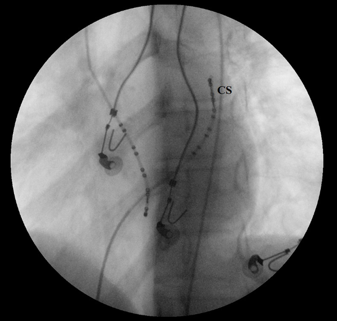
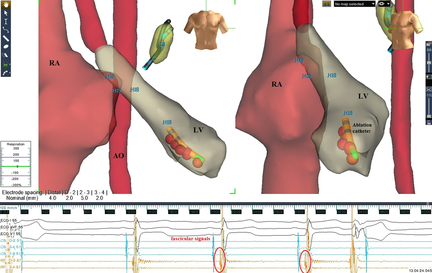
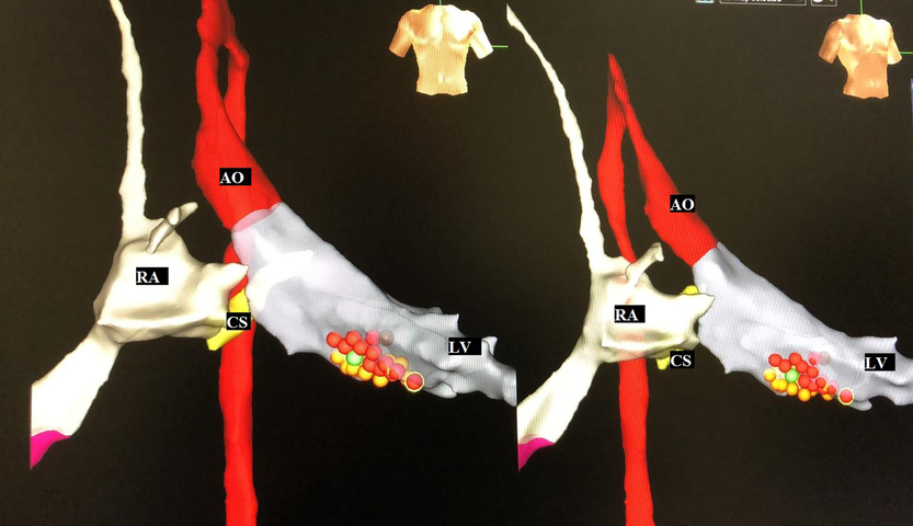
Before the patients underwent the procedure, their antiarrhythmic medications were withdrawn for a period of at least 5 half-lives. Written informed consent was obtained from the patient’s parents; then, an electrophysiological study was performed under general anesthesia. After inserting one sheath into the right femoral vein and the other into the left femoral vein, the endocardial right atrium and coronary sinus geometries were created without using fluoroscopy via three-dimensional electroanatomic mapping system guidance (EnSite NavX system; Abbott/St. Jude Medical Inc., St. Paul, Minnesota, United States of America). The superior caval vein, His potential, and inferior caval vein were marked on the right atrium map. After the basic measurements, atrium-His interval, His-ventricular interval, and QRS duration were evaluated; an induction of tachycardia was attempted via extrastimulation and burst pacing from the right atrium and right ventricle, with or without intravenous metaproterenol sulphate and/or dobutamine provocation. An additional sheath was inserted into the right femoral artery. Then, radiofrequency ablation and diagnostic catheters were introduced in a retrograde fashion into the left ventricle for mapping and ablation via fluoroscopy. However, fluoroscopy was not used in any of the cases, with the exception of this stage. At this stage, the patient was administered 70 units/kg (a maximum of 5000 units) of intravenous heparin. After creating the left ventricular geometry, the His and Purkinje potentials (fascicular signals) were marked on the left ventricular map and the mapping was performed during the sinus rhythm (Fig 1). The left ventricular mapping and His and Purkinje potential marking were performed with a 5 French 10-pole diagnostic catheter (Figs 2 and 3). Pace mapping was also used in some cases.
Marking anatomical and electrophysiological landmarks on created anatomy. Red dots show fascicular signals. AO = aorta; CS = coronary sinus; HIS = His potential; IVC=inferior caval vein; LMCA = left main coronary artery; LPF = left posterior fascicle; RA = Right atrium; SVC = superior caval vein.

Left anterior oblique view of a patient while inserting the 10-pole diagnostic catheter into the left ventricle. Mapping catheter had turned to the apical septum. CS = coronary sinus.

Markings are His and fascicular signals on the left ventricular map. Radiofrequency energy delivered on red points that indicate fascicular signals at the next stage. AO = aorta; CS = coronary sinus; HIS = His potential; LV = left ventricle; RA = right atrium.

For the ablation, a non-irrigated 7 French/5 French Marine multi-curve catheter (Medtronic Inc., Minneapolis, Minnesota, United States of America) was used. The radiofrequency generator (Atakr II; Medtronic Inc.) was set to deliver radiofrequency energy up to 50 W, and the temperature was not permitted to exceed 60°C. The energy was titrated to achieve an impedance drop of 10–15 Ω. The radiofrequency energy was delivered during the sinus rhythm, with or without premature ventricular contraction, at the most apical site at which a Purkinje potential was recorded. The ablation was applied where the Purkinje potentials were recorded up to the junction of one-third of the apical and mid-left ventricular septum. The ablation was terminated in the cases of junctional acceleration and a prolonged atrium-ventricle interval, and it was continued for 60 seconds in the absence of these conditions. Lesions were continued for 60 seconds, and we tried to apply radiofrequency ablation to all potentials remained at the site of ablation. Initially, we placed the lesions in a linear fashion along the fascicular signals. However, additional perpendicular lesions were applied to patients with recurrent inducible tachycardia and additional Purkinje potentials (Fig 4).
Additional perpendicular lesions (yellow dots) applied to patients with recurrent inducible tachycardia and additional Purkinje potentials. AO = aorta; CS = coronary sinus; LV = left ventricle; RA = right atrium.

The programmed/incremental stimulation was carried out from the atrium and ventricle, with and without metaproterenol and/or dobutamine, 30 minutes after the final ablation. Acute procedure success was defined as no further inducible or spontaneous left posterior fascicular ventricular tachycardia for 30 minutes after the final ablation lesion. After a waiting period, an additional ablation was performed in the sections where the Purkinje potentials were recorded in the mid-left ventricular septum in the patients with induced left posterior fascicular ventricular tachycardia.
Following the procedure, all of the patients were followed up for one night under continuous cardiac monitoring. All patients underwent an echocardiography, 12-lead electrocardiography, and 24-hour Holter monitoring before they were discharged. All patients were given 3–5 mg/kg of oral acetylsalicylic acid for 1 month, and the outpatient follow-ups were scheduled for 1 month, 6 months, and yearly.
Statistical analysis
The data were analysed using the Statistical Package for the Social Sciences version 17.0 (SPSS Inc., Chicago, Illinois, United States of America). They were presented as the count (%), mean ± standard deviation, and median ± interquartile range as appropriate. The distribution of the variables was analysed using the Kolmogorov–Smirnov test.
Results
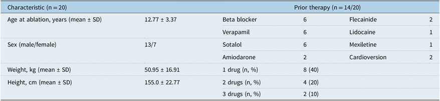
The demographic and clinical characteristics of the study population are presented in Table 1. The mean age of the patients (13 males and 7 females) was 12.7 years old (range 2–16 years). Most of the patients (n=19/20) had suffered from palpitations. The mean weight and height of the patients were 50.95 kg (range 11–84 kg) and 155 cm (range 77–184 cm), respectively. Only one patient had dilated cardiomyopathy (DCM). The echocardiographic examinations were normal in the other patients. Sustained left posterior fascicular ventricular tachycardia was documented at least once in all of the patients. Before the ablation, six of the patients were not receiving any antiarrhythmic medications. Of the remaining 14 patients, 8 (57%) were receiving monotherapy, 4 (29%) were receiving combination therapy with two drugs, and 2 (14%) were receiving combination therapy with three drugs. Two of the patients receiving antiarrhythmic therapy had undergone cardioversion due to tachycardia attacks leading to haemodynamic instability. One 2-year-old patient who weighed 11 kg had undergone more than one cardioversion before the ablation, and this patient had been given amiodarone, flecainide, lidocaine, propranolol, verapamil, and mexiletine as medical antiarrhythmic therapy.
Demographic and clinical characteristics of the study population.

SD = standard deviation
Values are given in counts (%) or mean ± SD as appropriate
Electrophysiological study, ablation, and fluoroscopy exposure
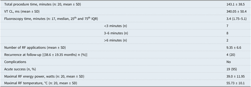
In all of the patients, the pre-procedural electrocardiographies demonstrated the sinus rhythm with normal axes and QRS durations. In addition, the atrium-His (67.0 ± 19.3 ms) and His-ventricular (49.1 ± 15.7 ms) intervals were within normal limits before the procedure. The patients’ procedural data are presented in Table 2, and the mean procedural time was 143.1 minutes (range 90–210 minutes). Fluoroscopy was not used in 3 patients, and the median fluoroscopy time was 3.4 minutes in the remaining 17 patients. There were only two patients in which the fluoroscopy was used for longer than 6 minutes. All patients had at least one documented left posterior fascicular ventricular tachycardia attack. Clinical tachycardia (left posterior fascicular ventricular tachycardia) was induced in the electrophysiological studies of 17 of the patients, and the mean tachycardia cycle length was 340 ms. Frequent ventricular extrasystoles and also couplets matched with clinical tachycardia were seen in the electrophysiological studies of remaining three patients. In these three patients, non-sustained left posterior fascicular ventricular tachycardia attacks (fascicular accelerations) were seen at the beginning of ablation, by the effect of radiofrequency energy.
Procedural data of the patients.

IQR = interquartile range; RF = radiofrequency, SD = standard deviation; VT CL = ventricular tachycardia cycle length
Values are given in counts (%), median with interquartile range, or mean ± SD as appropriate
A mean of 9.35 radiofrequency lesions were applied, and all patients underwent Purkinje potential-guided ablations. Pace mapping was used to determine the exit site of left posterior fascicular ventricular tachycardia in addition to the Purkinje potential-guided ablation in nine patients. The mean maximal radiofrequency energy power was 39 W, and the mean maximal radiofrequency temperature was 55.7°C. The procedure was unsuccessful in only one patient. A 5 French non-irrigated catheter was used in the 2-year-old patient, a 7 French non-irrigated catheter and an additional irrigated catheter were used in 2 patients, and 7 French non-irrigated catheters were used in the remaining 17 patients. None of the patients developed catheter ablation-related complications such as conduction abnormalities (left posterior hemi-block, etc.), vascular complications, or aortic valve abnormalities.
Follow-up
During a mean follow-up of 38.6 ± 19.35 months, sustained left posterior fascicular ventricular tachycardia attack recurred in four patients (20%). The earliest recurrence was within 1 month, and the latest was within 9.6 months (mean ± SD 4.6 ± 3.67). Repeat ablations were performed successfully in those patients who developed recurrences. Flecainide therapy was initiated in the patient with the failed ablation, who is currently being followed up without problems. The remaining 19 patients, with the exception of one patient with a failed procedure, are being followed up without antiarrhythmic therapy. The cardiac function of the patient who developed DCM normalised after successful ablation during the follow-up.
Discussion
The effectiveness and reliability of catheter ablation performed with the guidance of the three-dimensional electroanatomic mapping system and the decreased radiation exposure with this method have been shown in supraventricular arrhythmias and ventricular outflow tract tachycardia in children.Reference Tuzcu7–Reference Clark, Sumihara, McCarter, Berul and Moak10 However, there are insufficient studies regarding this method in children with left posterior fascicular ventricular tachycardia. This study demonstrated the effectiveness and reliability of three-dimensional electroanatomic mapping system-guided catheter ablation and the significant reduction in fluoroscopy usage with electroanatomic mapping systems in children with left posterior fascicular ventricular tachycardia. In our study, three-dimensional electroanatomic mapping was used in all the patients, no fluoroscopy was used in 3 of the patients, and the median fluoroscopy time was 3.4 minutes in the remaining 17 patients. We advance the ablation catheter to descending aorta under three-dimensional electroanatomic mapping system guidance without fluoroscopy. We try to put the tip of the catheter into J shape while the catheter is inside the descending aorta, under three-dimensional electroanatomic mapping system guidance without fluoroscopy. If there is a restriction to make the catheter as J shape at this stage, we use fluoroscopy in addition to the three-dimensional electroanatomic mapping system. We then cross the aortic valve with the J shaped catheter.
However, especially in older children, if the ablation catheter can be changed to J shape without any resistance while it is in the descending aorta, we push the catheter slowly and cross the aortic valve without fluoroscopy under three-dimensional electroanatomic system guidance.
A shift may occur in the created geometry because of the patient movement during the procedure. This may produce important errors on the three-dimensional map. Therefore, we performed the electrophysiology study and ablation under deep sedation. Fluoroscopy should be available to determine the catheter location if there is suspected localisation of the catheter within the cardiac chambers. In a similar population study with 14% three-dimensional electroanatomic mapping system usage, the median fluoroscopy time was 24 min (range 1–79 min).Reference Collins, Schaffer and Liberman6 In another study in the adult population with 71% three-dimensional electroanatomic mapping system usage, the mean fluoroscopy time was approximately 15 min.Reference Liu, Fang and Yang11 Fluoroscopy times can be further reduced by using intracardiac echocardiography in addition to the three-dimensional electroanatomic mapping system. Although there are no studies regarding this issue in pediatric left posterior fascicular ventricular tachycardia patients, successful ablations have been performed using the three-dimensional electroanatomic mapping system and intracardiac echocardiography without fluoroscopy in adult patients with left ventricular tachycardia.Reference Lamberti, Di Clemente and Remoli12
The acute success rate was 95% in our study in which left posterior fascicular ventricular tachycardia ablation was performed during the sinus rhythm based on the Purkinje potentials, and the mean follow-up duration was 3.3 years. In a multi-center study with a similar population with 14% three-dimensional electroanatomic mapping system usage, the acute success rate was 71%.Reference Collins, Schaffer and Liberman6 In our study, 4 of the 20 children developed recurrences during the follow-up after the left posterior fascicular ventricular tachycardia ablation. In one multi-centre study evaluating left posterior fascicular ventricular tachycardia ablation in children, recurrences were seen in 20%.Reference Collins, Schaffer and Liberman6 In another study of left posterior fascicular ventricular tachycardia ablations performed in 120 adult patients, the recurrence rate was found to be 15% at a median follow-up of 4.5 years.Reference Liu, Fang and Yang11 Although the recurrence rate was similar to the literature, it was rather significant. The lack of inducibility as an endpoint may be misleading. Therefore, different ventricular stimulation protocols or adrenergic drugs to induce posterior fascicular tachycardia may be used for the evaluation of post-ablation success.
Although, in general, left posterior fascicular ventricular tachycardia ablation during the sinus rhythm is recommended when tachycardia cannot be induced or in the case of haemodynamic instability during tachycardia, we prefer ablation during the sinus rhythm because it provides better catheter stability in children.Reference Kapa, Gaba, DeSimone and Asirvatham13 Ablation is performed during the sinus rhythm in all patients in our study. Fishberger et al. reported no recurrence during a 27-month mean follow-up in six children who underwent partial fascicular blocks during the left posterior fascicular ventricular tachycardia ablation.Reference Fishberger, Olen, Rollinson and Rossi14 Similarly, there have been adult population studies reporting that an ablation based on the Purkinje potentials performed by creating a left posterior fascicular block in the sinus rhythm is reliable and that a correction of the left posterior fascicular block during the follow-up is associated with a recurrence.Reference Luo, Zhou and Guo15 However, in another study evaluating 117 patients, it was reported that the development of a new left posterior fascicular block did not decrease the recurrence, and it was associated with basal ablation, which is riskier.Reference Liu, Fang and Yang11 Further studies are needed regarding this issue, especially in children. In the literature, new onset anterior fascicular ventricular tachycardia was reported in one-third of the fascicular ventricular tachycardia patients who developed recurrences.Reference Liu, Fang and Yang11 In our study, tachycardia recurred from the posterior fascicule in all four patients who developed recurrences; however, successful repeat ablations were performed in all of these patients. More definitive outcomes could be obtained with longer follow-up durations in children, who have a longer life expectancy.
In our study, a 14-year-old patient had dilated cardiomyopathy before the ablation. The improved cardiac functions following the successful ablation in this patient over time indicated tachycardia mediated cardiomyopathy.Reference Horenstein, Saarel, Dick and Karpawich16 Tachycardia mediated cardiomyopathy has been more commonly defined in supraventricular arrhythmias and less defined in ventricular arrhythmias.Reference Horenstein, Saarel, Dick and Karpawich16, Reference Nakazato17 It has been defined particularly in idiopathic ventricular outflow tract tachycardia.Reference Grimm, Menz, Hoffman and Maisch18, Reference Jaggarao, Nanda and Daubert19 The cases of left posterior fascicular ventricular tachycardia related tachycardia mediated cardiomyopathy, which was also seen in our study, have rarely been reported in the literature.Reference Velázquez-Rodríguez, Rodríguez-Piña, Pacheco-Bouthillier and Deras-Mejía20, Reference Arya, Haghjoo, Davari and Sadr-Ameli21
Calcium channel blockers are effective antiarrhythmic medications that are commonly used in the treatment of left posterior fascicular ventricular tachycardia. However, a recurrence rate of 28% has been reported with oral calcium channel blockers in the children group.Reference Yasui, Shibata and Yokoyama22 Combination treatments, including oral beta blockers, flecainide, and sotalol, can be effective, especially in resistant small left posterior fascicular ventricular tachycardia cases. Due to its side effects, amiodarone can be used during acute tachycardia and long term use should be avoided. When medical treatment failed, a successful ablation was performed in a 2-year-old patient with a history of more than one cardioversion. In the present study, a successful ablation was performed initially without any medication in 6 of the 20 patients who underwent ablations. Moreover, an ablation can be performed without any medical therapy in patients in whom a catheter ablation can be applied without wasting time, based on the centre’s experience. In our study, a successful ablation could not be achieved in only one patient, despite the use of an irrigated catheter. This patient is being followed up as asymptomatic with flecainide therapy. The usual medical therapy in left posterior fascicular ventricular tachycardia includes calcium channel blockers and beta blockers. Although there are new studies demonstrating that flecainide is effective and reliable in the treatment of ventricular arrhythmias, additional studies are warranted in pediatric left posterior fascicular ventricular tachycardia patients.Reference Wangüemert Pérez, Hernández Afonso and Groba Marco23, Reference Hyman, Mustin and Supple24
There is an association between the lifelong cancer risk and radiation dose.Reference Einstein25 This association is important for children in the somatic growth period, as well as for laboratory personnel.Reference Justino26 Therefore, fluoroscopy usage should be limited by using the three-dimensional electroanatomic mapping system in long cardiac electrophysiological procedures such as left posterior fascicular ventricular tachycardia ablation. However, fluoroscopy should be available to determine the catheter location if there is any suspicion. Fluoroscopy should also be easily available to perform transseptal puncture or coronary angiography when necessary.
The limitations of this study include its single centre design, a small number of patients (it is a rarely seen disease), and relatively short follow-up duration. In addition, our study did not include pediatric left posterior fascicular ventricular tachycardia patients who did not undergo the three-dimensional electroanatomic mapping system for a comparison with the low usage of fluoroscopy.
Catheter ablation of left posterior fascicular ventricular tachycardia with the three-dimensional electroanatomic mapping system can be performed with low complication, high success, and low recurrence rates in children. Ionizing radiation exposure, which has carcinogenic effects, can be significantly reduced with the three-dimensional electroanatomic mapping system.
Acknowledgements
None.
Financial Support
This research received no specific grant from any funding agency, commercial, or not-for-profit sectors.
Conflict of Interest
None.