Introduction
Spontaneous cerebrospinal fluid (CSF) leaks represent a clinical entity in which CSF rhinorrhea occurs in the absence of any inciting event.Reference Hubbard, McDonald, Pearson and Laws1, Reference Ommaya, Di Chiro, Baldwin and Pennybacker2 When CSF leakage occurs, it is usually unilateral but multiple osseous defects may occasionally be present. By classifying CSF leaks with a discernible cause, spontaneous CSF leaks can be uniformly evaluated and studied. Spontaneous CSF leaks were previously considered an infrequent entity, but this idea has recently changed.Reference Lopatin, Kapitanov and Potapov3 Patients with spontaneous CSF leaks may have elevated intracranial pressure (ICP) or have underlying idiopathic intracranial hypertension.Reference Jindal, Hiam, Raman and Rejali4–Reference Woodworth, Prince, Chiu, Cohen, Schlosser, Bolger, Kennedy and Palmer6 Skull base malformations and obesity are thought to be risk factors for spontaneous CSF leak development.Reference Schick, Prescher, Hofmann, Stegerwald and Draf7, Reference Lai, Kennedy and Bolger8
Patients with intracranial hypertension have elevated ICP without an identifiable cause, and may present with clinical symptoms of headache, tinnitus, imbalance and visual disturbances, along with imaging findings of a totally or partially empty sella.Reference Shetty, Shroff, Fatterpekar, Sahani and Kirtane9–Reference Friedman and Jacobson12 In this patient subset, successful repair rates are very low, with the recurrence rate reported to be anywhere from 25 per cent to 87 per cent over time.Reference Schick, Prescher, Hofmann, Stegerwald and Draf7, Reference Brazis10–Reference Schlosser and Bolger14 Obtaining a precise diagnosis for patients with spontaneous CSF leaks is critical for the successful repair of these defects because multiple studies have identified increased ICP as a risk factor for unsuccessful repair.Reference Woodworth, Prince, Chiu, Cohen, Schlosser, Bolger, Kennedy and Palmer6–Reference Lai, Kennedy and Bolger8 For these reasons, spontaneous CSF leak management requires procedures to supplement the surgical closure of the skull base bony defect including significantly reducing body weight, acetazolamide therapy or, in severe cases, fitting a ventriculoperitoneal shunt to reduce intracranial hypertension.Reference Brazis10
This retrospective study aimed to evaluate the effective closure rate of spontaneous CSF leaks with endoscopic endonasal surgery and identify patient characteristics that may be associated with a need for additional therapy.
Materials and methods
A retrospective review was performed of the clinical records of a consecutive series of patients who presented to the Endonasal Endoscopic Skull Base Unit, University Hospital Complex of Santiago de Compostela and underwent endoscopic endonasal surgery between December 2007 and December 2013. Spontaneous CSF leaks were defined as those without any determinable aetiology, such as head trauma, tumour, previous sinus or skull base surgery, congenital sinonasal or cranial malformation, head and neck radiation history, abnormal CSF composition, or obstructive hydrocephalus.
Patients underwent pre-operative evaluation according to the diagnostic algorithm of the Endonasal Endoscopic Skull Base Unit.Reference Martín-Martín, Martínez-Capoccioni, Serramito-García and Espinosa-Restrepo15 Pre-operatively, a combination of nasal endoscopy and high-resolution computed tomography (CT) can accurately delineate osseous defects; combining these with magnetic resonance imaging (MRI) may reveal the contents of osteodural defects. CSF identification was accomplished by testing for β2-transferrin and β-trace protein in the nasal secretions of all patients. These tests are highly sensitive and specific.
Complete data were available for analysing the following variables: age, gender, body mass index (BMI; patients with a BMI greater than 30 kg/m2 were considered obese; those with a BMI greater than 25 kg/m2 but less than 30 kg/m2 were considered overweight), spontaneous CSF leak location, skull base defect size and the presence of an encephalocele or meningoencephalocele at the defect site.
A diagnosis of intracranial hypertension was strictly based on currently accepted diagnostic criteria: (1) signs or symptoms of ICP; (2) documented elevated ICP greater than 25 cm H2O or greater than 20 cm H2O if a concurrent partially or totally empty sella is visible by MRI; (3) no evidence of a mass or structural or vascular lesion upon neuroimaging; and (4) no other identifiable cause of elevated ICP.Reference Friedman and Jacobson16 The presence of intracranial hypertension and the signs and symptoms of elevated ICP, such as headaches, tinnitus, diplopia, papilloedema and imbalance, were recorded when data were available. Patients underwent varying degrees of therapy and were categorised according to extent of intervention: (1) endoscopic endonasal surgery with multilayer closure; (2) endoscopic endonasal surgery with multilayer closure and oral acetazolamide therapy; and (3) endoscopic endonasal surgery with multilayer closure and a ventriculoperitoneal shunt.
A lumbar drain was placed in position before the application of general anaesthesia. ICP was measured by a lumbar drain at the time of surgery: after the subarachnoid space was punctured, ICP was measured using a standardised manometer. The lumbar drain was clamped throughout the surgical procedure.Reference Carrau, Snyderman and Kassam17 After defect closure, CSF drainage was maintained for 72 hours at 5–10 ml/hour.
Oral acetazolamide was administered 6–8 hours after endoscopic endonasal surgery (500 mg acetazolamide taken twice daily). This drug (a carbonic anhydrase inhibitor) is a diuretic that can reduce CSF production. Acetazolamide treatment was maintained for 6–8 months or until the resolution of any other clinical manifestation documented pre-operatively (headache, papilloedema or pulse synchronous tinnitus). Patient electrolytes were checked periodically to ensure there were no life-threatening abnormalities.
Multilayer closure was performed according to the leak location: a transethmoidal approach was used for leaks located in the cribriform plate and ethmoid roof. Ethmoid dissection allowed complete visualisation of the bony defect in the cribriform plate and complete review of the ethmoidal roof; this is important because the bone is very thin and fragile and there may be multiple bony defects. Ethmoidal dissection enabled the safe placement of a piece of cartilage and the mucoperiosteum flap overlay. A transethmoidal-pterygoidal-sphenoidal approach was used for leaks located in the lateral recess of the sphenoid sinus cavity.Reference Castelnuovo, Dallan, Pistochini, Battaglia, Locatelli and Bignami18
During surgery, precise localisation of the spontaneous CSF leak was generally achieved via an induced intra-operative increase in ICP. The intra-operative intrathecal fluorescein test was not used in this study. Once the leakage site was identified, the mucosa surrounding the bony defect was removed. It is important to emphasise that the bone of the ethmoid roof around the defect site is very fragile and easily fragmented. Bipolar cautery was used to reduce brain herniation. The leak size was measured using curettes during surgery.
When the bony defect is over 5 mm in the longest dimension, three-layer duraplasty was performed using an autologous material: fascia lata or a dural substitute, with a thin lamina of nasal cartilage for the intradural and intracranial extradural layers (underlay); and a nasal mucoperiosteum flap (pediculate nasoseptal flap or pediculate middle turbinate flap) was obtained from the contralateral fossa for the extracranial layer (overlay). If the bony defect was smaller than 5 mm, two-layer duraplasty was performed: a thin lamina of nasal cartilage was used for the intracranial extradural layer (underlay) and nasal mucoperiosteum flap.
The mucoperiosteum flap was stabilised with fibrin glue and Surgicel® for both duraplasty techniques. We used two layers intracranially because this technique reduces the risk of graft displacement and possible pneumocephalus and we believe that it provides a higher resistance to duraplasty. Nasal packing comprised Merocel® in a glove finger and was applied for four days. All patients were maintained on prophylactic antibiotics with activity against sinus flora for a total of seven days. The study was approved by the University Hospital Complex of Santiago de Compostela medical ethics board.
Results
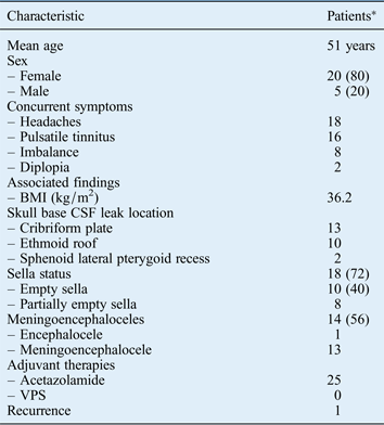
Twenty-five patients with spontaneous CSF leaks and evidence of intracranial hypertension were identified. Leak closure was accomplished in 24 patients after primary surgery; 1 patient required a second operation (detailed in Table I). The mean patient age was 51 years, 20 patients were female and the average BMI was 36.2 kg/m2 (range 24.5–66.4 kg/m2). All patients manifested CSF rhinorrhea, with headache (in 85 per cent) being the second commonest symptom. Other manifestations, including papilloedema, and visual disturbances, improved in 14 patients, as confirmed by ophthalmological testing. The mean hospitalisation period was 5.4 days.
Characteristics of patients with spontaneous CSF leaks

*n = 25; data presented as n (%) or n. CSF = cerebrospinal fluid; VPS = ventriculoperitoneal shunt
The most common leak location was the cribriform plate (13 patients; see Figure 1), followed by the ethmoid roof (10 patients; see Figure 2) and the lateral recess of the sphenoid sinus (2 patients; Figure 3). An encephalocele or meningoencephalocele at the defect site was found in 14 patients. The estimated defect size ranged from 3 to 8 mm, with most being less than 5 mm in the longest dimension: the defect size was less than 4 mm in seven patients and more than 5 mm in 18 patients.
Images showing spontaneous cerebrospinal fluid leaks in the posterior ethmoid roof and planum sphenoidale. (a) Coronal (upper-left side) and (d) sagittal (lower-left side) T2-weighted magnetic resonance imaging scans showing a hyperintense cystic lesion (arrows) from the posterior ethmoid roof, consistent with a meningocele. Intra-operative photographs showing (b) a bony defect and a meningeal defect (c) a dural substitute (intradural), (e) a dural substitute (extradural) and (f) a thin lamina of nasal cartilage (extradural). Dashed lines in (f) indicate a bony defect in the posterior ethmoid roof and planum sphenoidale. PEA = posterior ethmoidal artery
Images showing a spontaneous cerebrospinal fluid leak in the right cribriform plate. (a) Sagittal (upper-left side) and (d) coronal (lower-left side) T2-weighted magnetic resonance imaging scans showing a bony defect (arrows). Intra-operative photographs showing (b) a bony defect and a meningeal defect, (c) fascia lata (intradural), (e) a thin lamina of nasal cartilage (extradural) and (f) a nasal mucoperiosteum flap. Dashed lines in (b), (c) and (e) indicate a bony defect in the right cribriform plate.
Images showing a spontaneous cerebrospinal fluid leak in the sphenoid recess. (a) Coronal computed tomography image showing a bony defect in the lateral sphenoid recess (yellow arrow). (b) Coronal T2-weighted magnetic resonance imaging scans showing a hyperintense cystic lesion (arrow) extending from the subarachnoid space of the left middle fossa to the sphenoid body, consistent with a meningocele. Endoscopic views of a bony defect (left sphenoidal lateral recess) and a meningocele (c) and (d) fascia lata with cartilage achieving a gasket seal closure (dashed lines) of a leak in the lateral recess of the sphenoid; enlarged in (e).
For all patients, MRI scans were available for sella turcica evaluation. In 18 patients (72 per cent), there was evidence of an empty sella upon imaging (10 totally empty and 8 partially empty sellae).
All patients underwent endoscopic endonasal surgery for multilayer closure: 23 required a transethmoidal approach and 2 required a transethmoidal-pterygoidal-sphenoidal approach to repair CSF leaks in the lateral recess of the sphenoid.
A lumbar drain was inserted into each patient at the time of surgery. The mean ICP was 28.0 cm H2O (range 20–55 cm H2O) during the intra-operative period. Twenty-four spontaneous CSF leaks were successfully repaired at the first attempt. Acetazolamide therapy was used for all patients. Ventriculoperitoneal shunt placement was not performed for any patient. Spontaneous CSF leak recurrence at the primary site occurred in one patient on post-operative day four and was immediately repaired.
Patients' symptoms were followed closely in conjunction with endoscopic nasal examination one to three months after the repair. Follow-up periods ranged from one month to six years.
Discussion
Patients with spontaneous CSF leaks represent an important patient subset whose prevalence may be increasing;Reference Schuknecht, Simmen, Briner and Holzmann19 they are usually middle-aged, obese women who generally have elevated ICP and may complain of headache, pulsatile tinnitus and visual disturbances.Reference Friedman and Jacobson16 Spontaneous CSF leaks associated with elevated ICP have the highest recurrence rate (25–87 per cent) after surgical repair compared with most other types of CSF leaks (less than 10 per cent).Reference Schick, Prescher, Hofmann, Stegerwald and Draf7, Reference Lai, Kennedy and Bolger8 Technological developments and new research findings have increased the chances of achieving long-term surgical repair of spontaneous CSF leaks.
Skull base malformations or congenital dehiscence and obesity are thought to be risk factors for spontaneous CSF leak development.Reference Jindal, Hiam, Raman and Rejali4, Reference Schlosser, Wilensky, Grady and Bolger5, Reference Schuknecht, Simmen, Briner and Holzmann19 The current study indicates that spontaneous CSF leaks are more common in obese women, consistent with other reports.Reference Shetty, Shroff, Fatterpekar, Sahani and Kirtane20–Reference Rossi Izquierdo, Martín Martín and Labella Caballero23 Most participants qualified as obese (BMI > 30 kg/m2), with an average BMI of 36.2 kg/m2.
The thinnest areas of the anterior skull base are most susceptible to spontaneous CSF leak development, particularly the cribriform plate, tegmen tympani and sphenoid sinus (sphenoid lateral pterygoid recess).Reference Rossi Izquierdo, Martín Martín and Labella Caballero23–Reference Lloyd, DelGaudio and Hudgins25 In our experience, the bone around the defect is very fragile and easily fragmented.
High-resolution CT findings include thinning or attenuation of the skull base and evidence for arachnoid pits in up to 63 per cent of patients and the presence of multiple defects in up to 31 per cent.Reference Woodworth, Prince, Chiu, Cohen, Schlosser, Bolger, Kennedy and Palmer6, Reference Lloyd, DelGaudio and Hudgins25 MRI may reveal meningoceles and/or an encephalocele in up to 50 or 100 per cent of patients, as well as empty sellae and dilated optic nerve sheaths.Reference Schick, Prescher, Hofmann, Stegerwald and Draf7, Reference Lai, Kennedy and Bolger8, Reference Shetty, Shroff, Fatterpekar, Sahani and Kirtane9–Reference Friedman and Jacobson12 The presence of an empty sella upon MRI is strongly associated with both increased ICP in intracranial hypertension and spontaneous CSF leaks.Reference Brazis10, Reference Schlosser and Bolger13, Reference Badia, Loughran and Lund21–Reference Lopatin, Kapitanov and Potapov27 Empty sellae were identified in 62 per cent of participants and meningoceles and/or encephaloceles were identified in 72 per cent of participants who underwent MRI in the current study.
Penetration of the dura requires a watertight closure, if possible with a suitable graft material as well as a flap for secondary reinforcement. Spontaneous CSF leaks rarely resolve spontaneously and, if left untreated, increase the risk of meningitis. Patients with a skull base defect are at risk of ascending bacterial meningitis, independent of the defect size or location.Reference Bernal-Sprekelsen, Bleda-Vázquez and Carrau28, Reference Bernal-Sprekelsen, Alobid, Mullol, Trobat and Tomás-Barberán29 Pneumocephalus is another potential complication of CSF leakage; tension pneumocephalus is extremely unusual in this scenario, but has been reported.Reference Carrau, Snyderman and Kassam17, Reference Martínez-Capoccioni, Serramito-García, Cabanas-Rodríguez, García-Allut and Martín-Martín30
We believe that the graft technique used for defect repair in the current study provides a better seal and that cartilage provides the consistency necessary to achieve long-term stability at the reconstruction site. A mucoperiosteal flap allows the reconstruction area to be sealed and reduces the incidence of post-operative CSF fluid leaks. In addition, bipolar cautery was used to reduce brain herniation. Most authors agree that transection or resection of the pedicle is a potential source of intracranial infection if left unresected.Reference Zweig, Carrau, Celin, Snyderman, Kassam and Hegazy31, Reference Nyquist, Anand, Mehra, Kacker and Schwartz32
The most important factor in successful bony defect repair in patients with elevated ICP is reducing the ICP by nutritional, medical or/and surgical means.Reference Jindal, Hiam, Raman and Rejali4, Reference Schlosser, Wilensky, Grady and Bolger5, Reference Shetty, Shroff, Fatterpekar, Sahani and Kirtane9 Factors related to repair failure include obesity, leakage in the lateral recess of the sphenoid (which is difficult to access) and a large skull base defect.Reference Nyquist, Anand, Mehra, Kacker and Schwartz32, Reference McCarthy and Reed33 The first attempt to repair a spontaneous CSF leak in the ethmoid roof failed in a single patient with elevated ICP; this may be because a cartilage graft was not used in the reconstruction area and the mucoperiosteum flap was displaced by high ICP.
In our experience, indications for inserting a lumbar drain in patients undergoing skull base reconstruction include inadequate closure (ongoing CSF leakage following attempted skull base reconstruction), a high risk of post-operative leakage (obesity, high-volume pre-operative leak) and comorbidities associated with poor wound healing (chronic steroid use, diabetes, history of radiation therapy to the skull base). However, the use of a lumbar drain in managing patients with spontaneous CSF leaks remains controversial. Theoretical advantages include ICP reduction, decompression of the reconstruction area and a potentially reduced incidence of post-operative CSF leakage. Complications associated with lumbar drains include pneumocephalus and brain herniation; Reference Anand, Murali and Glasgold34–Reference Pepper, Lin, Sullivan and Marentette36 however, neither of these occurred in our study. Nevertheless, their impact on overall outcome remains poorly described. Some patients in this series underwent successful closure, supporting the primary importance of robust closure and post-operative lumbar drainage. In our experience, a lumbar drain is useful if kept open during extubation. Drainage during the first 72 hours allows the tissue sealant to harden, maintaining nasoseptal flap attachment to the base of the skull. Post-operatively, the most frequent complaint was headache. Although meningitis is a serious complication, its occurrence is more closely associated with the aetiology of the CSF leak and the surgery itself, rather than the use of a lumbar drain.Reference Bernal-Sprekelsen, Bleda-Vázquez and Carrau28, Reference Bernal-Sprekelsen, Alobid, Mullol, Trobat and Tomás-Barberán29
• Endoscopic endonasal surgery can be successfully used for spontaneous cerebrospinal fluid leak repair
• Imaging is used to confirm the diagnosis, identify the underlying cause or an associated meningoencephalocele, and characterise the osteodural defect
• Intracranial hypertension treatment coupled with endoscopic repair has a high success rate
To establish a diagnosis of intracranial hypertension, the lumbar CSF pressure should be greater than 25 cm H2O and the CSF composition should be normal. The best treatment for intracranial hypertension is currently not established but oral acetazolamide and placement of a ventriculoperitoneal shunt are the most common interventions. In the immediate post-operative period, acetazolamide therapy reduced ICP by a mean of 10 cm H2O in a limited set of patients with spontaneous CSF leak.Reference Schlosser, Wilensky, Grady, Palmer, Kennedy and Bolger37 The failure rate for spontaneous leak repair (one case) in this case series was significantly lower than in other reports;Reference Lai, Kennedy and Bolger8, Reference Schlosser and Bolger13, Reference Martín-Martín, Martínez-Capoccioni, Serramito-García and Espinosa-Restrepo15 this strongly suggests that elevated ICP treatment with acetazolamide and a post-operative lumbar drain would improve the long-term success rate. Prospective studies of obese intracranial hypertension patients found that weight loss leads to reduced symptoms, signs and ICP.Reference Thurtell and Wall38–Reference Sugerman, Felton, Sismanis, Kellum, DeMaria and Sugerman40 Therefore, weight loss should also be routinely recommended for all obese intracranial hypertension patients.
The relative rarity of spontaneous CSF leakage makes it difficult to obtain sufficient cohort sizes to perform a statistical analysis. However, the key determinants of clinical outcome included operative techniques and peri-operative protocols, as was found in other single institution reviews of skull base defects, complications and lesions.Reference Carrau, Snyderman and Kassam17, Reference Martínez-Capoccioni, Serramito-García, Cabanas-Rodríguez, García-Allut and Martín-Martín30 Thus, the results of this study seem relevant to managing this pathology. The low failure rate precludes the data reaching statistical significance.
Conclusion
Spontaneous CSF leaks represent a surgical challenge because of their high recurrence rate. This study indicates that treating underlying intracranial hypertension coupled with endoscopic repair can provide high success rates approaching those of other aetiologies of CSF leaks. The most important factor for successful repair in these patients is reducing their ICP through nutritional, medical or surgical means.
Acknowledgements
We would like to thank the Service of ENT–Head and Neck Surgery and Service of Neurosurgery, University Hospital Complex of Santiago de Compostela for their assistance in this study.