Tyrannosauroid dinosaurs are typically a rare component of even the most well studied Late Cretaceous faunas in western North America (e.g. Farlow & Pianka Reference Farlow and Pianka2002). Remains of these large predatory animals are particularly scarce in the Upper Cretaceous Aguja Formation of southwestern Texas (Lehman Reference Lehman, Wolberg and Stump1997). It is possible that their extreme rarity here could reflect competition in life with the giant crocodylian Deinosuchus, which may have been the top predator in coastal environments represented by the Aguja Formation (Schwimmer Reference Schwimmer2002). Even so, remains of tyrannosaurs have been reported from the Aguja Formation since the time these strata were first described (Udden Reference Udden1907). Such reports have, however, been based almost exclusively on detached teeth (Lehman Reference Lehman1985; Rowe et al. Reference Rowe, Cifelli, Lehman and Weil1992; Sankey Reference Sankey2001). While the size, serration count, and serration morphology of isolated teeth are sufficient to recognize that they pertain to tyrannosauroid theropods, the teeth do not provide much additional taxonomic information.
After nearly a century of prospecting, only a few non-dental tyrannosaur skeletal elements have been recovered from the Aguja Formation. In the past several years, the authors have collected a few isolated cranial and postcranial elements that provide additional morphological information about Aguja tyrannosaurs. The most common elements recovered (frontals, metatarsals, pedal phalanges, caudal vertebrae) represent very durable parts of the skeleton, and their selective preservation likely reflects a taphonomic bias. In view of the great rarity of tyrannosaur material and recognition that many taxa previously recovered from the Aguja represent species not known elsewhere in North America (e.g. Wagner & Lehman Reference Wagner and Lehman2009), it is useful to describe and illustrate these specimens.
The material described here was collected from the upper shale member of the Aguja Formation (informal member designations proposed by Lehman Reference Lehman1985). The upper shale member has produced most of the known vertebrate fauna of the Aguja (Lehman & Busbey Reference Lehman and Busbey2007). The lower shale member of the formation has thus far yielded scant vertebrate fauna, including tyrannosaurs, but this appears to be distinctly older than the upper shale member fauna, and will be described elsewhere. Ammonite biostratigraphy (Waggoner Reference Waggoner2006) and radiometric age determinations for the Aguja Formation (Befus et al. Reference Befus, Hanson, Lehman and Griffin2008) suggest that the upper shale member is early Judithian in age (e.g. correlative with the Oldman Formation of Alberta). Daspletosaurus torosus from Alberta (Russell Reference Russell1970), and Appalachiosaurus montgomeriensis from Alabama (Carr et al. Reference Carr, Williamson and Schwimmer2005) are the only well known tyrannosaurs from this time interval, and so the Aguja specimens are of interest in spite of their meagre nature. We refer to these specimens herein as representing the ‘Aguja tyrannosaur’. Although we consider it unlikely, it is possible that the specimens could pertain to more than one taxon, but lacking material sufficient for confident generic attribution, it is prudent to describe the specimens informally in this way.
Institutional abbreviations. AMNH, American Museum of Natural History, New York USA; BHl, Black Hills Institute, South Dakota, USA; CMNH, Cleveland Museum of Natural History, Cleveland, Ohio USA; FMNH, Field Museum, Chicago, Illinois USA; LACM, Los Angeles County Museum, Los Angeles, California USA; MOR, Museum of the Rockies, Bozeman, Montana USA; NHMUK, The Natural History Museum, London, UK; NMC, Canadian Museum of Nature, Quebec, Canada; NMMNH, New Mexico Museum of Natural History, Albuquerque, New Mexico USA; OMNH, Sam Noble Museum of Natural History, Oklahoma USA; ROM, Royal Ontario Museum, Toronto, Ontario, Canada; TMM, Texas Memorial Museum, Austin, Texas USA; TMP, Royal Tyrrell Museum, Drumheller, Alberta, Canada.
1. Description
There are varied hypotheses regarding the number of valid taxa included within Tyrannosauridae, or within the broader Tyrannosauroidea (e.g. Currie Reference Currie2003a; Sereno & Brusatte Reference Sereno and Brusatte2009; Carr & Williamson Reference Carr and Williamson2010). Whether some (Dryptosaurus, Appalachiosaurus, Bistahieversor) are properly considered sister taxa to a restricted Tyrannosauridae, or best included as basal members of a more broadly defined family, may also be considered. Further, there remains debate as to whether there are distinct clades among tyrannosaurids (Albertosaurinae and Tyrannosaurinae) and which taxa comprise them. The recovery and description of many new tyrannosaurs in recent years have revealed a broader range of morphological variation than previously recognised (e.g. Brusatte et al. Reference Brusatte, Carr, Erickson, Bever and Norell2009; Sereno et al. Reference Sereno, Tan, Brusatte, Kriegstein, Zhao and Cloward2009) and, as a result, hypotheses regarding phylogenetic relationships of Tyrannosauridae and close outgroups will likely be re-evaluated in the coming years.
The Aguja material is not informative in evaluating such hypotheses. Therefore, in general here we will follow the systematic and phylogenetic analysis of tyrannosaurs presented by Currie (Reference Currie2003a) and Currie et al. (Reference Currie, Hurum and Sabath2003), which we believe provides, at present, the most strongly supported argument for classification of these animals. Herein, we use the term “tyrannosaur” to refer broadly to all members of Tyrannosauroidea (e.g. as circumscribed recently by Carr & Williamson Reference Carr and Williamson2010), “less derived tyrannosauroid” to large-bodied tyrannosaurs excluded by some authors from Family Tyrannosauridae (e.g. Dryptosaurus, Appalachiosaurus, Bistahieversor) and “tyrannosaurid” for those taxa included by most authors within Tyrannosauridae (e.g. Currie et al. Reference Currie, Hurum and Sabath2003). Because nearly all tyrannosaur genera recognised today are monospecific, in the following discussion each taxon will be referred to only by its generic name, except for those few cases where more than one species may be assigned to a genus (e.g. Daspletosaurus torosus and Daspletosaurus sp., MOR 590).
Saurischia Seeley, Reference Seeley1887
Theropoda Marsh, Reference Marsh1881
Tyrannosauridae Osborn, Reference Osborn1906
Tyrannosaurinae Osborn, Reference Osborn1906
Tryrannosaurinae indeterminate
1.1. Teeth
Isolated teeth are the most common tyrannosaur remains recovered from the Aguja Formation. The same is true for correlative strata throughout most of western North America (e.g. Farlow & Pianka Reference Farlow and Pianka2002). As a result, substantial effort has been made to recognise morphological features of isolated teeth that could be useful for taxonomic purposes (Samman et al. Reference Samman, Powell, Currie and Hills2005; Smith et al. Reference Smith, Vann and Dodson2005). It may be helpful, therefore, to compare typical Aguja isolated teeth with those of better-known tyrannosaurs.
Most detached teeth recovered from the Aguja are broken and missing the base of the crown and/or the tip. Although these exhibit the typical size, form, and denticle morphology of tyrannosaurid teeth, they provide little additional information (e.g. Standhardt Reference Standhardt1986; Sankey Reference Sankey2001). Complete Aguja tooth crowns exhibit the expected variation in size, morphology and position within the jaw of juvenile and adult tyrannosaurids. The shed teeth show slight wear on the apex and apical part of the mesial carina. One large lateral tooth (TMM 42534-15) was broken in life and worn down by use in the manner described by Schubert & Ungar (Reference Schubert and Ungar2005). Most complete examples are distal lateral teeth; however, the collection includes one non-denticulate incisiform premaxillary tooth (of the sort formerly assigned to Aublysodon mirandus; see Currie Reference Currie2003a, Carr & Williamson Reference Carr and Williamson2004), and one sub-incisiform mesial maxillary tooth with markedly asymmetric carinae (TMM 42880-8 and 45908-3).
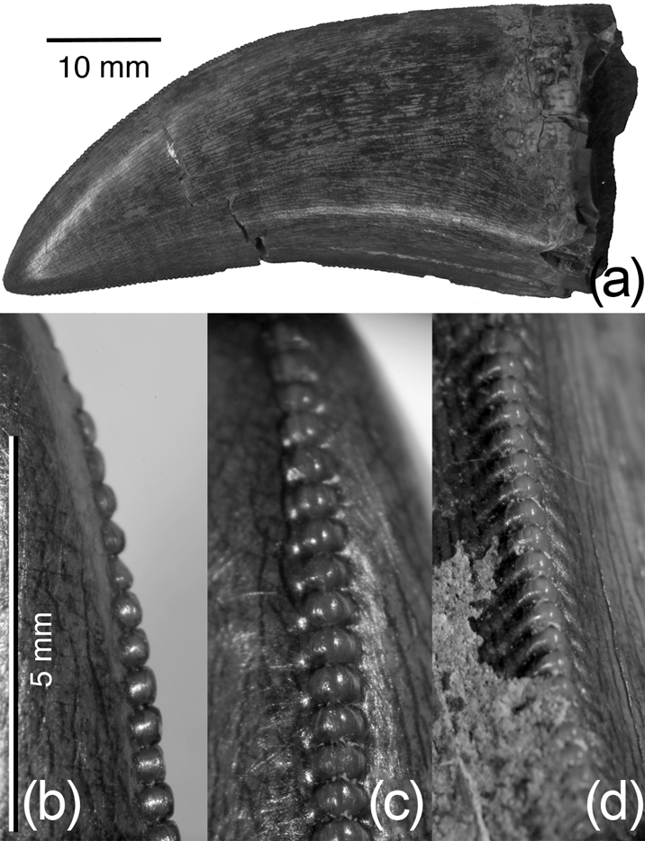
Two complete large lateral tooth crowns (TMM 42880-7 and 45908-2; Fig. 1) provide a basis for comparison with other tyrannosaurs. Both are large, laterally compressed, and distally curved (moreso at the apex compared to the base), with mesial and distal carinae having broad, chisel-shaped serrations typical of tyrannosaurids. The mesial carina is deflected lingually near its base, and terminates well above the base of the crown. The denticles (10–12 per 5 mm, apical to proximal) on the distal carina are slightly larger than those on the mesial carina. Well-developed ‘blood grooves’ arc away from the serrations only on the distal carina, and are most pronounced along the middle of the carina (Fig. 1). Together, these features suggest that TMM 42880-7 is the crown of a left dentary tooth, and TMM 45908-2 a right (Samman et al. Reference Samman, Powell, Currie and Hills2005). Comparison with incomplete crowns indicates that these are reasonably representative of large lateral teeth collected from the Aguja.
Aguja tyrannosaur left dentary tooth, TMM 42880-7: (a) lateral view; apical denticles on mesial carina in (b) lateral view and (c) anterior view; (d) middle denticles on distal carina in posterior view.

Farlow et al. (Reference Farlow, Brinkman, Abler and Currie1991), Samman et al. (Reference Samman, Powell, Currie and Hills2005), Smith et al. (Reference Smith, Vann and Dodson2005) and Buckley et al. (Reference Buckley, Larson, Reichel and Samman2010) conducted exhaustive morphometric studies of tyrannosaurid teeth, and found that much of their morphological variation is a result of varied tooth size and position within the jaws. Teeth of less derived tyrannosauroids have not been as extensively studied, but some appear to have higher denticle densities than the 8–16 per 5 mm typical of tyrannosaurids (17–18 in Dryptosaurus; up to 16 in Appalachiosaurus; as high as 18–21 in Bistahieversor) particularly along the basal third of the carinae (Carr et al. Reference Carr, Williamson and Schwimmer2005; Brusatte et al. Reference Brusatte, Benson and Norell2011); and the teeth of Dryptosaurus are much more laterally compressed than those typical of more derived tyrannosauroids, where the basal width typically exceeds 60% of the mesiodistal length. Although isolated teeth belonging to different taxa may be difficult to identify confidently on the basis of these studies, they do reveal differences in tooth morphology among tyrannosaurids.
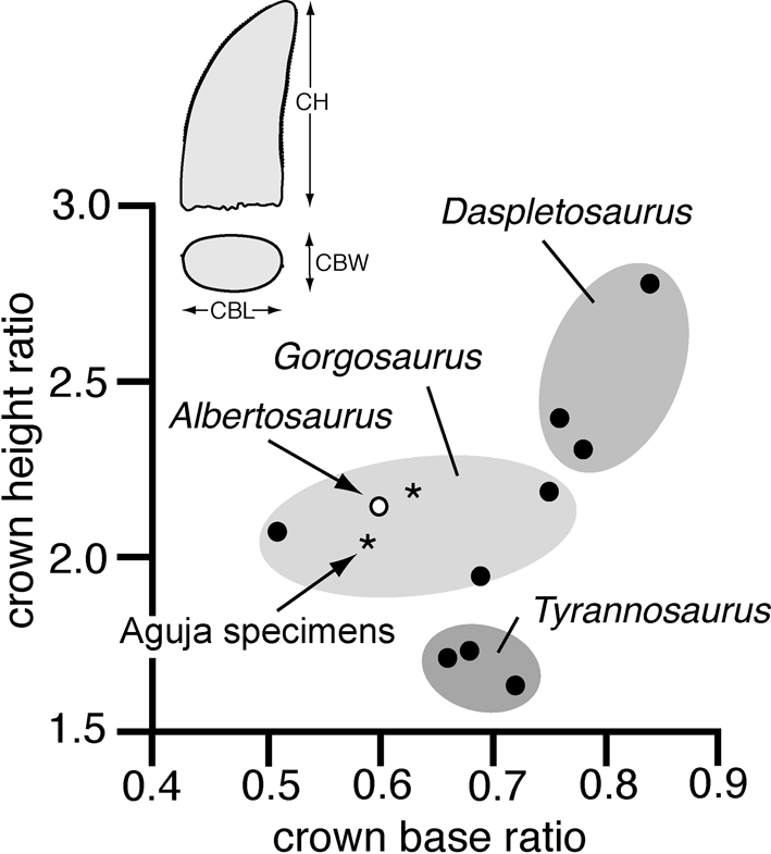
For comparison with the Aguja specimens and to control for variation in tooth size, measurements of large lateral dentary or maxillary teeth with crown heights of about 55 mm (±5 mm) were selected from those listed by Samman et al. (Reference Samman, Powell, Currie and Hills2005) and Smith et al. (Reference Smith, Vann and Dodson2005). These show that it may be possible to distinguish similarly sized teeth of Gorgosaurus, Daspletosaurus sp. (MOR 590) and Tyrannosaurus, primarily on the basis of their crown base and crown height ratios (Fig. 2). Teeth of Albertosaurus are proportionally similar to those of Gorgosaurus, and teeth of the Aguja tyrannosaur are most like these two taxa. In contrast, teeth with comparable crown heights belonging to Daspletosaurus sp. tend to be relatively wider and taller, while those of Tyrannosaurus are wider and shorter for their basal length (Fig. 2).
Bivariate plot comparing crown height (CH/CBL) versus crown base (CBW/CBL) ratios for large lateral teeth (CH±55 mm) in several tyrannosaurs. Data for Daspletosaurus (MOR 590), Gorgosaurus (ROM 1247, NHMUK R4863) and Tyrannosaurus (AMNH 5027, MOR 555, BHI 3033) are from Smith et al. (Reference Smith, Vann and Dodson2005); data for Albertosaurus (TMP 81.10.1) are from Sammon et al. (2003). Abbreviations: *=Aguja specimens (TMM 42880-7 and 45908-2); CBL=crown basal length; CBW=crown basal width; CH=crown height, measured in the manner shown by Smith et al. (Reference Smith, Vann and Dodson2005).
1.2. Frontal
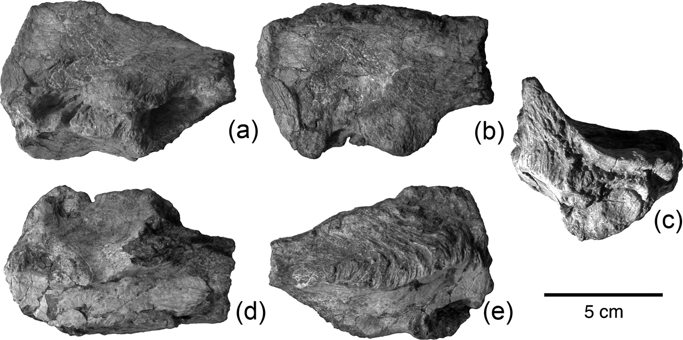
An isolated right frontal (TMM 45905-1; Figs 3, 4) is well preserved, but lacks most of the rostral process for articulation with the nasal. The sutural surfaces are sharply defined and the bone had separated freely from neighbouring elements. It is similar in size to those of other large tyrannosaurs. The length (95+ mm), width (55 mm), and depth (35 mm) measured in the manner described by Currie (Reference Currie2003a, table 2) are comparable to some individuals of Gorgosaurus (TMP 91.36.500) and Daspletosaurus sp. (TMP 94.143.1), but slightly smaller than is typical for either of these (average lengths=108 and 125 mm, respectively), Albertosaurus (117 mm), or Tyrannosaurus (138 mm), based on the specimens measured and tabulated by Currie (Reference Currie2003a, table 2).
Aguja tyrannosaur right frontal, TMM 45905-1: (a) lateral view; (b) dorsal view; (c) posterior view; (d) ventral view; (e) medial view.
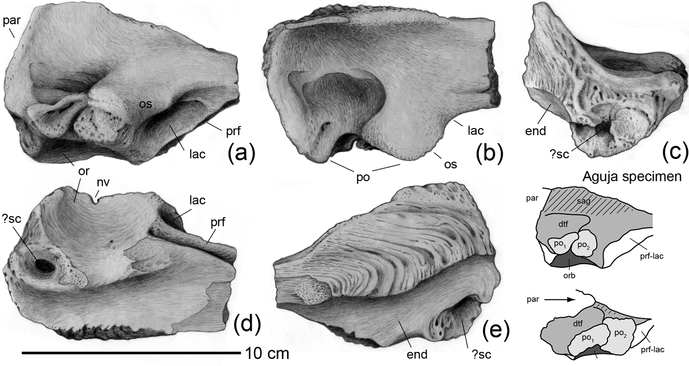
Aguja tyrannosaur right frontal, TMM 45905-1: (a) lateral view; (b) dorsal view; (c) posterior view; (d) ventral view; (e) medial view. Inset diagram compares right frontal of TMM 45905-1 with Bistahieversor sealeyi (OMNH 10131; from Lehman and Carpenter Reference Lehman and Carpenter1990) in lateral view, reduced to common length. Abbreviations: dtf=dorsotemporal fossa; end=endocranial cavity; lac=suture for lacrimal; nv=neurovascular foramen; or=orbit; orb=inner wall of orbit; os=orbital slot; par=parietal wedge; po=postorbital suture; po1=posterior postorbital suture; po2=anterior postorbital suture; prf=suture for prefrontal; prf-lac=suture for prefrontal and lacrimal; sag=sagittal crest; ?sc=sinus cavity.
The dorsal surface of the Aguja specimen is markedly concave, both mediolaterally and rostrocaudally, with a fractured oval depression over the skull roof that may indicate the presence here of a collapsed sinus cavity within the interior of the bone beneath (Figs 3, 4). The anterior shelf of the supratemporal fossa (attachment area for temporal musculature; ‘dorsotemporal fossa’ of some authors) appears to extend in an arc along the anterior edge of this oval depression. However, its anterior limit is not marked by the distinct ridge found in specimens of some tyrannosaurs (e.g. Gorgosaurus, Albertosaurus, Daspletosaurus; Currie Reference Currie2003b). A tall sagittal crest extends along the border with the adjacent frontal from the parietal suture to the base of the rostral process. The sagittal crest ends abruptly anteriorly where the surface of the frontal instead turns downward medially, to form a narrow trough along the interfrontal suture. The sagittal crest is particularly long and high in the Aguja specimen, a feature typical of the tyrannosaurines Daspletosaurus and Tyrannosaurus, in contrast to less derived tyrannosaurs, where the sagittal crest is restricted to the parietal or extends only a short distance onto the frontal. A fragmentary left frontal from a juvenile individual (TMM 41918-24) approximately three-fourths the size of TMM 45905-1, is identical in form and has a sagittal crest proportionally as well developed (about 80 percent of the height, measured above the endocranial cavity). This indicates that the sagittal crest may have developed earlier ontogenetically in the Aguja tyrannosaur than in some others (e.g. Tyrannosaurus; Carr & Williamson Reference Carr and Williamson2004).
There is a deep well-developed socket for insertion of the lacrimal. Ventral and medial to the socket for the lacrimal is a prominent groove for insertion of the prefrontal; the placement of this suture suggests that the prefrontal may have had little exposure on the skull roof. The remnant of the orbital rim is weakly expressed as a posterolaterally inclined inflection between the lacrimal and postorbital sutures. The strong interdigitating suture for the postorbital overlaps slightly onto the dorsal surface of the frontal, and is divided into two distinct parts, an anterior bulbous vertical buttress and a posterior simple horizontal tabular joint. The two sutural faces are separated by a small neurovascular foramen in the orbital wall, visible in ventral view, but this foramen does not appear to pass entirely through to the dorsal surface of the bone, as it does in some tyrannosaurids (Tyrannosaurus; Brochu Reference Brochu2003). The nature and distribution of this foramen among individuals or in other tyrannosaurids has received no comment from previous authors.
The configuration of the frontoparietal suture indicates that the parietals extend only slightly forward along the midline to partially separate the frontals. The foramen that pierces the interfrontal suture near its junction with the parietal, noted particularly in young tyrannosaurids (e.g., Tyrannosaurus), is not obvious here; however, its path may be evident as a forwardly deflected depression on the interfrontal suture (Fig. 4). Traced laterally along the wall of the supratemporal fenestra, the frontoparietal suture is depressed below the level of the postorbital suture and deflected slightly anteriorly, rather than extending directly transversely. The frontoparietal sutural surface itself is a deeply interdigitate serrate joint, and unlike some (e.g. Gorgosaurus, Albertosaurus; Currie Reference Currie2003b) where the suture is marked by a prominent raised ridge, here it is only slightly raised above the surface of the frontal where it ascends along the side of the sagittal crest. The interfrontal suture exhibits a striking sigmoidal serrate morphology. On the ventral surface of the frontal, the endocranial cavity is strongly bowed anteroposteriorly, rising toward the parietal suture. A prominent articulation for the laterosphenoid separates the endocranial cavity from the inner wall of the orbit. There appears to be a tubular pneumatic opening which passes through the laterosphenoid buttress into the body of the frontal, directly beneath the collapsed sinus cavity evident on the dorsal skull roof (Figs 3, 4). Alternatively, this opening may represent a vascular feature or one that resulted from post-mortem decay of the cancellous interior of the bone.
1.3. Vertebrae
Only isolated mid and distal caudal vertebrae of tyrannosaurs have been recovered from the Aguja. One of these (TMM 43057-505) is comparable in size and form to caudals 21 through 24 of Gorgosaurus (Lambe Reference Lambe1917), another (TMM 45909-1) to caudals 30 through 33. These specimens both have amphicoelous centra lacking pneumatopores, with quadrangular intercentral articulation surfaces depressed above the midpoint but bulging out below. Chevron facets are present both front and back, but more pronounced posteriorly. The neurocentral sutures are completely closed, neural canals are very constricted (6 mm diameter), and the rod-shaped neural spines are strongly inclined posteriorly.
1.4. Tibia
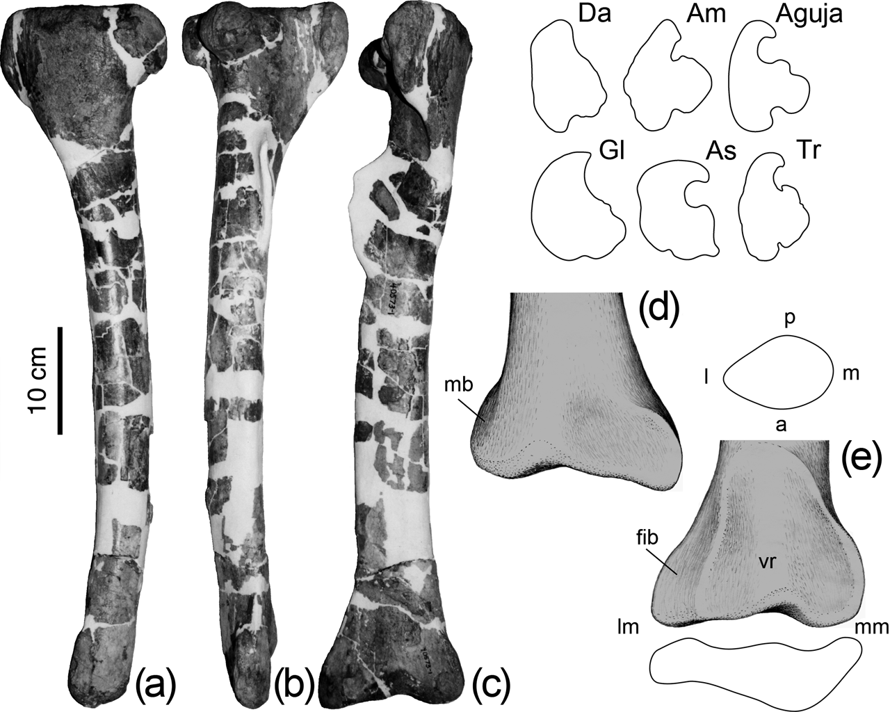
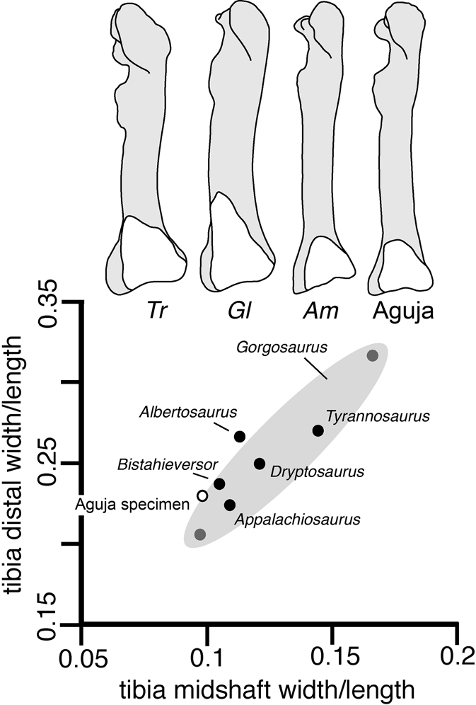
A right tibia (TMM 40573-1; Fig. 5) is nearly complete, although the fibular crest is broken. Its length (610 mm) is less than in typical large adult tyrannosaurs (e.g. 775 mm in Dryptosaurus; up to 1143 mm in Tyrannosaurus; Carpenter et al. Reference Carpenter, Russell, Baird and Denton1997; Brochu Reference Brochu2003) and so this specimen may be from an immature individual. Similarly, its form is slender (ratio of midshaft transverse width to length=0·10) compared to adult Gorgosaurus (0·17 in NMC 2120; Lambe Reference Lambe1917) or Tyrannosaurus (0·14 in FMNH PR2081; Brochu Reference Brochu2003); also in keeping with an immature condition. However, both Dryptosaurus (0·12) and Appalachiosaurus (0·11) have similarly slender tibiae, even as adults. In the Aguja specimen, the shaft is nearly circular in cross-section at midlength (ratio of anteroposterior width to transverse width=0·83) and close to that in Appalachiosaurus (0·85; Carr et al. Reference Carr, Williamson and Schwimmer2005) compared to Gorgosaurus (0·71 in ROM 1247; 0·66 in NMC 2120) or Albertosaurus (0·68 in ROM 807) where the mediolateral width substantially exceeds the anteroposterior width.
Aguja tyrannosaur right tibia TMM 40573-1: (a) medial view; (b) lateral view; (c) anterior view showing detail of distal end; (d) posterior view; (e) anterior view with cross-section of shaft and outline of distal end. Inset diagrams compare outlines of the proximal end of the Aguja specimen with Am=Appalachiosaurus (from Carr et al. Reference Carr, Williamson and Schwimmer2005), As=Albertosaurus (from Parks Reference Parks1928), Da=Dryptosaurus (from Carpenter et al. Reference Carpenter, Russell, Baird and Denton1997), Gl=Gorgosaurus (from Lambe Reference Lambe1917) and Tr=Tyrannosaurus (from Brochu Reference Brochu2003), all reduced to common length. Abbreviations: a=anterior: fib=fibular facet; l=lateral; lm=lateral malleolus; m=medial; mb=medial buttress; mm=medial malleolus; p=posterior; vr=vertical ridge dividing two parts of astragalar facet.
The proximal end of the tibia is relatively more elongate anteroposteriorly, and not as stout mediolaterally as is Gorgosaurus or Albertosaurus, but it has a strong laterally deflected cnemial crest (Fig. 6). The lateral femoral condyle is stout mediolaterally and separated from the medial condyle by a deep flexor fossa, as in Appalachiosaurus (Fig. 5). The cranial process of the lateral femoral condyle, lacking in Dryptosaurus but found in Appalachiosaurus and more derived tyrannosaurs, is present in the Aguja specimen. The cnemial crest does not rise abruptly above the level of the proximal articulation surface as it does in Tyrannosaurus (Brochu Reference Brochu2003), a characteristic evident even in juvenile individuals (e.g. Molnar Reference Molnar1980).
Bivariate plot of tibia distal width/length ratio versus shaft width/length ratio for several tyrannosaurs. Diagram compares outline of Aguja tyrannosaur right tibia in anterior view with astragular articulation facet highlighted with Am=Appalachiosaurus (from Carr et al. Reference Carr, Williamson and Schwimmer2005), Gl=Gorgosaurus (from Lambe Reference Lambe1917) and Tr=Tyrannosaurus (from Brochu Reference Brochu2003), reduced to common length.
The distal end of the Aguja tibia is wider than the astragalus, as in other tyrannosaurs. However, compared to others, the distal end is not as expanded transversely (e.g. ratio of distal width to length=0·23 compared to 0·32 in Gorgosaurus NMC 2120; Lambe Reference Lambe1917). The articulation surface for the ascending process of the astragalus is shallow and also extends only a short distance up the shaft (ratio of ascending process height to length of the tibia=0·20) and closer to Appalachiosaurus (0·21 to 0·23; Carr et al. Reference Carr, Williamson and Schwimmer2005) than in Gorgosaurus (0·32). As in other tyrannosaurs, the articulation facet for the ascending process of the astragalus extends higher on the lateral side of the shaft than on the medial, and it has a subtle vertical ridge along the midline; this ridge divides the contact into medial and lateral facets (Fig. 5). Sharp angulations define the medial and lateral sides of the articulation, but the dorsal edge grades smoothly onto the shaft of the tibia. In other tyrannosaurs, the fibular flange and lateral malleolus typically extends distally, notably farther than the medial malleolus; however, in the Aguja specimen, the two have nearly the same extent (e.g. as in Alioramus; Brusatte et al. Reference Brusatte, Carr, Erickson, Bever and Norell2009). The facet that backs the fibula is marked and occupies a substantial part of the distal end of the tibia; more so than in Tyrannosaurus for example (Brochu Reference Brochu2003).
Some tyrannosauroids (Dryptosaurus, Appalachiosaurus) were comparatively gracile, with relatively slender limb elements, even as adults. However, most tyrannosaurids known from population samples rather than single individuals, show substantial ontogenetic change in the proportions and robustness of their limb elements (Currie Reference Currie2003a). Standardized relative to the length of the tibia, both the width of the shaft and width of the distal end increase proportionally with body size (Fig. 6). Therefore, it is difficult to interpret isolated limb elements such as the Aguja tibia; its features could reflect those of a young animal or a gracile adult. Among tyrannosauroid tibiae illustrated in the literature, this specimen is much like that in Appalachiosaurus (Fig. 6).
1.5. Astragalus
An isolated right astragalus (TMM 45910-1) is compatible in size, with a tibia slightly larger than that described above, but is missing most of the ascending process; it shows a weak vertical slot on the posterior articulation surface for the tibia. The ventrolateral buttress of the ascending process is prominent, with a deep adjacent tendon fossa and a marked horizontal groove separating it from the condylar area. The trochlea is constricted dorsoventrally, less so anteroposteriorly, but the medial condyle is broken in this specimen. The lateral condyle and surface for the calcaneum lack the ‘peg-and-socket’ articulation shown in Appalachiosaurus (Carr et al. Reference Carr, Williamson and Schwimmer2005) and also lacks the prominent lip separating the condyle from the ascending process in Dryptosaurus (Brusatte et al. Reference Brusatte, Benson and Norell2011).
1.6. Metatarsus
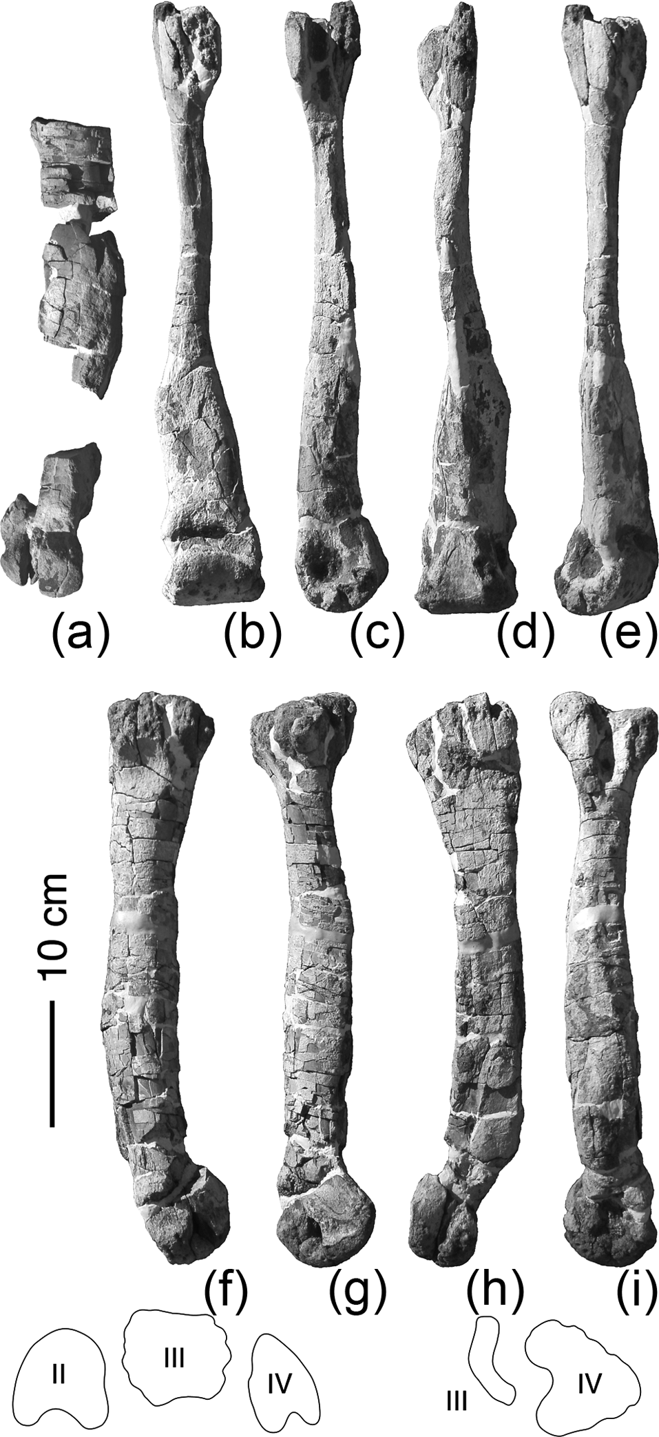
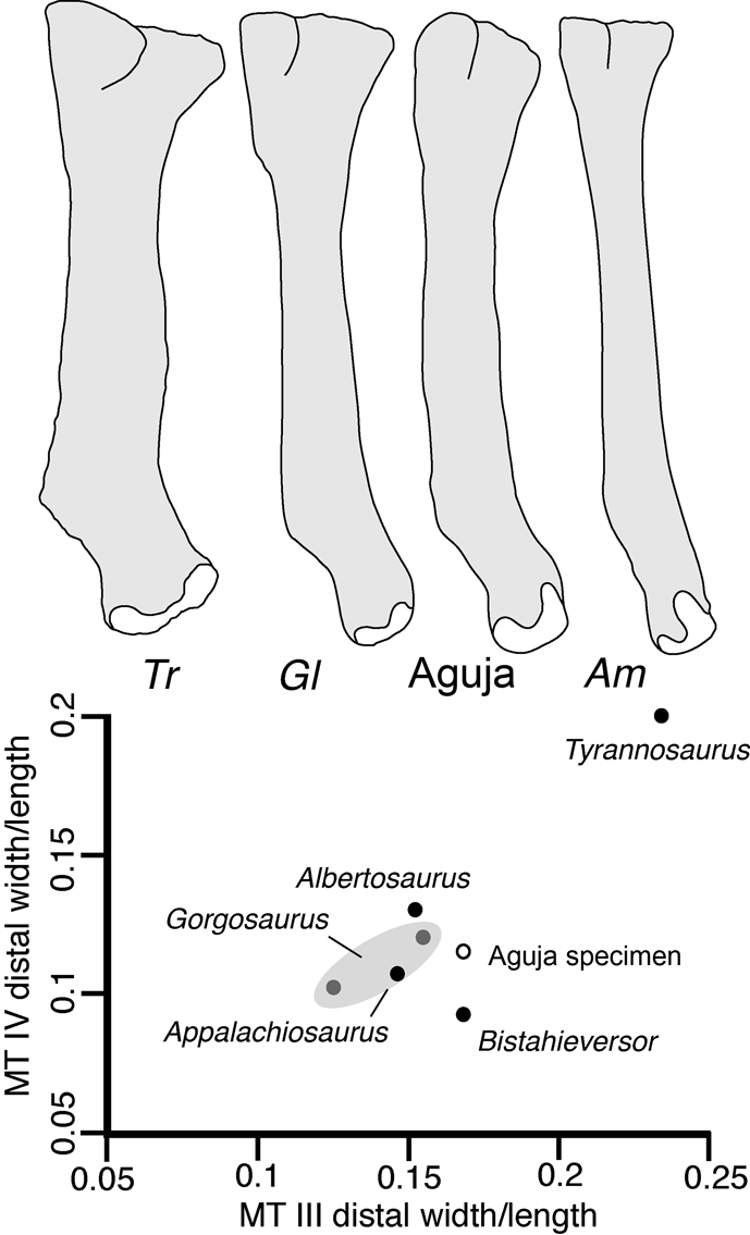
An associated set of metatarsals (MT) and fragmentary phalanges (TMM 45906-1; Fig. 7) pertains to the left foot of a single individual. Metatarsals III and IV are complete, but II is missing the proximal end and parts of the shaft. These clearly exhibit the arctometatarsalian condition (Holtz Reference Holtz1995). In their size, slender form and proportions, the metatarsals are much like those of Appalachiosaurus (Carr et al. Reference Carr, Williamson and Schwimmer2005). Their lengths (488 mm for MT III, 456 for MT IV) and form are also compatible with younger individuals of Gorgosaurus and Albertosaurus, for example with individuals 4, 5 and 6 from the Albertosaurus bone bed described by Currie (Reference Currie1998), but substantially smaller than in adults of these species or in Tyrannosaurus. Relative to their lengths, the distal widths of the metatarsals are proportionally much broader in Tyrannosaurus than in the Aguja tyrannosaur or in other tyrannosauroids for which data are available (Fig. 8).
Aguja tyrannosaur left metatarsus, TMM 45906-1: (a) MT II in anterior view; (b–e) MT III in (b) anterior view, (c) lateral view, (d) posterior view and (e) medial view; (f–i) MT IV in (f) anterior view, (g) lateral view, (h) posterior view and (i) medial view. Inset diagrams show outlines of the distal condyles II–IV (left) and proximal articulation surfaces III–IV (right).
Bivariate plot of metatarsal IV distal width/length ratio versus MT III distal width/length ratio for several tyrannosaurs. Diagram compares outline of Aguja tyrannosaur MT IV in posterior view with distal articulation condyle highlighted with Am=Appalachiosaurus (from Carr et al. Reference Carr, Williamson and Schwimmer2005), Gl=Gorgosaurus (from Lambe Reference Lambe1917) and Tr=Tyrannosaurus (from Brochu Reference Brochu2003), reduced to common length.
The proximal half of MT III is dramatically constricted, and exhibits the ‘L-shaped’ proximal articulation surface, with the top of the ‘L’ directed posteriorly, typical of tyrannosaurids. The shaft of MT III is triangular in section distally, with the apex of the triangle directed posteriorly. The proximal part of the shaft is slightly twisted laterally so that here the apex of the triangular section is directed posterolaterally. The entire shaft is curved such that the midline is shifted laterally. The distal articulation surface is much larger than in either MT II or IV, and quadrangular in form viewed distally, with anterior and medial sides slightly broader than the posterior and lateral sides (Fig. 7). The phalangeal articulation surface extends higher anteriorly than posteriorly, and more so on the medial than on the lateral side. The collateral ligament pit is also deeper and more pronounced on the medial side. There is a deep transverse groove on the anterior face of the shaft above the phalangeal articulation surface, lacking on the posterior face.
The proximal end of MT IV is triangular with a medial indentation to receive the L-shaped process of MT III. The proximal articulation surface flares outward away from the shaft only slightly on the posterior and lateral sides (unlike Tyrannosaurus; Brochu Reference Brochu2003). The proximal half of the shaft is triangular in section, with the sharpest corner directed laterally. The posterior face of the shaft is flat while the anterior face is broadly rounded; it is much broader anteroposteriorly than in Dryptosaurus and compatible instead with more derived tyrannosauroids (Brusatte et al. Reference Brusatte, Benson and Norell2011). A deep groove extends along the posterolateral edge of the shaft above midlength; this is the ‘flexor groove’ of Carr et al. (Reference Carr, Williamson and Schwimmer2005). At midlength, the shaft begins to curve slightly laterally and the curvature is more pronounced at the distal end, much more so than in Dryptosaurus but not so markedly as in Tyrannosaurus (Brusatte et al. Reference Brusatte, Benson and Norell2011; Fig. 8). The articulation facet for MT III extends from the midpoint of the shaft, broadens distally, and terminates above the point where the distal condyle turns outward. The distal condyle is triangular, narrow on the anterior side and bifurcating into two distinct articulation surfaces posteriorly. The lateral articulation extends farther on the posterior surface, as in Appalachiosaurus, and the medial articulation is broader transversely (Fig. 8). The collateral ligament insertion pit is pronounced on the medial side, but only weakly expressed on the lateral side.
The articulation surfaces and parts of the shaft of metatarsal II are preserved. The shaft is stouter than that of MT IV but similar in form, with flat posterior and rounded anterior surfaces, and a sharply angled posterolateral edge. The distal condyle narrows anteriorly, extends higher on the lateral side, and broadens posteriorly where it divides into two articulation surfaces; the lateral one is broad transversely but the medial one is longer anteroposteriorly. Collateral ligament insertion pits are pronounced on both medial and lateral sides.
1.7. Phalanges
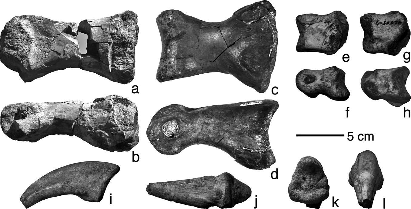
Apart from teeth, isolated phalanges are the most common remains of tyrannosaurs found in the Aguja, probably because their dense compact forms are durable sedimentary ‘particles’. The pedal phalanges exhibit strong ginglymoid interphalangeal joint surfaces and deep collateral ligament insertion pits. A proximal pedal phalanx of digit III (TMM 43057-142; Fig. 9) is smaller but comparable in form to that in Gorgosaurus and other relatively small tyrannosaurids. It is substantially smaller than in Tyrannosaurus and its proximal articulation surface is not so expanded dorsoventrally (Brochu Reference Brochu2003). A proximal pedal phalanx of digit II (Fig. 9, TMM 45906-1·4; associated with the metatarsus described above) is also similar in size and proportions to those of smaller tyrannosaurids, and lacking the marked proximal dorsoventral expansion. Likewise, several examples of phalanges 3 and 4 from digit IV (Fig. 9; TMM 42533-5, 42545-7, 45908-1) are similar in form, but slightly smaller than in adult Gorgosaurus (e.g. NMC 2120; Lambe Reference Lambe1917).
Aguja tyrannosaur pedal phalanges: (a–b) right PII-1 (TMM 45906-1·4) in (a) dorsal view and (b) medial view; (c–d) right PIII-1 (TMM 43057-142) in (c) ventral view and (d) medial view; (e–f) right PIV-3 (TMM 42545-7) in (e) dorsal view and (f) medial view; (g–h) left PIV-4 (TMM 42545-7) in (g) dorsal view and (h) lateral view; (i–l) right PII-3 (TMM 45907-1) in (i) medial view, (j) ventral view, (k) proximal view and (l) distal view.
An isolated pedal ungual phalanx (TMM 45907-1; Fig. 9) is relatively narrow, triangular in transverse section, with a flat ventral surface and relatively strong ventral curvature. The ginglymus is not very pronounced, and there are only low rugosities on the adjacent medial and lateral sides. The nutrient grooves are not very deep; the lateral one has a more dorsal position compared to the medial one, and this, along with the slight asymmetry of the proximal articulation surface, suggests that this is the ungual from digit II on the right foot. Although smaller, its form is comparable to the same ungual in Tyrannosaurus (Brochu Reference Brochu2003; length 175 mm). If the missing tip was restored, the length of this specimen would be close to the same ungual in adult Gorgosaurus (NMC 2120, Lambe Reference Lambe1917; length 120 mm). The posterodorsal margin is not particularly elongate, nor concave in lateral view, and so lacks the ‘lipped’ morphology shown in Appalachiosaurus (Carr et al. Reference Carr, Williamson and Schwimmer2005).
1.8. Bone histology and growth history
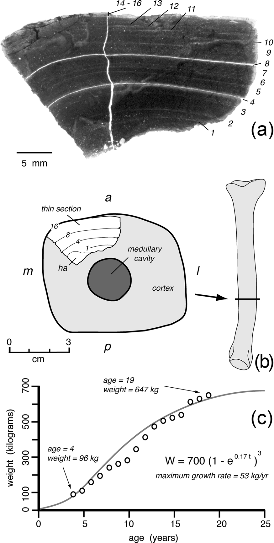
The fragmentary condition of metatarsal II in TMM 45906-1 allowed for preparation of a histologic section from the cortex of the anteromedial side of the shaft (Fig. 10). This section displays the dense laminar fibro-lamellar tissue with narrow vascular canal lumina typical of primary bone tissue in the diaphyses of theropod limb bones. Secondary osteons are sparsely distributed in the innermost third of the cortex; densely spaced secondary osteons and a few large endosteal resorption cavities are present only in the innermost compacta on the medial side (Fig. 10). The primary tissue preserves distinct growth modulations marked by thin intervals of avascular lamellar tissue. Sixteen growth lines may be readily distinguished, several of these (numbers 4, 8; Fig. 10) are more pronounced, and the sample has parted along these. The outermost three growth lines (numbers 14 to 16; Fig. 10) are very closely spaced lines of arrested growth, indicating that growth had nearly ceased at the time of this individual's death.
Aguja tyrannosaur histologic section taken from left MT II (TMM 45906-1·1): (a) photomicrograph in plane polarised light showing growth modulations (1–16); (b) location of the section on the anterolateral side of MT II showing a=anterior, l=lateral, m=medial, and p=posterior sides of shaft cross-section, and ha=areas of extensive remodeling; (c) growth history based on the 16 preserved growth increments, using the method of Lehman & Woodward (Reference Lehman and Woodward2008) with equation describing best-fit growth curve.
The method of growth curve fitting presented by Lehman & Woodward (Reference Lehman and Woodward2008) is used here to reconstruct the growth history for TMM 45906-1 based on the sampled histologic section (Fig. 10). In this case, allometric relationships between length of MT III and femur given by Currie (Reference Currie2003a, fig. 13) were used to estimate the femur length (765 mm) and femur circumference (263 mm) so that the body mass (647 kg) could be estimated from the metatarsus in TMM 45906-1. The preserved annual growth increments and body mass estimates fit best to a growth curve, indicating that this individual died approaching an adult weight of 700 kg at age 19 and attained a maximum growth rate of about 53 kg/year during its lifetime (Fig. 10). If this growth curve is correct, then expansion of the medullary cavity and endosteal remodelling had engulfed the first three growth increments, and the earliest preserved growth line was produced at age four.
The c. 20 years required to reach adult weight is comparable to that estimated for other tyrannosaurids (Erickson et al. Reference Erickson, Makovicky, Currie, Norell, Yerby and Brochu2004). However, the asymptotic adult weight and maximum growth rate in the Aguja tyrannosaur are approximately half that estimated by Erickson et al. (Reference Erickson, Makovicky, Currie, Norell, Yerby and Brochu2004) for most tyrannosaurids (1200–1400 kg adult weight, 122–180 kg/year growth rate) and dramatically lower than for Tyrannosaurus (5550 kg, 767 kg/year). This may be in keeping with the hypothesis that tyrannosaurids that attained ‘giant’ proportions did so by accelerating the exponential growth stage, and not by extending their life span (Erickson et al. Reference Erickson, Makovicky, Currie, Norell, Yerby and Brochu2004). Because the Aguja specimen is from an individual that had nearly reached adult size at the time of its death, these estimates are also in accordance with evidence that the Aguja tyrannosaur was relatively small compared to most Late Cretaceous tyrannosaurs.
2. Affinities of the Aguja tyrannosaur
Collectively, the isolated skeletal elements recovered from the Aguja Formation indicate the presence here of a relatively small slender tyrannosaur comparable in size to Appalachiosaurus or subadult Gorgosaurus and Albertosaurus. None of the elements found thus far pertain to individuals as large or robust as in adult Daspletosaurus or Tyrannosaurus.
2.1. Postcranial skeleton
The limb bones of the Aguja tyrannosaur are compatible with all tyrannosaurs less derived than tyrannosaurines. Some subtle features of the postcranial elements are close to Appalachiosaurus (e.g. circular shaft section and low height of the astragular facet on the tibia, and deep proximal intercondylar groove) but the Aguja ungual phalanx lacks the lipped morphology seen in Appalachiosaurus.
2.2. Cranial skeleton
Teeth of the Aguja tyrannosaur are closest in form to those of the albertosaurines Gorgosaurus and Albertosaurus, but teeth of less derived tyrannosauroids such as Appalachiosaurus and Bistahieversor have yet to be analyzed morphometrically, and are likely very similar. Teeth of Dryptosaurus are, however, much more laterally compressed (Brusatte et al. Reference Brusatte, Benson and Norell2011). The Aguja tyrannosaur lateral teeth lack the stout crowns typical of the derived tyrannosaurines Daspletosaurus and Tyrannosaurus, and likely associated with their fully developed bone biting “puncture and pull” feeding behaviour (e.g. Erickson & Olson Reference Erickson and Olson1996).
Currie (Reference Currie, Currie and Koster1987, p. 52) remarked that “the frontal is one of the more useful isolated bones of theropods as there is only a single pair in any individual, they tend to be found relatively frequently and they are diagnostic at the species level.” Therefore, it is fortunate, and perhaps not unexpected, that frontals are the only tyrannosaurian cranial elements thus far recovered from the Aguja, warranting its thorough comparison with other species.
The Aguja tyrannosaur frontal (Fig. 11) exhibits two features shared by all tyrannosaurids included in the phylogenetic analysis presented by Currie et al. (Reference Currie, Hurum and Sabath2003); the frontoparietal suture is extended anteriorly along the midline (Currie et al. Reference Currie, Hurum and Sabath2003, character 15), and the area for temporal muscular attachment is extensive on the dorsal surface of the frontals (Currie et al. Reference Currie, Hurum and Sabath2003, character 64). Holtz (Reference Holtz, Tanke and Carpenter2001, character state 62.1) elaborated likewise that tyrannosaurids share a supratemporal fossa that extends on to at least the posterior third of the frontal. Together these features suggest that the Aguja tyrannosaur is a member of Tyrannosauridae.
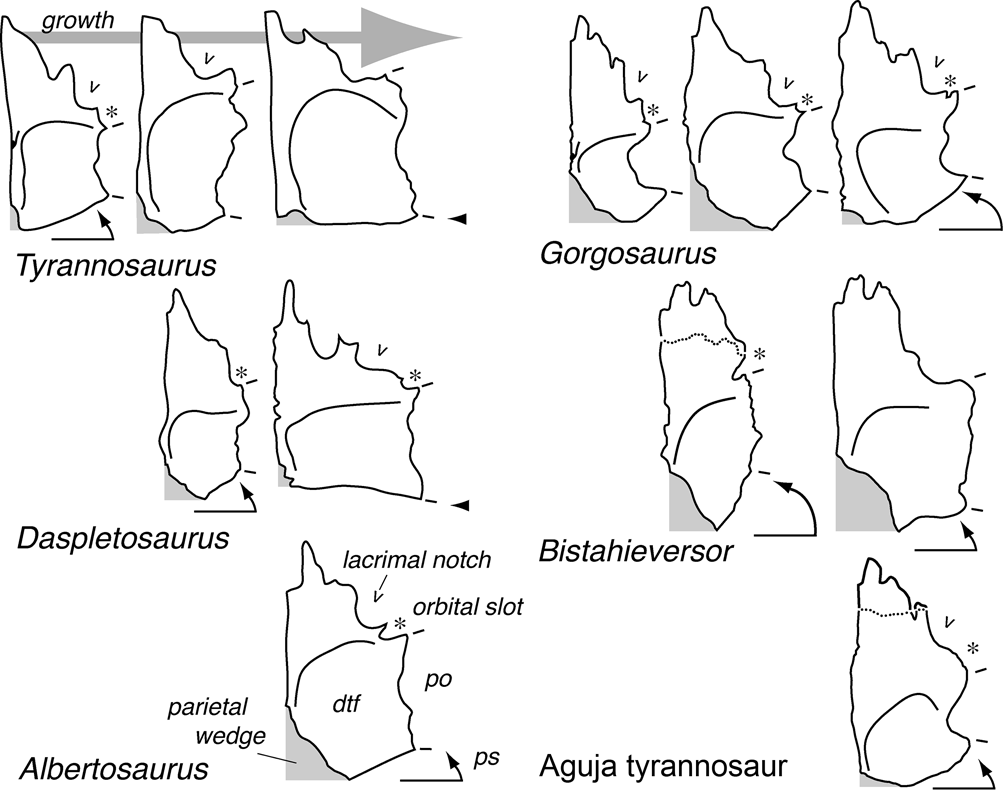
Comparison of tyrannosaur right frontals in dorsal view, reduced to common length: Albertosaurus (TMP 81.10.1; from Currie Reference Currie2003b); Bistahieversor (OMNH 10131; from Lehman & Carpenter Reference Lehman and Carpenter1990; NMMNH P-27469 modified from Carr & Williamson Reference Carr and Williamson2010); Daspletosaurus (TMP 94.143.1, NMC 8506; from Currie Reference Currie2003b, Russell Reference Russell1970); Gorgosaurus (TMP 91.36.500, AMNH 5664, TMP 80.16.924; from Currie Reference Currie, Currie and Koster1987, Reference Currie2003b; Carr Reference Carr1999); Tyrannosaurus (CMNH 7541, LACM 23845, FMNH PR2081; from Carr & Williamson Reference Carr and Williamson2004, Brochu Reference Brochu2003). Abbreviations: dtf=dorsotemporal fossa; po=postorbital suture (between tic marks); ps=angle of parietal suture; *=orbital slot.
Holtz (Reference Holtz, Tanke and Carpenter2001) also indicated that tyrannosaurids (excluding ‘Aublysodon’) have a frontal with the posterior end expanded laterally (Holtz Reference Holtz, Tanke and Carpenter2001, character state 61.2), and in the most derived tyrannosaurids (e.g. Tyrannosaurus) the body of the frontal is rectangular, with only a small rostral prong remaining (Holtz Reference Holtz, Tanke and Carpenter2001, character state 61.3). However, Currie (Reference Currie2003a, Reference Currieb) demonstrated that the shape of the frontal varies ontogenetically and/or with body size in at least some tyrannosaurids, from narrow and triangular in juveniles to broad and rectangular in adults, as the skull widens posteriorly with growth (Fig. 11). He regarded specimens referred to Aublysodon as simply juvenile individuals of other tyrannosaurids. Therefore, the shape of the frontal may be of limited use in assessing relationships if maturity of the individual cannot be established. Even so, some tyrannosaurs (e.g. Bistahieversor and the albertosaurines Gorgosaurus, Albertosaurus) as adults do not attain a broad quadrangular frontal (Fig. 11).
In Gorgosaurus, the anterior limit of the supratemporal shelf covers only the posterior part of the frontal, and extends from the midline junction with the parietals in an anteriorly curved arc to the anterior buttress for the postorbital (Russell Reference Russell1970). In Daspletosaurus torosus, this shelf extends laterally over a broader area of the frontal, and in Tyrannosaurus the supratemporal shelf covers most of the frontal. In the Aguja specimen, the supratemporal shelf covers even less of the frontal than in Gorgosaurus and Albertosaurus, and is closest to Bistahieversor in this feature. However, in Bistahieversor and albertosaurines, the parietals extend significantly forward on the midline along the sagittal crest to form a wedge separating the frontals, and traced laterally the frontoparietal suture forms a posteriorly curved arc. In Daspletosaurus and Tyrannosaurus, the parietals only slightly divide the frontals on the midline and the frontoparietal suture is transversely oriented (Fig. 11). The Aguja specimen is closer to the tyrannosaurines in this feature. Carr & Williamson (Reference Carr and Williamson2010) assessed the same basic features of the frontal with three characters: length of the ‘forehead’ (character 131); extent of the dorsotemporal fossa (character 134); and form of the frontoparietal suture (character 145). The Aguja specimen retains the long ‘forehead’ and short dorsotemporal fossa found in less derived tyrannosaurids, but rather than the markedly wedge-shaped frontoparietal suture present in these, here the suture is close to the transverse condition found in tyrannosaurines (Fig. 11).
The presence and anterior extent of the sagittal crest on the frontals (Holtz Reference Holtz, Tanke and Carpenter2001, character state 61·3) varies among tyrannosaurids, and also ontogenetically or with body size in a given species; but in most, the dorsal surface of the frontals is relatively flat anterior to the orbital ‘slot’. In the Aguja specimen, the sagittal crest is very pronounced and extends forward of the postorbital suture. This feature, plus a proportionally similar crest developed on a juvenile individual (TMM 41918-24), suggests that the large sagittal crest is a diagnostic characteristic of the Aguja tyrannosaur. Although the upper edge of the sagittal crest is abraded, the forward deflection of the frontoparietal suture along the midline suggests that it may have been only slightly overlapped by the forward-tilting leading edge of the parietal part of the crest. The entire crest was probably more pronounced than that in Bistahieversor (Carr & Williamson Reference Carr and Williamson2010; Fig. 4).
Even as adults, albertosaurine tyrannosaurids do not develop a strong sagittal crest on the frontal. The form of the sagittal crest is one of the key features used by Carr & Williamson (Reference Carr and Williamson2010) to diagnose the tyrannosaur Bistahieversor. However, here the ‘peaked’ crest appears to be developed mostly on the parietal; the extent of the sagittal crest on the frontal itself is comparable to that in most tyrannosaurids. If OMNH 10131 (Lehman & Carpenter Reference Lehman and Carpenter1990) is referable to Bistahieversor, as indicated by Carr & Williamson (Reference Carr and Williamson2010), then the frontal portion of the sagittal crest is much less developed than in the Aguja specimen, and the frontoparietal suture is so strongly deflected anteriorly that the parietal dorsally overlaps the frontal to a much greater extent (Fig. 4). In neither Gorgosaurus nor Albertosaurus does the sagittal crest extend significantly onto the frontal from the parietal (Currie Reference Currie2003b). However, in Daspletosaurus and Tyrannosaurus, the sagittal crest extends forward on the frontal medial to the anterior end of the postorbital suture, as in the Aguja specimen and Bistahieversor. Carr & Williamson (Reference Carr and Williamson2010) scored three features of the sagittal crest on the frontal: whether it is lacking, single or paired (character 137); its height (character 138); and length (character 139). The Aguja specimen has a tall and long sagittal crest as in Bistahieversor and tyrannosaurines, but the crest is not divided along the midline (in contrast to Tyrannosaurus). The tall sagittal crest is considered a derived tyrannosaurine feature by Carr & Williamson (Reference Carr and Williamson2010, character 138). It is interesting that the sagittal crest in the Aguja tyrannosaur appears to extend forward of the dorsotemporal fossa, suggesting that it may have been at least in part ornamental rather than solely for expanded attachment of the temporal musculature.
In tyrannosaurids, two or three prongs of the nasal overlap and interdigitate with the anterior end of the frontal; in some specimens (TMP 80.16.924; Currie Reference Currie, Currie and Koster1987) there are as many as four such prongs. In Bistahieversor, the prongs are said to be “supernumerary” (Carr & Williamson Reference Carr and Williamson2010, character 44) but it is not clear that this condition is particularly unusual. Brochu (Reference Brochu2003) did not place much systematic value on the configuration of the suture. In the Aguja specimen, the nasal process is broken and the cleft for only the most lateral of the nasal prongs is evident; its posterior limit is medial to the lacrimal socket as in Gorgosaurus (Fig. 11). As a result, the missing nasal process was relatively wide (Carr & Williamson Reference Carr and Williamson2010, character 133), as in albertosaurines.
The orbital slot (‘frontal cleft’ of Russell Reference Russell1970 or ‘supraorbital incisure’ of Molnar Reference Molnar1978) varies in its expression among tyrannosaurids (Holtz Reference Holtz, Tanke and Carpenter2001, character 58). Gorgosaurus has a marked posteriorly-curved cleft in the frontal above the orbit that separates the lacrimal and postorbital. This cleft remains present, but does not penetrate into the frontal in Daspletosaurus torosus (Russell Reference Russell1970). In Tyrannosaurus, the cleft is absent entirely and the lacrimal and postorbital are in contact, excluding the frontal from the dorsal border of the orbit. However, the orbital slot is known to have closed ontogenetically in at least some tyrannosaurs, and as a result, this character has not been considered useful in some phylogenetic analyses (Currie et al. Reference Currie, Hurum and Sabath2003; Carr & Williamson Reference Carr and Williamson2004). The Aguja specimen has a weakly expressed and relatively wide orbital slot, but no sulcus on the dorsal surface of the frontal and no distinct lateral cleft.
A pneumatic opening of the sort found within the frontal in the Aguja specimen, has not been described in other tyrannosaurs. Nor is a cavity evident in the form of postmortem collapse of the skull roof in this position in other specimens (although Currie Reference Currie2003b, fig. 20 shows crushing of the skull roof in this area on a young individual of Daspletosaurus sp.). Computerized tomographic scans of the skull in Tyrannosaurus (Brochu Reference Brochu2003) do not reveal a sinus in this position, although cranial pnuematicity has been extensively delineated in other skull bones in most tyrannosaurs (e.g. Currie Reference Currie2003b; Brusatte et al. Reference Brusatte, Carr, Erickson, Bever and Norell2009). The sinus cavity in the larger Aguja frontal (TMM 45905-1) is clearly not present in the smaller specimen (TMM 41918-24), and so it may have developed ontogenetically, or as a result of pathology. The cavity could even be a taphonomic artifact, and as a result it is of doubtful systematic value.
In the Aguja specimen, the prefrontal contact is a deep groove along the ventrolateral edge of the bone. The contact rises anteriorly, but the prefrontal must have been exposed on the skull roof only in a limited area lateral to the anterior nasal process of the frontal. Currie (Reference Currie, Currie and Koster1987) described the prefrontal suture as a socket overlapped posteriorly by the frontal, and interpreted this condition as a primitive one for tyrannosaurids, present only in Gorgosaurus and Albertosaurus. In tyrannosaurines Daspletosaurus and Tyrannosaurus, the prefrontal/frontal contact is instead an ‘hourglass-shaped’ serrate immobile joint (Molnar Reference Molnar1980, Reference Molnar1991). However, Molnar (Reference Molnar1980) described a specimen (LACM 23845) with the prefrontal ‘socket’ condition that is identified as a juvenile individual of Tyrannosaurus by recent authors (Currie Reference Currie2003a; Carr & Williamson Reference Carr and Williamson2004). As a result, variation in this feature may be regarded as mostly ontogenetic and it is not considered in recent phylogenetic analyses. The prefrontal articulation in the Aguja tyrannosaur is closer to the ‘hourglass-shaped’; joint in tyrannosaurines, and is overlapped posteriorly and laterally by the frontal and lacrimal to such an extent that the prefrontal may have been exposed only in a limited area on the dorsal surface of the skull.
The form of the joint surface for the lacrimal was regarded as particularly significant by Carr & Williamson (Reference Carr and Williamson2010). They assessed three features (Carr & Williamson Reference Carr and Williamson2010, characters 140, 141 and 142) including the length of the contact in dorsal view (none, long, or short), the width of the contact in dorsal view (none, narrow, or wide), and whether the lacrimal articulation is with the prefrontal, rostromedial, or a distinct notch. The ‘notch’ articulation is shared by all tyrannosaurids in their analysis, and is present in the Aguja tyrannosaur. The short articulation is found only in derived tyrannosaurines (Daspletosaurus, Tyrannosaurus) and is lacking in the Aguja tyrannosaur.
One feature of the Aguja specimen is shared only with albertosaurines (Gorgosaurus and Albertosaurus) in the phylogenetic analysis of Currie et al. (Reference Currie, Hurum and Sabath2003); the suture for the postorbital is divided into a distinct vertical anterior portion and a horizontal posterior shelf (Currie et al. Reference Currie, Hurum and Sabath2003, character 14). Other tyrannosaurids included in their analysis lack this distinction. This feature suggests that the Aguja tyrannosaurid is a member of Albertosaurinae. However, if OMNH 10131 is correctly referred to Bistahieversor (Carr & Williamson Reference Carr and Williamson2010), then this sutural configuration is also found in less derived tyrannosauroids and it might, therefore, be a primitive condition retained in Albertosaurines. Carr & Williamson (Reference Carr and Williamson2010, character 143) scored this condition as a derived one present in Bistahieversor and all tyrannosaurids instead. However, in adult Daspletosaurus and Tyrannosaurus, the postorbital suture is substantially expanded posteriorly, resulting in the wide frontal and transverse frontoparietal suture characteristic of tyrannosaurines (Fig. 11). In Tyrannosaurus, a neurovascular foramen pierces the union between the frontal and postorbital at the junction between the two distinct postorbital sutural facets (Brochu Reference Brochu2003). This foramen pierces the inner wall of the orbit in the Aguja specimen, but does not appear to extend to the skull roof. The configuration of the postorbital suture in the Aguja specimen is similar to Bistahieversor and albertosaurines; however, the entire postorbital sutural surface is relatively much smaller than in any other tyrannosaur (Figs 4, 11).
The frontal of the Aguja tyrannosaur is distinctly different from that in other species. It is narrow, has a wide nasal prong, a wide orbital slot, a small postorbital articulation divided into two distinct facets, and a frontoparietal suture that extends only slightly anterolaterally but not directly laterally. In most of these features, the frontal is like that in albertosaurines; however, the frontal is unknown in the less derived tyrannosauroids Dryptosaurus and Appalachiosaurus, and could also be similar. The tall sagittal crest on the frontal, and very weakly embayed midline suture with the parietal, is similar to that in highly derived tyrannosaurines Daspletosaurus and Tyrannosaurus. Although Bistahieversor also has a pronounced sagittal crest, if OMNH 10131 is correctly referred, its frontal is quite different from that in the Aguja tyrannosaur. The parietal suture on the sagittal crest in Bistahieversor is much more strongly deflected anteriorly, and the crest is developed almost entirely on the forwardly inclined parietal wedge inserted between the frontals on the midline. Hence, the only diagnostic cranial element thus far recovered of the Aguja tyrannosaur exhibits a mosaic of features, but the tall sagittal crest suggests that it is a tyrannosaurine.
2.3. Summary
The postcranial elements teeth and most features of the frontal in the Aguja tyrannosaur suggest that it is either an albertosaurine or less derived tyrannosauroid. In contrast, the pronounced frontal sagittal crest and frontoparietal suture are most similar to those of derived tyrannosaurines. The tall sagittal crest is so distinctive it is regarded by some (e.g. Carr & Williamson Reference Carr and Williamson2010; Carr et al. Reference Carr, Williamson, Britt and Stadtman2011) as a synapomorphic character of Tyrannosaurinae; and so on that basis, we have referred the Aguja tyrannosaur to this clade.
2.4. Fragmentary specimens and systematics
Although features of its frontal, the only cranial element thus far known of the Aguja tyrannosaur suggest that this may be an unknown tyrannosaurine species; past experience makes it clear that it would be unwise to provide a name for this, or assign it to what may be a near relative. This philosophy is best illustrated by several examples.
Aublysodon was initially a taxon based solely on distinctive premaxillary teeth (e.g. Molnar Reference Molnar1980). On the basis of a fragmentary skull (LACM 28471), a case was made by Molnar & Carpenter (Reference Molnar and Carpenter1989) that Aublysodon was a distinct small tyrannosaur. Currie (Reference Currie, Currie and Koster1987) suggested that isolated slender tyrannosaur frontals might be referable to Aublysodon, and Lehman & Carpenter (Reference Lehman and Carpenter1990) referred a fragmentary tryrannosaur specimen from New Mexico (OMNH 10131) to Aublysodon on the basis of its premaxillary teeth and frontal. Carr & Williamson (Reference Carr, Williamson, Lucas and Heckert2000) later identified OMNH 10131 as Daspletosaurus sp. However, recently Carr & Williamson (Reference Carr and Williamson2010) referred the specimen to a new genus, Bistahieversor. Following the recovery and study of juvenile tyrannosaur specimens, Currie (Reference Currie2003a) determined that material referred to Aublysodon represented juvenile specimens of other tyrannosaurids, and he referred OMNH 10131 to Tyrannosaurus. Hence, over the past decade this single fragmentary specimen has been referred to four different genera (Aublysodon, Daspletosaurus, Bistahieversor, and Tyrannosaurus).
Another fragmentary specimen (LACM 23845) from Montana was believed to represent a slender Lancian tyrannosaur distinct from Tyrannosaurus. Initially, it was referred to Albertosaurus lancensis (Molnar Reference Molnar1980) but later made the type of a new species, A. megagracilis (Paul Reference Paul1988) and, ultimately, a distinct new genus Dinotyrannus (Olshevsky Reference Olshevsky1995). The type of A. lancensis (CMNH 7541) was itself later assigned to its own genus Nanotyrannus (Bakker et al. Reference Bakker, Williams and Currie1988). Most recent authorities suggest that both Dinotyrannus and Nanotyrannus are simply juvenile individuals of Tyrannosaurus rex (Currie Reference Currie2003a; Carr & Williamson Reference Carr and Williamson2004). Hence, over the past decade these specimens have been referred to four different genera.
The past conservative tendency was to refer isolated fragmentary specimens from new geographic provinces to better-known genera found in other areas. More recently, the predominant philosophy has been to refer such specimens to their own monotypic genera. Depending on the prevailing taxonomic paradigm and the degree of individual variation that one is likely to regard as intraspecific, ontogenetic, or sex-associated, even relatively complete specimens may be subject to markedly different interpretations. So, in the case of the ‘Aguja tyrannosaur’, although the meagre material suggests that it may represent an unknown species, it seems unwise to provide a formal name for it, and it is advised to await the discovery of more complete material that may help resolve its relationships.
3. Conclusions
The scarce remains of tyrannosauroid dinosaurs from the Aguja Formation record the presence here of a relatively gracile species, comparable in size and form to Appalachiosaurus known from Alabama, or subadult individuals of the Canadian albertosaurines, Gorgosaurus and Albertosaurus. Histologic analysis of one of the specimens indicates that the Aguja tyrannosaur attained an adult size (700 kg, 6·5 m body length) substantially smaller than adult albertosaurines. The frontal bone is narrow, exhibits a wide orbital slot and bipartite joint for the postorbital, features thought to be diagnostic of Albertosaurinae, but a pronounced sagittal crest and reduced midline parietal wedge generally thought to be diagnostic of Tyrannosaurinae. Because the tall sagittal crest may be a synapomorphy of Tyrannosaurinae, we refer the Aguja tyrannosaur to that clade. However, the unique combination of character states exhibited by the frontal prevents its confident attribution to any known species.
If the Aguja tyrannosaur is indeed a member of Tyrannosaurinae, then it appears to lack some features typical of derived members of the group (e.g. Daspletosaurus, Tyrannosaurus), such as their stout teeth and robust limb elements, but this may simply be in keeping with its relatively small adult body size. There are other small tyrannosaurines. The Aguja tyrannosaur is comparable in size to Teratophoneus (c. 700 kg; Carr et al. Reference Carr, Williamson, Britt and Stadtman2011) and both are approximately twice the size of Alioramus, which is generally regarded as a basal tyrannosaurine (Brusatte et al. Reference Brusatte, Benson and Norell2011). The Aguja tyrannosaur also retains narrow frontals, similar to Alioramus. This may reflect its small size, and correspond with a relatively narrow skull, unlike the posteriorly widened skull in larger derived tyrannosaurines (Daspletosaurus, Tyrannosaurus).
The Aguja tyrannosaur provides further evidence that the predatory tyrannosauroids that inhabited the shores of ‘Laramidia’ may have been as diverse and as provincial as their large herbivorous dinosaurian prey (Lehman Reference Lehman, Wolberg and Stump1997; Carr et al. Reference Carr, Williamson, Britt and Stadtman2011). Furthermore, if the Aguja tyrannosaur is a tyrannosaurine, then this most derived clade was more widespread geographically than previously recognised. By the middle Campanian, representatives of the group were present in Texas as well as Alberta. Although the Western Interior Sea provides a plausible mechanism for geographic isolation of ‘Appalachia’ where less derived tyrannosauroids held sway through the end of Cretaceous time (Brusatte et al. Reference Brusatte, Benson and Norell2011), it is puzzling that less derived forms also remained (or were reintroduced) later in New Mexico and that others (albertosaurines) remained in Canada. If current taxonomy is upheld, then no Campanian tyrannosauroid found in any one area of North America is found in any other. In the absence of significant geographic barriers to dispersal, such marked provinciality seems inexplicable, particularly in light of the fact that modern large mammalian carnivore species typically have ranges that span most, or all, of a continent (e.g. Lehman Reference Lehman, Wolberg and Stump1997). This high degree of endemism in such large terrestrial predators may reflect specialisation in prey selection, or other ecological constraints on these animals, unlike those that govern the geographic distribution of large mammalian carnivores.
4. Acknowledgements
The authors thank W. Langston, Jr. for his longtime support of our work in the Aguja Formation and for many hours of entertaining and often intellectually stimulating conversations over the years. We also thank T. Rowe and L. Murray of the Texas Memorial Museum for their kind assistance in studying and curating specimens at the Vertebrate Paleontology Laboratory, D. Corrick of the Science and Resource Management staff of Big Bend National Park for his support of our fieldwork in the Park, J. Schubert for his help with photography and J. Browning for thin section preparation. P. Currie offered helpful advice, and T. Carr and S. Brusatte provided useful comments that substantially improved the manuscript. Drawings are the work of T. Lehman.