INTRODUCTION
Glottic cancer is the most common malignant tumour of the larynx, with approximately 30–40% tumours presenting in the early stages.Reference Mendenhall, Mancuso, Amdur and Werning 1 , Reference Petrakos, Kontzoglou, Nikolopoulos, Papadopoulos and Kostakis 2 Radiotherapy has been frequently chosen as the primary treatment because of its superiority in preserving the voice quality.Reference Mendenhall, Mancuso, Amdur and Werning 1 , Reference Teshima, Chatani, Hata and Inoue 3
The conventional radiotherapy technique, opposed lateral field technique, creates a uniform dose distribution on the beam pathways. This feature causes high radiation dose exposure in unnecessary areas. Thus, the risk of adverse effects can be increased in organs lying in the beam paths, such as the carotid artery, spinal cord, pharyngeal constrictor muscle, all designated as organs at risk (OAR). The potential risk of long-term complications caused by radiation exposure to the carotid artery during head and neck cancer treatment has been reported in a previous study. Gujral et al.Reference Gujral, Shah, Chahal, Senior, Harrington and Nutting 4 who reported that the thickness of carotid intima-media can be increased by radiation exposure, which causes an increase in carotid artery stenosis risk, thereby consequently increasing the risk of a cerebrovascular accident.
In our study, our first objective was to decrease the dose of radiation received by the carotids; the second objective was to compare the planning target volume (PTV) coverage as well as the dose received by the spinal cord by undertaking a comparison of intensity-modulated radiotherapy (IMRT) and 3D conformal radiotherapy (3DCRT) planning techniques.
PATIENTS and METHODS
This study was undertaken at Gharbia Cancer Society, Egypt, where six patients with early stage glottis carcinoma (stage T1-2 N0M0) were included in the study. These patients were staged with laryngoscopy, computed tomography (CT) scans, tissue biopsy and chest CT.
SIMULATION
All patients were immobilised in the supine position with a thermoplastic mask. CT simulation was undertaken for all patients with a 3-mm slice thickness and CT scans were obtained from the top of the skull to the top of aortic arch. The CT images were imported into Eclipse treatment planning system, version 10.0.34 (CA, USA).
CONTOURING
Clinical target volume (CTV) was delineated; superiorly, to include the most cranial extent of the arytenoid cartilage, inferiorly, 1·5 cm below the level of the true vocal cords, posteriorly, to include the arytenoid cartilage, posterior commissure, and anteriorly and laterally, to include the anterior commissure and to exclude the thyroid cartilage; if the carotid artery overlapped with CTV.
CTV was modified manually to exclude the carotid artery. The PTV volume had 0·5 cm margin from the CTV. Dose of 66 Gy/33 fractions was prescribed to the PTV.
The carotid arteries and the spinal cord were delineated as OAR. The carotid artery volumes and spinal cord were expanded by 3 mm to create planning target at risk volume (PRV).
Two plans were created on the Eclipse treatment planning with pencil beam algorithm for each patient.
The first plan was 3D plan which consisted of two laterally opposed wedged fields and normalised such that D 95% for the PTV≥95% of the prescription dose (PD). The second plan was an IMRT plan which had multiple approaches with different numbers of beams, angles and segments that were evaluated. The final approach selected for standardised planning was a seven-field IMRT plan. The optimal gantry angle degrees were 180, 231·5, 283, 334·5, 25·5, 77 and 128·5°. The direct posterior beam allowed better coverage for the PTV close to the sensitive structures (spinal cord and carotid) and decreased the dose received to these structures.Reference Doaa, Ehab, Shady and Wafaa 5
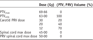
The optimisation parameters were performed by lowering the desired mean dose to the carotid as shown in Table 1.
Intensity-modulated radiotherapy optimisation parameters

Abbreviations: PTV, planning target volume; PRV, planning target at risk volume.
The IMRT plan was normalised so that 100% of the PTV received more than 95% of the PD. To spare the carotid artery, V 25 Gy, V 35 Gy, V 50 Gy and the mean dose were calculated for PRV to carotid artery.
In addition, the conformity and homogeneity indices were calculated for both 3D and IMRT plans using the following formula according to the definition of ICRU 83. 6
 $${\rm Conformity}\,{\rm index{\equals}}{{{\rm Treatment}\,{\rm volume}} \over {{\rm PTV}}},$$
where treatment volume was defined as the volume irradiated to more than 95% of the PD.
 $${\rm Homoginity}\,{\rm index}{\equals}{{D_{2} {\minus}D_{{98}} } \over {D_{{50}} }},$$
where D 2%, D 98%, D 50% were defined as the minimum doses delivered to 2 and 98 of PTV, and median dose to PTV.
 $${\rm Over}\,{\rm dose}\,{\rm index{\equals}}{{V_{{105}} } \over {{\rm PTV}}}$$
Treatment was delivered by 6 MV (Varian medical system 600c, California, USA) with 40 paired multi-leaf collimators, the leaf width at the isocentre was 1 cm.
For IMRT plans, the actual dose delivered to the patient was verified before treatment using a 2D detector array.
Data were analysed using Origin 6 and summarised as mean and standard deviation. Probability (p-value) <0·05 is considered significant.
RESULTS
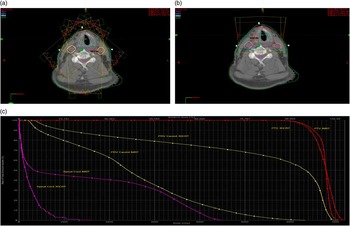
Figure 1 demonstrates an example of dose distributions in (a) IMRT, (b) 3D plans and (c) comparison of dose–volume histograms for the corresponding IMRT and 3D plans.
The transverse view dosimetry comparison between (a) seven-beam intensity-modulated radiotherapy (IMRT) treatment and (b) two-beam conventional 3D conformal radiotherapy treatment at the isocentre level for one patient. The clinical target volume and the planning target volume (PTV) were represented by the solid redline. The 95, 100,105 and107% of the prescription dose are shown by the green, blue, cyan and purple colour isodose lines. A better dose conformity to the PTV can be achieved in the IMRT treatment. (c) Shows dose–volume histogram for 3D and IMRT planning techniques. Contours for PTV (red lines), planning targets at risk volume of carotid arteries (yellow line) and spinal cord (purple lines).

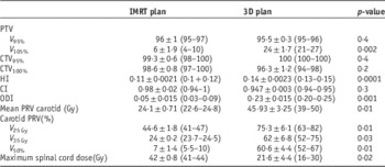
Table 2 shows the mean, standard error of the mean and range for the dosimetric data for the tumour coverage, conformity index (CI), homogeneity index (HI), over dose index (ODI) and normal structure parameters of the two different plans.
Dosimetric data for target volume and organs at risk in intensity-modulated radiotherapy (IMRT) and 3D conformal radiotherapy plans

Abbreviations: PTV, planning target volume; CTV, clinical target volume; HI, homogeneity index; CI, conformity index; ODI, over dose index; PRV, planning target at risk volume.
PTV coverage
The dosimetric data for PTV coverage (V 95%, V 105%), CI, HI and ODI are shown in Table 1. The mean V 95% values for IMRT and 3DCRT plans were 96±1 (range, 95–97) and 95·5±0·3 (range, 95–96), respectively, with no significant difference (p=0·4). However, the V 105% values which represent the hot spots were higher in the 3DCRT plans 24·1±1·7 (range, 21–27) compared with the IMRT plans 6±1·9 (range, 4–10), respectively, with statistical significance (p=0·002). The mean CI was 0·98±0·02 (range, 0·94–1) in the IMRT plans compared with 0·947± 0·003 (range, 0·94–0·95) in the 3DCRT plans (p=0·3). The mean homogeneity indices were 0·11±0·0021 (range, 0·1–0·12) and 0·14±0·0023 (range, 0·13–0·15) in the IMRT and 3DCRT plans, respectively, with statistical significance (p=0·0001).
Carotid artery PRV dose
The doses for the carotid artery PRV with the two different plans are shown in Table 1. The mean carotid artery PRV dose was 24·1±0·7 Gy (range, 22·6–24·8 Gy) for the IMRT plan and 45·93±3·25 Gy (range, 39–50 Gy) for the 3DCRT plan; thus, the dose was significantly lower in the IMRT plan compared with the 3DCRT plan (p=0·01). Carotid artery PRV V 25 Gy, V 35 Gy and V 50 Gy were 44·6%±1·8 (range, 41–47%), 24%±0·2 (range, 23·7–24·5%) and 7%±1·4 (range, 5·5–10%) for the IMRT plan compared with 75·3%±6·1 (range, 63–82%), 62%±6·8 (range, 52–75%) and 60·4%±4·4 (range, 52–67%), respectively, for the 3DCRT plan. There was a statistically significant reduction in the volume of the carotid artery PRV irradiated when using IMRT than 3DCRT at different dose levels (p=0·01, 0·03 and 0·01), respectively.
Spinal cord dose
The spinal cord dose was significantly higher in the IMRT plan. The maximum spinal cord dose was mean 42±0·8 Gy (range, 41–44 Gy) for the IMRT plans, and 21·6±4·4 Gy (range, 16–30 Gy) for the 3DCRT plans. However, all values were below the spinal cord threshold dose of 45 Gy.
DISCUSSION
Carotid arteries lying in the beam pathway of conventional radiotherapy for early glottic cancer are exposed to relatively high doses almost identical to the target dose. There are several reports showing the relation between radiotherapy and carotid artery stenosis. Brown et al.Reference Brown, Foote and McLaughlin 7 reported that the incidence of carotid artery stenosis was higher in the irradiated side of neck than that in the contralateral unirradiated neck. In addition, some reports showed that radiotherapy increased the risk of cerebrovascular accident. Smith et al.Reference Smith, Smith and Buchholz 8 reported that the 10-year incidence of cerebrovascular events was increased by 9% in head and neck cancer patients when they were treated with radiotherapy. Dorresteijn et al.Reference Dorresteijn, Kappelle and Boogerd 9 also reported that the 15-year cumulative risk of a stroke after radiotherapy to the neck was 12% and there was a 5·6 times higher chance of stroke in patients who had undergone neck radiotherapy than those who had not undergone neck radiotherapy.
Hence, in order to reduce the carotid artery dose for early glottic cancer treatment, some studies have adopted the carotid-sparing techniques by using modified oblique field, IMRT, arc therapy or tomotherapy instead of conventional radiotherapy. Rosenthal et al.Reference Rosenthal, Fuller and Barker 10 first reported their carotid-sparing IMRT technique for early glottic cancer. They treated 11 patients with T1-2 glottic cancer, and suggested that IMRT significantly reduced radiation dose to the carotid arteries compared with conventional techniques. They concluded that IMRT techniques are necessary, especially for younger patients.
Recently, Zumsteg et al.Reference Zumsteg, Riaz and Jaffery 11 reported the dosimetric as well as the clinical results of 48 patients who were treated with carotid-sparing IMRT. They found that the 3-year local control rate was 88%, which was similar to that for conventional radiotherapy. They suggested that carotid-sparing IMRT was a safe and effective treatment method.
Our study also demonstrated that the carotid artery dose was much lower with carotid-sparing IMRT than with 3DCRT for early glottic cancer.
In our study, the difference in the mean dose to PRV of carotid arteries between IMRT and 3DCRT was very large, ~21 Gy and the dose in the IMRT plan was lower at all dose levels.
Our study also demonstrated that there was no difference in the PTV coverage for V 95% between IMRT and 3DCRT but the hot spot was much lower in the IMRT than the 3DCRT. In addition, we found that there was no difference in the conformity between IMRT and 3DCRT plans, where as the IMRT plan showed significant favourable homogeneity than the 3DCRT plan.
This was similar to what was reported by Hoon et al.Reference Hoon, Bae and Hojin 12 who reported that there was no difference regarding PTV coverage between IMRT and 3DCRT plans with better homogeneity and conformity for the IMRT plan.
For the IMRT plan, spinal cord dose was significantly higher than that for the 3DCRT plan, but all values were below the threshold dose of 45 Gy.
CONCLUSION
IMRT significantly reduces the radiation dose to the carotid arteries compared with 3DCRT while maintaining CTV coverage. Such an outcome assists in decreasing the incidence of radiation-induced carotid stenosis, thus improving the quality of life for these patients.