Introduction
A laryngopyocoele is an infected laryngocoele containing pus. It is a rare diagnosis and can present as an acute airway emergency. We present such a case, in which a simple intervention avoided the need for endotracheal intubation or a tracheostomy.
Case report
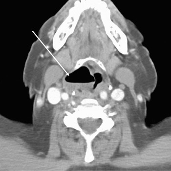
A 71-year-old woman was referred to the out-patient department with a three-month history of dysphonia. Laryngoscopy revealed mild right supraglottic swelling. A computed tomography (CT) scan of the neck identified an air-filled cavity consistent with a right-sided laryngocoele (Figure 1).
Transverse computed tomography scan of the neck showing a right-sided laryngopyocoele (arrow) at the level of the thyrohyoid membrane.

Six weeks later, the patient was admitted as an emergency with a three-day history of increasing dyspnoea and stridor.
On arrival, the patient was distressed but had a Glasgow coma score of 15. Her tympanic temperature was 37°C, with a respiratory rate of 35 breaths per minute, heart rate 120 beats per minute and oxygen saturation 92 per cent on air. The patient was dysphonic, with a marked inspiratory stridor.
Neck examination revealed a large, right-sided, soft neck mass, centred at level III, measuring approximately 10 × 5 cm. Fibre-optic laryngoscopy revealed a large, smooth, inflamed, right supraglottic mass obscuring the laryngeal inlet.
Nebulised adrenaline and high flow oxygen were commenced immediately. One hundred milligrams of intravenous hydrocortisone was given.
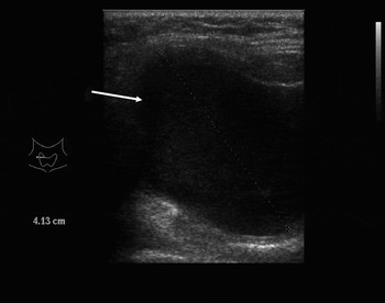
The patient was taken directly to the radiology department. Ultrasound imaging confirmed a laryngopyocoele (Figure 2). Under ultrasound guidance, a 21-G needle was directed into the cyst and 30 ml of pus was aspirated. The patient's dysphonia and stridor resolved immediately. The right neck mass was no longer palpable. Repeat fibre-optic laryngoscopy confirmed resolution of the mass, with a clear view down to the glottis and upper trachea.
Ultrasound image of the right-sided laryngopyocoele (arrow) prior to aspiration.

Two days later, the patient underwent internal deroofing of the laryngocoele. This was performed using microlaryngeal instruments, with an endotracheal tube in situ.
Six weeks later, the patient underwent definitive surgical excision of the laryngocoele, using an external approach. The external portion of the laryngocoele dissected clear to identify the neck at the thyrohyoid membrane. A limited lateral thyrotomy was performed to ensure complete excision of the internal portion, with careful preservation of the superior laryngeal nerve. An elective tracheostomy was not required.
Discussion
The laryngeal ventricle has a horizontally arranged, elliptical orifice extending from the notch in the thyroid cartilage to the anterior edge of the arytenoid cartilage. At the base of the ventricle, there is an extension called the saccule, which varies in size and is lined by respiratory epithelium. A laryngocoele is an abnormal dilatation of the saccule.
The estimated incidence of laryngocoeles is 1:2 500 000 population per annum in the UK, and the sex incidence is 5:1 in favour of men.Reference Stell and Maran1, Reference Rashid and Warltier2 The aetiology of laryngocoeles remains uncertain. It has been postulated that the laryngocoele represents a remnant, in the human, of a structure found normally in some apes.Reference Broyles3 The high intraglottic pressure associated with activities such as glass-blowing and playing certain wind instruments has been proposed as a risk factor. The rarity and unilateral tendency of laryngocoeles makes this unlikely. An underlying congenital predisposition may be a factor.Reference Canalis, Maxwell and Hemenway4 There have been a number of reports documenting the association between laryngocoele and laryngeal carcinoma.Reference Canalis5–Reference Scott, Johnson, Curtin and Barnes7 It is certainly conceivable that pathology involving the ventricle could cause obstruction of the saccule neck, with dilatation and a resulting laryngocoele. The presence of a laryngocoele should always prompt a detailed examination of the ventricle to exclude an underlying carcinoma.
The commonest presenting symptoms of a laryngocoele are dysphonia and neck swelling. Approximately 10 per cent become infected and present as a laryngopyocoele.Reference Stell and Maran1, Reference Rashid and Warltier2 Laryngopyocoele typically presents with rapidly progressive respiratory obstruction. A neck mass is usually present and may be accompanied by dysphonia, dysphagia, odynophagia and fever. Emergency airway management is paramount, including oxygen, nebulised adrenaline and steroid medication. Attempts at endotracheal intubation may fail and could result in rupture of the cyst and purulent aspiration. Tracheostomy has been reported to be the preferred management option, one report stating it to be mandatory.Reference Iversen8, Reference Sutay, Guneri and Gunbay9
We present the first reported case of ultrasound-guided aspiration of a laryngopyocoele in a patient with an obstructed airway. It is a quick and straightforward technique and gives immediate resolution of the airway crisis. Laryngopyocoele is particularly suited to this technique as it has a single cavity without loculations. The ultrasound is a diagnostic aid, and allows precise entry of the needle into the cyst, avoiding major structures. The need for endotracheal intubation or a tracheostomy is avoided.
• A laryngopyocoele is an infected laryngocoele containing pus, and is a rare diagnosis
• This study is the first reported case of ultrasound-guided aspiration of a laryngopyocoele in a patient with an obstructed airway, avoiding the need for a tracheostomy
Close observation is required post-aspiration to detect signs of re-accumulation. In the case presented, controlled deroofing of the cyst helped avoid re-accumulation of the cyst and allowed the patient to go home for a period before definitive surgery. The authors recommend excising laryngocoeles soon after diagnosis, especially in symptomatic patients or in those with a laryngopyocoele. This could avoid a potentially life-threatening situation.