Implications
Deer farming has a turnover of 3000 million dollars in the United States of America and hundreds of millions in New Zealand and other countries. Previous results from our research group demonstrated that copper (Cu) supplementation tended to increase the cortical thickness of red deer antlers, which may increase their weight and value as trophies, in addition to their resistance to fracture. However, Cu supplementation could influence meat quality and increase its concentration on meat to excessive levels for humans. Current results indicate that supplementation with Cu bolus is safe for consumption of venison and does not impair meat quality of different muscles.
Introduction
The main deer products for most countries are antlers as a trophy and meat. In addition, immature antlers are also an important product for Asian traditional medicine in China, New Zealand and some regions of Russia. Deer meat is considered an excellent product from the nutritional point of view and may be considered a very healthy food among red meats. In fact, deer meat has a low intramuscular fat content (Volpelli et al., Reference Volpelli, Valusso, Morgante, Pittia and Piasentier2003), and a high content of minerals (iron, calcium, phosphorus, magnesium and zinc) and polyunsaturated fatty acids (FA; Fisher et al., Reference Fisher, Bayntun, Enser and Elliot1998; Bureš et al., Reference Bureš, Barton, Kotrba and Hakl2015).
One of the common practices in deer management is Cu supplementation because its lack is one of the most frequent and severe trace mineral deficiencies in deer. Cattle are also usually supplemented with Cu because it improves offspring growth and, therefore, its profitability (Cashman, Reference Cashman2006). This practice led to the European Union authorities to establish the safe levels of Cu in meat for human consumption, and thus they set the maximum content of Cu allowed in the food of animal origin to 5 mg/kg of fresh weight (Council Regulation EC No 149, 2008). Thus, considering the recent studies showing the positive effects of Cu supplementation in deer antlers (Gambín et al., Reference Gambín, Serrano, Gallego, García, Capelli, Ceacero and Landete Castillejos2017) and the fact that venison is consumed, even from animals shot for trophy purposes, we set to study the effects of Cu supplementation in deer meat using one of the methods most widely used to supplement this mineral, rumen boluses. The ultimate reason was to assess if Cu contents may reach harmful levels for human consumption. For such study, we assessed the Cu content and the meat quality traits of two red deer muscles: sternocephalicus (ST) and rectus abdominis (RA). Because the aim of such supplementation was to improve antler quality rather than preventing deficiency, we set out to assess the effect on deer fed with a balanced diet.
Material and methods
Experimental design
At 3 months of age (23 days after weaning as average), Cu bolus (Agrimin 24.7; Agrimin Limited, Arlanda Way, Lincolnshire, UK), containing 3.4 g of Cu (4 g copper oxide), was administered orally and deposited at ruminal level using a dosing gun. All deer (n=10 and 16 for Cu bolus supplemented and control groups, respectively) were slaughtered with 6 months of age (as average). At the beginning of the trial (before being subjected to Cu treatment), deer were assigned to the experimental treatments so that the average (mean±SEM) BW and body condition in Cu supplemented and control groups were statistically the same (BW: 50.0±5.00 and 46.9±4.69 kg for Cu treatment and control, respectively; body condition: 3.1±0.13 and 2.9±0.16 for Cu treatment and control, respectively). Animals were weighed using a ±50-g electronic balance. Body condition was measured by palpation of rump, scoring a range from one (very poor condition) to five (very good condition), with the scale divided in quarters of units, according to what is widely used by deer managers (Audigé et al., Reference Audigé, Wilson and Morris1998).
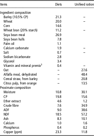
The study was performed at a commercial farm where deer were reared in captivity with individuals kept in a 5000-m2 open door enclosure on an irrigated mixed pasture. In addition, feed and water were offered ad libitum throughout the trial. The feeding programme (diet and unifeed ration as shown in Table 1) was the same for all deer and met or exceeded the nutrient requirements for cervids (National Research Council, 2007) with and average daily intake (on DM) of diet and unifeed ration (calculated from the amount used to replenish the feeders daily) of 1.8 and 0.8 kg, respectively. The ingredients and determined nutrient content (Association of Official Analytical Chemists, 2000) of the diet and unifeed ration are shown in Table 1. Diet and unifeed ration mineral contents were analysed by inductively coupled optical emission spectrometry as indicated by Gambín et al. (Reference Gambín, Serrano, Gallego, García, Capelli, Ceacero and Landete Castillejos2017).
Ingredient composition (% DM) and proximate composition (% DMFootnote 1 ) of diets and unifeed rationFootnote 2 for red deer yearling males

1 Unless otherwise indicated.
2 Unifeed rations were homogenized and cut into small portions in a tractor-driven commercial mixer.
3 Supplied per kg of diet: vitamin A (trans-retinyl acetate), 10 000 IU; vitamin D3 (cholecalciferol), 2 000 IU; vitamin E (all-rac-tocopherol-acetate), 15 IU; manganese (MnSO4·H2O), 75 mg; iron (FeCO3), 50 mg; zinc (ZnSO4·H2O), 115 mg; copper (CuSO4·5H2O), 7.5 mg; iodine (KI), 2 mg; cobalt (2CoCO3·3Co(OH)2·H2O), 0.83 mg; selenium (Na2SeO3), 0.22 mg; ethoxiquin, 0.025 mg; butilhidrotoluen, 0.18 mg; butylhydroxyanisole, 0.016 mg; sepiolite, 950 mg.
Slaughtering and samples collection
Blood samples were taken from resting deer after an overnight fast as indicated by Gambín et al. (Reference Gambín, Serrano, Gallego, García, Capelli, Ceacero and Landete Castillejos2017) on the 1st day of the trial (basal level) and the 90th day after the first Cu injection (final level). Blood samples were drawn by jugular venepuncture using Vacutest Clot activators and were coagulated in the tubes (Kima S.A., Arzegrande, Italy). Serum was separated using a centrifugal separator (Jouan® CR23-22; 2848×g at 4°C for 15 min), and samples were kept in a freezer at −20°C until the analysis of mineral content. Immediately later, deer were fasted at the farm for 14 h and transported to an accredited slaughterhouse (Incarsa, Burgos, Spain). At slaughterhouse, deer had a 12-h rest period with full access to water but not to feed. Deer from Cu supplemented and from control groups were not mixed at any time, and stress was minimized as much as possible. Animals were stunned with a captive bolt, slaughtered and dressed according to Council Regulation EC No 1099 (2009). Carcasses were split in two longitudinally along the centre of the vertebral column and weighed. The head was then removed at the atlanto-occipital junction and, immediately after, carcasses were chilled at 4°C in a cold chamber for 24 h.
After cooling, samples (300±25 g) of ST and RA muscles were excised from the left side of each chilled carcass. At 24 h postmortem, pH (pH24) was measured using a Crison pH meter (Crison 507; Crison Instruments S.A., Barcelona, Spain) equipped with a glass electrode (model no. 52-11; Crison Instruments S.A., Barcelona, Spain) according to Pateiro et al. (Reference Pateiro, Lorenzo, Díaz, Gende, Fernández, González, García, Rial and Franco2013). The incidence of dark, firm and dry (DFD) meats (pH24 above 6 as indicated by Smith and Dobson, Reference Smith and Dobson1990) was calculated for each muscle studied. Meat samples were transported to the laboratory (Centro Tecnolóxico da Carne, Ourense, Spain) in refrigerated conditions. After measuring the colour, mincing the meat and assessing moisture, the rest of the samples were vacuum packaged and kept at −20°C for further analysis. In addition, livers were removed and were vacuum packaged and frozen at −20°C until analysis.
Meat, liver and serum analysis
Colour parameters and chemical composition were measured at 24 h postmortem according to Pateiro et al. (Reference Pateiro, Lorenzo, Díaz, Gende, Fernández, González, García, Rial and Franco2013). For determination of total cholesterol, samples were treated following the procedure described by Domínguez et al. (Reference Domínguez, Crecente, Borrajo, Agregán and Lorenzo2015a). The content of total cholesterol was calculated in duplicate for each muscle sample, based on the external standard technique, from a standard curve of peak area v. concentration. For the analysis of FA methyl esters, total fat was extracted from 10 g of ground meat sample, according to Bligh and Dyer (Reference Bligh and Dyer1959) method. Total FA was quantified according to Domínguez et al. (Reference Domínguez, Crecente, Borrajo, Agregán and Lorenzo2015a) procedure. Separation and quantification of the methyl esters was carried out using a gas chromatograph (GC-Agilent 6890N; Agilent Technologies Spain, S.L., Madrid, Spain), equipped with a flame ionization detector and an automatic sample injector HP 7683, and using a Supelco SPTM-2560 fused silica capillary column (100 m, 0.25 mm internal diameter, 0.2 μm film thickness; Supelco Inc., Bellafonte, PA, USA), following the chromatographic conditions described by Domínguez et al. (Reference Domínguez, Crecente, Borrajo, Agregán and Lorenzo2015a). Individual FA methyl esters were identified by comparing their retention times with those of authenticated standards. In addition, lipid quality indices were calculated, that is nutritional value according to Estévez et al. (Reference Estévez, Morcuende, Ramírez, Ventanas and Cava2004); hypocholesterolemic/hypercholesterolemic ratio according to Santos-Silva et al. (Reference Santos-Silva, Bessa and Santos-Silva2002) and index of atherogenicity and index of thrombogenicity according to Ulbricht and Southgate (Reference Ulbricht and Southgate1991). To analyse the protein amino acid (AA) profile, the hydrolysis, derivatization and identification of hydrolysed AA was carried out following the procedure described by Domínguez et al. (Reference Domínguez, Borrajo and Lorenzo2015b). The mineral elements were quantified by inductively coupled optical emission spectrometry for meat and liver according to Lorenzo et al. (Reference Lorenzo, Bermúdez, Domínguez, Guiotto, Franco and Purriños2015) and for serum as indicated by Gambín et al. (Reference Gambín, Serrano, Gallego, García, Capelli, Ceacero and Landete Castillejos2017). The final value for the content of each element was calculated as the average of three determinations for each sample.
Statistical analysis
Prerequisites to run each test were checked, and data normality was tested with the Shapiro–Wilk test. All data were normally distributed. A GLM test was performed to study the effects of Cu supplementation and muscle type on meat quality characteristics and DFD incidence, chemical composition, FA profile, AA composition and mineral content. The model included the main effects of Cu supplementation and muscle, as well as the interaction between these variables. Interaction was not significant for any trait studied and, in consequence, was removed from the analysis. In addition, the influence of Cu supplementation on the mineral content of liver was analysed using a GLM test. Carcass weight was included as a covariate for all traits studied. The model was validated testing the goodness-of-fit to the data with R 2 (Supplementary Tables from S1 to S4). On the other hand, a general linear mixed procedure was performed to study the effects of Cu supplementation on the mineral content of serum at basal (day 0) and at final (day 90) levels. In all cases, the experimental unit was the sample excised from each individual animal (n=10 and 16 for Cu bolus supplemented and control groups, respectively, and n=13 for each muscle). To determine liver mineral composition, samples were excised from five, and eight carcasses from Cu bolus supplemented and control groups, respectively. All analyses were carried out with SPSS software, version 22.0 for Windows (Copyright; SPSS Inc., NY, USA).
Results
Effects of supplementation with copper bolus
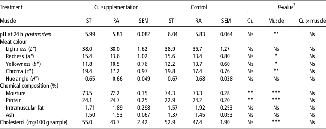
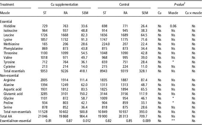
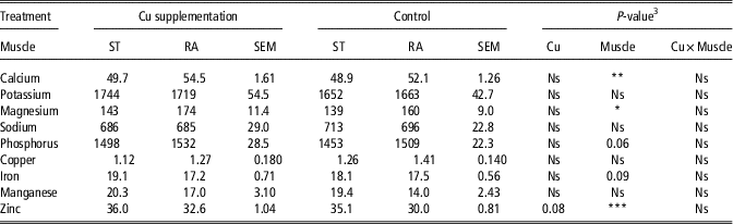
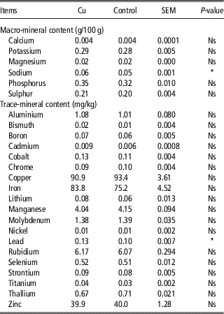
Supplementation with Cu increased protein (P<0.01) and decreased moisture (P=0.01; Table 2) contents of meat but did not affect pH24, colour or cholesterol content. In addition, Cu supplementation did not affect the incidence of DFD meats (30% of samples for Cu supplemented deer and 50% of samples for control deer). Although no effect was observed for intramuscular fat content, fat from deer supplemented with Cu tended to have higher content of lauric acid (C12:0) and had higher content of DHA (C22:6n-3, P=0.01) but lower of elaidic acid (9t-C18:1, P=0.05) and linolenic (C18:3n-3, P<0.05) acids than fat from deer of control group (Table 3). However, no difference was observed for total saturated, monounsaturated and polyunsaturated FA contents and lipid quality traits studied. In addition, Cu supplementation did not influence the AA profile of meat (Table 4) and had a little effect on the mineral profile of meat (Table 5), liver (Table 6) and serum. The only differences observed were that Cu supplemented deer tended to have a higher content of zinc in meat and had a significantly higher content of sodium (P<0.05) and lead (P<0.05) in liver than those found in control deer. Copper supplementation did not modify the content of this mineral on serum (0.73±0.058 v. 0.72±0.031 ppm for Cu and control groups, respectively). In fact, the only serum mineral affected by Cu supplementation was sulphur which was lower (P<0.01) in the experimental group (0.08±0.003 g/100 g) than in the control group (0.09±0.002 g/100 g). Mineral content of serum varied from the basal to the final level (P<0.05) with an increase of sulphur and zinc contents, whereas the contents of potassium, magnesium, boron and rubidium decreased in the same period. Moreover, the serum content of iron tended to increase whereas the contents of sodium and strontium tended to decrease from the basal to the final level.
Influence of copper (Cu) supplementation with ruminal bolus and muscle (sternocephalicus (ST) v. rectus abdominis (RA)) on meat quality characteristics and chemical composition of red deer yearling malesFootnote 1

1 The experimental unit was the sample (n=10 and 16 for Cu bolus supplemented and control groups, respectively and n=13 for ST and RA).
2 No significant interactions between Cu supplementation and muscle were detected for any of the traits studied.
The P-values are indicated by *, ** and *** for P<0.05, P<0.01 and P<0.001, respectively.
Influence of copper (Cu) supplementation with ruminal bolus and muscle (sternocephalicus (ST) v. rectus abdominis (RA)) on the fatty acid profile (mg/g of fat) of red deer yearling malesFootnote 1

SFA=saturated fatty acids; MUFA=monounsaturated fatty acids; PUFA=polyunsaturated fatty acids.
1 The experimental unit was the sample (n=10 and 16 for Cu bolus supplemented and control groups, respectively and n=13 for ST and RA).
2 No significant interactions between Cu supplementation and muscle were detected for any of traits studied.
3 Nutritional value=Σ(C12:0+C14:0+C16:0)/Σ(C18:1n-9+C18:2n-6).
4 Hypocholesterolemic/hypercholesterolemic ratio=[Σ(C18:1n-9+C18:1n-7+C18:2n-6+C18:3n-3+C20:3n-6+C20:4n-6)/Σ(C14:0+C16:0)].
5 Index of atherogenicity=[C12:0+(4×C14:0)+C16:0]/[(∑MUFA)+(∑PUFA)].
6 Index of thrombogenicity=[C14:0+C16:0+C18:0]/[(0.5×∑MUFA)+(0.5×n-6)+(3×n-3)+(n-3/n-6)].
Influence of copper (Cu) supplementation with ruminal bolus and muscle (sternocephalicus (ST) v. rectus abdominis (RA)) on amino acid (AA) content of meat (mg/100 g of sample) of red deer yearling malesFootnote 1

1 The experimental unit was the sample (n=10 and 16 for Cu bolus supplemented and control groups, respectively and n=13 for ST and RA).
2 No significant interactions between Cu supplementation and muscle were detected for any of the traits studied.
The P-values are indicated by * and ** for P<0.05 and P<0.01, respectively.
Influence of copper (Cu) supplementation with ruminal bolus and muscle (sternocephalicus (ST) v. rectus abdominis (RA)) on mineral compositionFootnote 1 (mg/kg except µg/100 g for manganese) of red deer yearling malesFootnote 2

1 Next minerals were detected at concentrations lower than 0.01 mg/kg: arsenic, beryllium, bismuth, cadmium, cobalt, molybdenum, lead, antimony and vanadium.
2 The experimental unit was the sample (n=10 and 16 for Cu bolus supplemented and control groups, respectively and n=13 for ST and RA).
3 No significant interactions between Cu supplementation and muscle were detected for any of the traits studied.
The P-values are indicated by *, ** and *** for P<0.05, P<0.01 and P<0.001, respectively.
Influence of copper (Cu) supplementation with ruminal bolus on mineral contentFootnote 1 of liver from red deerFootnote 2

1 Next minerals were detected at concentrations lower than 0.01 mg/kg: arsenic, beryllium, antimony and vanadium.
2 The experimental unit was the sample (n=5 and 8 for Cu supplemented and control groups, respectively).
The P-values are indicated by * for P<0.05.
Effects of muscle type
Meat quality varied with muscle. In fact, ST muscle presented higher values of pH24 (P<0.01), redness (P<0.05), yellowness (P<0.05) and chroma (P<0.01) than RA muscle. The percentage of samples from RA which had the pH24 higher than 6.0 was 23%, whereas this percentage was 61.5% for ST samples (P<0.05). In consequence, a higher incidence of DFD meats would be expected for ST than for RA muscle. In addition, ST muscle had higher (P=0.001) contents of moisture and cholesterol and lower (P<0.001) of protein than RA muscle without that differences between muscles were observed for the fat content. However, RA muscle had higher (P<0.05) content of mono- and lower (P=0.001) of polyunsaturated FA than ST muscle. In consequence, ST muscle presented a higher polyunsaturated/saturated FA ratio (P=0.001), total n-6 content (P<0.01), total n-3 content (P=0.001) and hypocholesterolemic/hypercholesterolemic ratio (P<0.001) but lower nutritional value (P<0.001), index of atherogenicity (P<0.05) and index of thrombogenicity (P=0.05) than RA muscle. In addition, differences were observed for essential and non-essential AA content of the two muscles studied. The RA muscle tended to have a higher content of histidine and had (P<0.05) higher content of tyrosine than ST muscle. However, ST muscle presented higher content of glycine (P<0.05) and proline (P<0.05) than RA muscle. In consequence, the proportion between essential and non-essential AA was higher (P<0.01) for RA than for ST muscle. Differences were observed for mineral profile between muscles with ST tending to have a higher content of iron and having a higher content of zinc (P<0.001) but lower of calcium (P=0.01) and magnesium (P<0.05) than RA muscle. In addition, ST muscle tended to have a lower content of phosphorus than RA muscle.
Discussion
Effects of supplementation with copper bolus
Despite Cu supplementation is a common nutritional strategy on farmed deer (Gambín et al., Reference Gambín, Serrano, Gallego, García, Capelli, Ceacero and Landete Castillejos2017), to our knowledge, there is no study in the literature regarding the effects of Cu supplementation in deer meat characteristics. In general, no differences were detected for pH24, colour, cholesterol content, AA profile and mineral composition. Current results suggest that Cu supplementation might decrease the incidence of DFD meats in deer from 50% to 30%. However, likely due to the small sample size of our study, the differences did not reach statistical significance.
Regarding chemical composition, only an increase of protein content (with the consequent reduction of moisture content) on meat from Cu supplemented deer was observed which agrees with results reported by Solaiman et al. (Reference Solaiman, Shoemaker, Jones and Kerth2006) for goat kids. The potential influence of Cu supplementation on composition and/or deposition of fat, with subsequently changes in meat quality, has been widely noticed for large ruminants (Engle and Spears, Reference Engle and Spears2000; Engle et al., Reference Engle, Spears, Armstrong, Wright and Odle2000a and Reference Engle, Spears, Fellner and Odle2000b), whereas Saran Netto et al. (Reference Saran Netto, Zanetti, Del Claro, Garcia Vilela, Pires de Melo, Correa and Piccoli Pugine2013) did not find any effect. However, this does not seem to be the case for deer. In fact, in the current trial, no effects of Cu supplementation were observed for the fat content of meat. In general, Cu supplementation induces the modification of lipid metabolism decreasing the fat deposition (Engle et al., Reference Engle, Spears, Armstrong, Wright and Odle2000a and Reference Engle, Spears, Fellner and Odle2000b). Thus, high Cu levels might reduce the activity of the enzyme FA synthase as observed by Konjufca et al. (Reference Konjufca, Pesti and Bakalli1997) in poultry and, consequently, the synthesis and deposition of fat. Considering that deer has one of the lowest fat content in red meats, it is not surprising that Cu could not reduce it further.
In the current study, no differences were observed for cholesterol content of Cu supplemented and control animals, which agrees with results reported by Engle et al. (Reference Engle, Spears, Armstrong, Wright and Odle2000a) for steers and by Huang et al. (Reference Huang, Wang, Lin and Guo2014) for goats. In this sense, it has been observed that total plasma cholesterol concentrations increase in steers when organic sources of Cu are used v. inorganic sources as copper sulphate (Johnson and Engle, Reference Johnson and Engle2003). However, current results showed an influence of Cu supplementation on FA profile of fat similar to that reported by Engle and Spears (Reference Engle and Spears2000). The FA composition is the result of lipogenesis in adipose tissue and the dietary lipids with the subsequent ruminal biohydrogenation. In this sense, Cu promotes the decrease of ruminal biohydrogenation with the consequent increase of intestinal absorption of unsaturated FA. However, results can vary with the fat source of diet. In fact, Cu supplementation combined with diets containing soya bean oil seems to inhibit this effect (Engle et al., Reference Engle, Spears, Fellner and Odle2000b; Saran Netto et al., Reference Saran Netto, Zanetti, Del Claro, Garcia Vilela, Pires de Melo, Correa and Piccoli Pugine2013). In the current trial, the increase of C12:0 and C22:6n-3 and the decrease of 9t-C18:1 and C18:3n-3 contents of fat were observed without that Cu supplementation affected the total contents of mono- and polyunsaturated FA. Therefore, some interaction with the fat source of diet could have taken place, so that palm oil (included on the diet supply during the current trial) had a similar effect to that of soya bean inhibiting the reduction of biohydrogenation caused by Cu supplementation.
In this research, average values observed for Cu content in deer meat were of 1.3 mg/kg of fresh meat. These values are lower than 1.7 mg/kg reported by Schlichting et al. (Reference Schlichting, Sommerfeld, Müller-Graf, Selhorst, Greiner, Gerofke, Ulbig, Gremse, Spolders, Schafft and Lahrssen-Wiederholt2017) and lower than 3.3 mg/kg reported by Jarzyńska and Falandysz (Reference Jarzyńska and Falandysz2011). The cause of differences among authors for mean Cu content of meat is unknown, but it might be related with the housing regime as Jarzyńska and Falandysz (Reference Jarzyńska and Falandysz2011) and Schlichting et al. (Reference Schlichting, Sommerfeld, Müller-Graf, Selhorst, Greiner, Gerofke, Ulbig, Gremse, Spolders, Schafft and Lahrssen-Wiederholt2017) analysed meat from free-ranging deer whereas, in the current trial, meat samples were excised from farmed deer. In any case, values for deer meat Cu content obtained in the current trial were much lower than 5 mg/kg of fresh weight permitted by European Council Regulation EC No 149 (2008) for food of animal origin. Therefore, Cu supplementation did not increase the Cu concentration of meat to levels that could cause human toxicity, making it suitable for consumption from this perspective. Health risk for the consumer due to an average consumption of farmed deer meat with the reported content of Cu is, therefore, extremely unlikely. In addition, the hepatic and the serum Cu concentrations did not vary with supplementation either. According to the criteria of Wilson and Grace (Reference Wilson and Grace2001), even deer in the control group had adequate serum Cu levels in this study. It is possible that if this study had been conducted using deer suffering from Cu deficiency, the results would have been very different from those reported here for Cu content of serum on supplemented deer.
Effects of muscle type
In general, muscles as longissimus and semimembranosus (Volpelli et al., Reference Volpelli, Valusso, Morgante, Pittia and Piasentier2003; Daszkiewicz et al., Reference Daszkiewicz, Hnatyk, Dabrowski, Janiszewski, Gugołek, Kubiak, Smiecinska, Winarski and Koba-Kowalczyk2015; Razmaitè et al., Reference Razmaitė, Šiukščius, Šveistienė, Bliznikas and Švirmickas2017) have been used to evaluate cervid meat quality. The ST and RA characteristics were studied in the current trial because their consumption is common due to their lower prices comparing with loin or other cuts. However, their quality has not been assessed. The ST muscle presented a higher pH24 than RA muscle. Variation in ultimate pH between different muscles is probably due to the way in which the muscle is affected by physical activity and by the fibre type distribution within the muscle. A large proportion of oxidative fibres (types I and II) entails a high ultimate pH value and an increase of DFD incidence. In fact, a higher incidence of DFD meats were observed for ST (with higher content of fibres I and II) than for RA muscle (61.5% v. 23%, respectively). The average incidence of DFD meat observed in the current trial was high but lower than the 57% reported by Daszkiewicz et al. (Reference Daszkiewicz, Hnatyk, Dabrowski, Janiszewski, Gugołek, Kubiak, Smiecinska, Winarski and Koba-Kowalczyk2015) on farmed animals. It is possible that consumers do not associate the characteristics of this type of meat as a defect if not as part of the intrinsic quality of venison meat.
Current results showed a similar content of saturated, mono- and polyunsaturated FA than those reported for farmed red deer by Bureš et al. (Reference Bureš, Barton, Kotrba and Hakl2015) for longissimus muscle and by Razmaitè et al. (Reference Razmaitė, Šiukščius, Šveistienė, Bliznikas and Švirmickas2017) for longissimus dorsi and semimembranosus muscles. On the other hand, the cholesterol content was similar to that reported by Razmaitè et al. (Reference Razmaitė, Šiukščius, Šveistienė, Bliznikas and Švirmickas2017) for longissimus dorsi and semimembranosus muscles from farmed red deer but lower than that reported by Polak et al. (Reference Polak, Rajar, Gašperlin and Zlender2008) for semitendinosus and triceps braquii muscles from wild red deer. Previous studies have showed that the FA composition varies according to muscle type in red deer (Polak et al., Reference Polak, Rajar, Gašperlin and Zlender2008; Razmaitè et al., Reference Razmaitė, Šiukščius, Šveistienė, Bliznikas and Švirmickas2017), according with results from the current trial where a higher content of mono- and lower of polyunsaturated FA (particularly, n-3 FA) were observed for RA than for ST. Therefore, in this sense, consumption of ST muscle would be healthier for a human. However, this muscle had 14% higher content of cholesterol and a lower content of some essential AA with a worse essential/non-essential AA ratio than the RA muscle.
In conclusion, Cu supplementation did not impair meat quality and did not increase meat Cu levels on farmed deer. Despite of the lower content of polyunsaturated FA, RA muscle was characterized by a better quality compared with ST muscle due to the higher content of protein (with more essential AA and a better essential/non-essential AA ratio) and lower cholesterol content and pH24.
Acknowledgements
This work was supported by INCYDEN Project (grant no. RTC-2016-5327-2) (Ministerio de Economía y Competitividad and the European Union Fund for Regional Development) and Project 01110AB020 funded by Excma. Diputación Provincial de Albacete. J. M. Lorenzo is a member of the MARCARNE network, funded by CYTED (ref. 116RT0503). Funders had no role in study design, data collection and analysis, decision to publish or preparation of the manuscript.
Declaration of interest
The authors have no conflict of interest to declare.
Ethics statement
This study was carried out in accordance with the Spanish legislation regarding the use of animals in research (Boletín Oficial del Estado, 2013) and with the approval of the Ethical Committee in Animal Experimentation at the Universidad de Castilla-La Mancha (permit number: 1002.04).
Software and data repository resources
Data and models are not deposited in any official repository.
Supplementary material
To view supplementary material for this article, please visit https://doi.org/10.1017/S1751731118002173