Introduction
Despite recent advances, the efficiency of in vitro production of sheep embryos is still low compared with that observed in vivo (Souza-Fabjan et al., Reference Souza-Fabjan, Panneau, Duffard, Locatelli, Figueiredo, Freitas and Mermillod2014). One of the most crucial factors affecting the embryo development potential is oocyte quality, which is gradually acquired during folliculogenesis and is directly related to nuclear and cytoplasmic maturation (Rizos et al., Reference Rizos, Lonergan, Ward, Duffy and Boland2002). According to Krisher (Reference Krisher2004), the support of cumulus cells is also of vital importance for the development of a competent oocyte.
In most mammals, oocytes are kept at the diplotene stage of prophase I from fetal life until ovulation, when meiosis is resumed in response to a luteinizing hormone (LH) surge (Mehlmann, Reference Mehlmann2005). During this meiotic arrest, structural and molecular changes that are essential to the acquisition of developmental competence occur (Ferreira et al., Reference Ferreira, Vireque, Adona, Meirelles, Ferriani and Navarro2009). In vivo, these nuclear and cytoplasmic events progress synchronously, ensuring that only competent oocytes reach ovulation (Mermillod et al., Reference Mermillod, Dalbies-Tran, Uzbekova, Thelie, Traverso, Perreau, Papillier and Monget2008). In this context, cumulus cells play a crucial role as oocyte maturation is basically controlled by low-molecular-weight peptides transferred to oocytes through gap junction communications (Tanghe et al., Reference Tanghe, Soom, Nauwynck, Coryn and De Kruif2002).
However, when oocytes are removed from the follicle and cultured in a suitable medium, meiosis spontaneously resumes independently of the cytoplasmic maturation stage (Pincus & Enzmann, Reference Pincus and Enzmann1935). This premature meiotic resumption is also associated with disruption of the intimate contact between oocyte and cumulus cells (Gharibi et al., Reference Gharibi, Hajian, Ostadhosseini, Hosseini, Forouzanfar and Nasr-Esfahani2013). Furthermore, the oocytes destined to in vitro maturation are usually retrieved from follicles at diverse stages of folliculogenesis and, therefore, the vast majority of them has not completed in vivo all required changes to acquire developmental competence (Watson, Reference Watson2007). So, the temporary arrest of meiosis has been proposed as alternative to provide additional time for oocyte capacitation and, consequently, to improve the quality of in vitro matured oocytes (Marchal et al., Reference Marchal, Tomanek, Terqui and Mermillod2001; Han et al., Reference Han, Lan, Wu, Han, Wang and Tan2006).
Among the pharmacological inhibitors that act on synthesis or phosphorylation of proteins, cAMP transduction and transcriptional activity, roscovitine, a potent and selective inhibitor of cyclin-dependent kinases, is recognized due to its specific action on M-phase promoting factor (MPF), a cell cycle regulator (Donnay et al., Reference Donnay, Faerge, Grøndahl, Verhaeghe, Sayoud, Ponderato, Galli and Lazzari2004). Studies performed with bovine (Mermillod et al., Reference Mermillod, Tomanek, Marchal and Meijer2000; Lagutina et al., Reference Lagutina, Ponderato, Lazzari and Galli2002), pig (Coy et al., Reference Coy, Romar, Ruiz, Cánovas, Gadea, Vázquez and Matás2005), horse (Franz et al., Reference Franz, Choi, Squires, Seidel and Hinrichs2003), sheep (Crocomo et al., Reference Crocomo, Marques Filho, Ulian, Branchini, Silva, Ackermann, Landim-Alvarenga and Bicudo2015) and goat (Han et al., Reference Han, Lan, Wu, Han, Wang and Tan2006) oocytes have demonstrated that roscovitine at 25 μM, 50 μM, 66 μM, 75 μM and 200 μM, respectively, was efficient to reversibly arrest the meiosis for 20–24 h. However, the effect of this inhibitor on oocyte competence is still discrepant among authors and species studied.
In goat, Han et al. (Reference Han, Lan, Wu, Han, Wang and Tan2006) reported that the reduction of exposure time of oocytes to roscovitine from 24 to 8 h improved the rates of cumulus cell expansion and blastocyst formation. According to these same authors, meiosis inhibitors used at high concentrations and/or for an extended period can be harmful to oocytes and compromise its developmental competence. In fact, our previous studies have demonstrated that treatment of sheep oocytes with 100 μM roscovitine for 24 h was excessive and promoted irreversible ultrastructure alterations (Crocomo et al., Reference Crocomo, Marques Filho, Sudano, Paschoal, Alvarenga and Bicudo2013). Based on that finding, the present study aimed to evaluate the ability of roscovitine to reversibly arrest the meiosis of sheep oocytes at different times of culture in order to establish the optimal incubation duration. In addition, the effect of roscovitine on cumulus cell expansion was also examined.
Materials and methods
All chemicals were purchased from Sigma Chemical Co. (Sigma-Aldrich Corp., St. Louis, MO, USA), unless otherwise indicated.
Cumulus–oocyte complexes collection
Ovaries from adult sheep were collected at a slaughterhouse and transported to the laboratory within 1–2 h in sterile saline solution (0.9% NaCl) at 32ºC. All visible follicles with a diameter of 2–6 mm were aspirated with a 20-gauge needle attached to a 10-ml syringe containing 0.5 ml pre-incubated HEPES-buffered TCM199 medium (12340–030; Gibco, Invitrogen Co., USA) supplemented with 50 IU/ml heparin. Only COCs with several compact cumulus cell layers and a homogeneous ooplasm were selected under stereomicroscopy (Shirazi et al., Reference Shirazi, Shams-Esfandabadi, Ahmadi and Heidari2010).
Meiosis inhibition and reversibility
After washes in HEPES-buffered TCM199 medium, the selected COCs were cultured for 6, 12 or 20 h in basic maturation medium comprised of TCM199 with Earle's salts (11150059; Gibco, Invitrogen Co., USA), 0.3 mM sodium pyruvate, 75 μg/ml penicillin, 100 μM cysteamine and 10% fetal calf serum (10437; Gibco, Invitrogen Co., USA) (Control) or supplemented with 75 μM roscovitine (Rosco). For reversion of meiotic inhibition, after 6, 12 or 20 h of culture, COCs from each treatment were washed in HEPES-buffered TCM199 medium and matured in vitro for a further 18 h in basic maturation medium supplemented with 0.1 IU/ml follicle stimulating hormone (FSH) (Follitropin®, Bioniche Co., Bellevile, ON, Canada) and 0.1 IU/ml LH (Lutropin-V®, Bioniche Co.).
About 20 COCs per 100 μl droplet of medium were cultured in 96-well plates without oil overlay at 38.5ºC and in 5% CO2 in air. The inhibitor concentration and the culture conditions were based on our preliminary studies (Crocomo et al., Reference Crocomo, Marques Filho, Ulian, Branchini, Silva, Ackermann, Landim-Alvarenga and Bicudo2015). The stock solution of roscovitine (1 mg/ml) was prepared in dimethylsulphoxide, aliquoted and stored at –20ºC until use.
Assessment of cumulus cell expansion
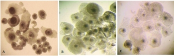
In order to evaluate the possible effects of roscovitine on cumulus cells and the relationship between cumulus cell expansion and meiotic arrest, COCs were examined under a stereomicroscope and classified according to the degree of cumulus cell expansion (Heidari Amale et al., Reference Heidari Amale, Zare Shahne, Abavisani and Nasrollahi2011) as: total cumulus cell expansion (all layers of cells expanded); partial cumulus cell expansion (subtle expansion of outer layers of cells); and absence of cumulus cell expansion (cells strongly adhered to each other and to the pellucid zone) (Fig. 1).
Cumulus cell expansion of sheep COCs evaluated under a stereomicroscope. (A) Absence of cumulus cell expansion. (B) Partial cumulus cell expansion. (C) Total cumulus cell expansion (×100 magnification).

Assessment of oocyte chromatin organization
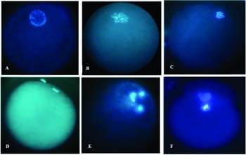
Oocytes were stripped from their cumulus cells by repeated pipetting in HEPES-buffered TCM-199 medium, fixed for 30 min in 4% paraformaldehyde, and transferred to droplets of Hoechst 33342 stain in glycerol (10 μg/ml) on a glass slide. The oocytes were examined under a fluorescence inverted microscope (Leica® DMIRB) and classified according to the stage of nuclear maturation as: germinal vesicle (GV), germinal vesicle breakdown (GVBD), metaphase I (MI) or metaphase II (MII) (Shirazi et al., Reference Shirazi, Shams-Esfandabadi, Ahmadi and Heidari2010). The oocytes with the extrusion of one or two polar body and the presence of one or two pronucleus were classified as parthenogenetic (PG) (Marshall et al., Reference Marshall, Wilton and Moore1998). Those oocytes with altered nuclear structure were classified as degenerate (DEG) (Fig. 2)
Nuclear status of sheep oocytes stained with Hoechst 33342 and evaluated under a fluorescence inverted microscope: (A) GV: germinal vesicle; (B) GVBD: germinal vesicle breakdown; (C) MI: metaphase I; (D) MII: metaphase II; (E) DEG: degenerate; (F) PG: parthenogenetic (×200 magnification).

Experimental design
Experiment 1 – Inhibitory action of roscovitine at different times of culture
This experiment was conducted according to a completely randomized design with eight treatments (Control groups 0, 6, 12, or 20 h, and Rosco groups 0, 6, 12, or 20 h) and five replicates for each parameter assessed. At the end of each treatment, the cumulus cell expansion degree and the nuclear maturation stage of COCs were evaluated under a stereomicroscope and by Hoechst 33342 staining, respectively. The treatments Rosco 0 h and Control 0 h corresponded to COCs evaluated immediately after aspiration with and without 75 μM roscovitine, respectively, aiming at the determination of the nuclear status of immature oocytes and the analysis of the effectiveness of meiotic inhibition during the interval between oocyte recovery and the culture
Experiment 2 – Reversibility of inhibitory action of roscovitine
This experiment was conducted according to a completely randomized design with six treatments (Control groups 6 + 18, 12 + 18, 20 + 18 h and Rosco groups 6 + 18, 12 + 18, 20 + 18 h) and five replicates for each parameter assessed. In each treatment, the cumulus cell expansion degree and nuclear maturation stage were evaluated at the end of additional culture for 18 h in inhibitor-free medium supplemented with gonadotropins.
Statistical analysis
The data were transformed into square root of x + 0.5 and subjected to analysis of variance (ANOVA software) according the factorial system with eight treatments (Control groups 0, 6, 12, 20 h, and Rosco groups 0, 6, 12, 20 h), in the first experiment, and six treatments (Control groups 6 + 18, 12 + 18, 20 + 18 h and Rosco groups 6 + 18, 12 + 18, 20 + 18 h), in the second experiment, three parameters in the case of cumulus cell expansion (total, partial and absence) and six parameters in the case of nuclear maturation (GV, GVBD, MI, MII, DEG, PG). Five replicates were performed for each parameter assessed and the means were compared by Tukey test at 5% probability.
Results
Experiment 1
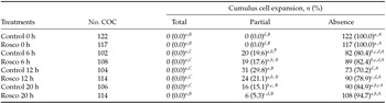
According to Table 1, in all treatments, the proportion of COCs with compact cumulus cells was significantly high and no oocyte showed total expansion (P < 0.05). At 0 h, the absence of cumulus cell expansion prevailed in all COCs aspirated with (Rosco) and without (Control) roscovitine (P < 0.05). At 20 h, the rate of COCs from the Control groups (n = 90, 84.9%) and the Rosco group (n = 108, 94.7%) with compact cumulus cells was similar to that observed for Rosco 6 h (n = 89, 82.4%) and Control 6 h (82, 80.4%) (P > 0.05), but significantly differed from that recorded in the Control 12 h (n = 73, 70.2%, P < 0.05). In contrast, no significant difference was observed among Control group 12 h (n = 73, 70.2%), Rosco group 12 h (n = 90, 78.9%), Rosco group 6 h (n = 89, 82.4%) and Control group 6 h (n = 82, 80.4%) with respect to the rate of COCs with compact cumulus cells (P > 0.05). The proportion of COCs with partial cumulus cell expansion was similar among Rosco group 6 h (n = 19, 17.6%), Control group 6 h (n = 20, 19.6%), Rosco group 12 h (n = 24, 21.1%) and Control group 12 h (n = 31, 29.8%), but significantly differed from that observed in the Rosco groups at 20 h (n = 6, 5.3%) and at 0 h (0%) (P < 0.05).
Cumulus cell expansion degree of sheep COCs in vitro cultured for 6, 12 or 20 h in basic maturation medium (Control) or supplemented with 75 μM roscovitine (Rosco). 0 h: COCs were aspirated with (Rosco) or without (Control) roscovitine and immediately evaluated

Standard error (SE) = 0.036 and coefficient of variation (CV) = 4.10% for the analysis.
Significant differences are indicated by lowercase letters within columns and capital letters within rows (P < 0.05).
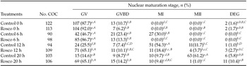
As shown in Table 2, almost all oocytes from Rosco group 0 h (n = 104, 92.0%) and Control group 0 h (n = 107, 87.7%), evaluated immediately after the follicular aspiration, were at GV. At 6 h, an important decrease of the GV rate was observed in the Control group (n = 42, 46.7%) while significantly higher proportion of oocytes treated with roscovitine (n = 85, 86.7%) remained at this stage (P < 0.05). In comparison, the rate of oocytes from Control group 6 h at GVBD (n = 21, 23.4%) and MI (n = 27, 30%) was significantly higher than that observed in the Rosco 6 h for these same stages (P < 0.05). A significant decrease of the GV rate occurred in the Rosco group 12 h (n = 71, 65.1%, P < 0.05) but the value reached was similar to that observed in the Rosco group 20 h (n = 69, 65.1%, P > 0.05). In the Control group, there was a continuous decrease of GV rate at 12 h (n = 24, 25.5%) and 20 h (n = 15, 14.6%) (P < 0.05).
Nuclear configuration of sheep oocytes cultured in vitro for 6, 12 or 20 h in basic maturation medium (Control) supplemented with 75 μM roscovitine (Rosco). 0 h: COCs were aspirated with (Rosco) or without (Control) roscovitine and immediately stained

Standard error (SE) = 0.037 and coefficient of variation (CV) = 4.54% for the analysis.
Significant differences indicated by lowercase letters within columns and capital letters within rows (P < 0.05). GV: germinal vesicle; GVBD: germinal vesicle breakdown; MI: metaphase I; MII: metaphase II; DEG: degenerate.
Still in Table 2, it is possible to note an evident increase in the MI rate in the Control group 12 h (n = 51, 54.3%) which was significantly higher than that observed in the in the Rosco group 12 h (n = 11, 18.4%), Control group 20 h (n = 10, 9.7%) and Rosco group 20 h (n = 10, 9.4%) (P < 0.05). At 20 h, significantly higher proportion of oocytes from Control group reached the MII (n = 63, 61.2%) while only 1% (n = 1.0) of oocytes from Rosco were at this same stage (P < 0.05). The rate of degenerate oocytes was similar among treatments (P > 0.05) and there was no parthenogenetic oocyte in any treatment.
Experiment 2
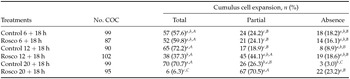
As shown in Table 3, in vitro culture for a further 18 h in inhibitor-free medium supplemented with gonadotropins allowed total cumulus cell expansion at a significantly high rate of COCs compared with the Control group 12 h (n = 65, 72.2%) and 20 h (n = 70, 70.7%) (P < 0.05), which was similar to the Control group 6 h (n = 57, 57.6%) and Rosco group 6 h (n = 52, 59.8%) (P > 0.05), but significantly differed from that recorded in the Rosco group 12 h (n = 38, 37.3%) and Rosco group 20 h (n = 6, 6.3%) (P < 0.05). The rate of COCs with partial cumulus cell expansion was significantly higher in the Rosco group 20 h (n = 67, 70.5%) in comparison to Control group 20 h (n = 26, 26.3%), Control group 6 h (n = 24, 24.2%), Rosco group 6 h (n = 21, 24.1%) and Control group 12 h (n = 17, 18.9%) (P < 0.05), but did not significantly differ from that observed in the Rosco group 12 h (n = 45, 44.1%) (P > 0.05). There was no important difference among treatments with respect to the rate of COCs with compact cumulus cells (P > 0.05).
Cumulus cell expansion of sheep COCs after in vitro culture for 6, 12 or 20 h in basic maturation medium (Control) supplemented with 75 μM roscovitine (Rosco) followed by 18 h of in vitro maturation in inhibitor-free medium

Standard error (SE) = 0.071 and coefficient of variation (CV) = 7.85% for the analysis.
Significant differences indicated by lowercase letters within columns and capital letters within rows (P < 0.05).
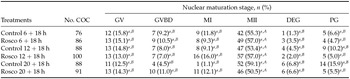
As shown in Table 4, the rate of oocytes at MII after in vitro culture for a further 18 h in inhibitor-free medium was similar among treatments (P > 0.05) and significantly higher than that observed in the other meiotic stages (P < 0.05). In addition, the percentages of GV, GVBD and MI oocytes, oocyte degeneration and parthenogenesis were similar among these and did not significantly difference among treatments (P > 0.05).
Meiotic progression in sheep oocytes in vitro cultured for 6, 12 or 20 h in basic maturation medium (Control) supplemented with 75 μM roscovitine (Rosco) followed by 18 h of in vitro maturation in inhibitor-free medium

Standard error (SE) = 0.057 and coefficient of variation (CV) = 5.92% for the analysis.
Significant differences indicated by lowercase letters within columns and capital letters within rows (P < 0.05). GV: germinal vesicle; GVBD: germinal vesicle breakdown; MI: metaphase I; MII: metaphase II; DEG: degenerate; PG: parthenogenetic.
Discussion
The asynchrony between nuclear and cytoplasmic maturation observed in in vitro matured oocytes is the most crucial aspect affecting the effectiveness of systems of in vitro embryo production (Rizos et al., Reference Rizos, Lonergan, Ward, Duffy and Boland2002). In this context, inhibitors of cyclin-dependent kinases, such as roscovitine, have been tested in several animal species in order to maintain meiotic arrest in vitro and, consequently, provide additional time for the oocyte to complete its capacitation (Mermillod et al., Reference Mermillod, Tomanek, Marchal and Meijer2000; Franz et al., Reference Franz, Choi, Squires, Seidel and Hinrichs2003; Coy et al., Reference Coy, Romar, Ruiz, Cánovas, Gadea, Vázquez and Matás2005; Han et al., Reference Han, Lan, Wu, Han, Wang and Tan2006; Sananmuang et al., Reference Sananmuang, Techakumphu and Tharasanit2010). In sheep, however, this strategy to improve the quality of in vitro matured oocytes has been investigated infrequently.
Based on this situation and considering that the efficiency of meiosis inhibition depends upon not only the drug concentration but also on the exposure time of oocyte to inhibitor (Han et al., Reference Han, Lan, Wu, Han, Wang and Tan2006), the present study aimed to evaluate the ability of roscovitine to reversibly arrest meiosis in sheep oocytes at different times of culture in order to establish the optimal incubation duration. The possible interference of roscovitine on cumulus cell expansion and its relationship with the meiotic arrest were also examined.
For this study, only COCs with compact cumulus cells were selected, indicating those cells that probably, have not undergone LH stimulation in vivo (Dekel et al., Reference Dekel, Lawrence, Gilula and Beers1981). Our findings demonstrated that follicular aspiration with roscovitine was not necessary, as the GV rates in the Control group 0 h and Rosco group 0 h were significantly high. These findings also suggested that time interval between recovery and selection of COCs was adequate and did not induce meiotic resumption before in vitro culture. Similar results have been observed in goat (Han et al., Reference Han, Lan, Wu, Han, Wang and Tan2006) and bovine (Barretto et al., Reference Barretto, Castro, Garcia and Mingoti2011) oocytes.
The absence of cumulus cell expansion observed in the Control groups at 6, 12 and 20 h is, probably, related not only to culture time but also to medium composition. As commonly performed in most similar studies, gonadotropins were not inserted into the basic maturation medium to avoid any interference with roscovitine action (Mermillod et al., Reference Mermillod, Tomanek, Marchal and Meijer2000; Franz et al., Reference Franz, Choi, Squires, Seidel and Hinrichs2003; Han et al., Reference Han, Lan, Wu, Han, Wang and Tan2006). However, based on preliminary studies, serum supplementation was maintained due to its benefits to the culture system and to cellular viability (Natsuyama et al., Reference Natsuyama, Noda, Narimoto and Mori1993). Our results demonstrated, therefore, that only serum was not enough to promote cumulus cell expansion, as observed by Mattioli et al. (Reference Mattioli, Bacci, Galeati and Seren1991) and Accardo et al. (Reference Accardo, Dattena, Pilichi, Mara, Chessa and Cappai2004) in pig and sheep COCs, respectively.
According to Kito & Bavister (Reference Kito and Bavister1997) the synergistic effect of gonadotropins and serum is required for cumulus cell expansion. This finding was also demonstrated in our study, as a significant proportion of COCs from Control groups 6, 12 and 20 h had total cumulus cell expansion after additional culture for 18 h in the presence of LH, FSH and serum. Usually, in appropriate supplemented medium, the cumulus cells remain compacted until about 12 h, when expansion becomes evident and continuously increases up to end of culture (Hyttel et al., Reference Hyttel, Xu, Smith and Greve1986; Berg et al., Reference Berg, Thompson and Asher2002). In the present study, this kinetics of cumulus cell expansion was not observed due to the absence of gonadotropin supplementation during the first 20 h of culture. We believe that the slight difference observed at 6, 12 and 20 h with respect to cumulus cell expansion is probably due to the heterogeneity of ovaries obtained at the slaughterhouse (Coy et al., Reference Coy, Romar, Ruiz, Cánovas, Gadea, Vázquez and Matás2005).
The cumulus cell expansion pattern observed in the Rosco groups 6, 12 and 20 h treatments after additional culture for 18 h in inhibitor-free medium demonstrated that roscovitine acted on cumulus cells and the reversibility of this action was time exposure dependent. Likewise, Han et al. (Reference Han, Lan, Wu, Han, Wang and Tan2006) reported that the rate of goat COCs with total cumulus cell expansion increased when the exposure time to roscovitine was reduced from 24 to 8 h. However, despite the interference of roscovitine on cumulus cell expansion evidenced in our study, it is not well established if this inhibitory action occurs via the oocyte or directly via cumulus cells (Schoevers et al., Reference Schoevers, Bevers, Roelen and Colenbrander2005). According to Mermillod et al. (Reference Mermillod, Tomanek, Marchal and Meijer2000), the inhibition of cumulus cell expansion even under EGF stimulation indicates that the mucification process may be dependent on the MPF or another roscovitine-sensitive kinase such as MAPK.
The kinetics of nuclear maturation observed in the Control group with a significant rate of GVBD (23.4%) at 6 h, MI (54.3%) at 12 h, and MII (61.2%) at 20 h of culture is consistent with that reported by Moor & Crosby (Reference Moor and Crosby1985) in sheep oocytes. Furthermore, the significant decrease of GV rate during the first 6 h of culture without inhibitor is also in accordance with that recorded in sheep oocytes by Gharibi et al. (Reference Gharibi, Hajian, Ostadhosseini, Hosseini, Forouzanfar and Nasr-Esfahani2013). However, in contrast with these authors, the meiotic progression observed in our Control groups at 6, 12 and 20 h occurred in the absence of gonadotropins. This result reinforces the evidence that LH and FSH are not essential to resumption and progression of nuclear maturation in vitro (Sanbuissho & Threlfall, Reference Sanbuissho and Threlfall1990). Despite this result, medium supplementation with these substances not only facilitates and accelerates oocyte meiosis but also favours cumulus cell expansion and improves cytoplasmic maturation (Mattioli et al., Reference Mattioli, Bacci, Galeati and Seren1991; Cotterill et al., Reference Cotterill, Catt and Picton2012). As already observed in bovine (Sirard et al., Reference Sirard, Parrish, Ware, Leibfried-Rutledge and First1988) and goat (Han et al., Reference Han, Lan, Wu, Han, Wang and Tan2006), our findings also demonstrated that nuclear maturation was not dependent upon cumulus cell expansion in sheep COCs.
The significantly high rate of oocytes kept at GV stage in the presence of 75 μM roscovitine in comparison with the continuous decrease of GV rate observed in the Control groups at 6, 12 and 20 h suggested that roscovitine was efficient in preventing meiosis resumption of sheep oocytes at different times of culture. However, according to our results, the maximal efficiency of meiotic inhibition promoted by roscovitine was reached at 6 h of culture. Similar GV rate were also reported in bovine, porcine and cat oocytes treated with different doses of roscovitine for 22–24 h (Mermillod et al., Reference Mermillod, Tomanek, Marchal and Meijer2000; Marchal et al., Reference Marchal, Tomanek, Terqui and Mermillod2001; Sananmuang et al., Reference Sananmuang, Techakumphu and Tharasanit2010). We presumed that the profile of meiotic inhibition observed in the present study is related to the action mode of roscovitine, which prevents the activation of MPF by competing with the ATP-binding site on the catalytic subunit of this M-phase kinase (Meijer et al., Reference Meijer, Borgne, Mulner, Chong, Blow, Inagaki, Inagaki, Delcros and Moulinoux1997).
Our findings also demonstrate that, independent of time exposure, meiotic inhibition promoted by roscovitine was reversible, as a significant proportion of sheep oocytes reached the MII stage after additional culture for 18 h in inhibitor-free medium. Similar MII rates were reported in porcine and cat oocytes that had been matured in vitro for 22–24 h after treatment with different concentrations of roscovitine (Marchal et al., Reference Marchal, Tomanek, Terqui and Mermillod2001; Sananmuang et al., Reference Sananmuang, Techakumphu and Tharasanit2010). However, better MII rates were recorded by Mermillod et al. (Reference Mermillod, Tomanek, Marchal and Meijer2000) and Han et al. (Reference Han, Lan, Wu, Han, Wang and Tan2006) in bovine and goat oocytes, respectively, pre-matured with roscovitine. This discrepancy among authors is directly related to the inhibitor dose, time of incubation, culture conditions, quality of material obtained at the slaughterhouse beyond the particularities of the species studied (Coy et al., Reference Coy, Romar, Ruiz, Cánovas, Gadea, Vázquez and Matás2005; Han et al., Reference Han, Lan, Wu, Han, Wang and Tan2006; Crocomo et al., Reference Crocomo, Marques Filho, Sudano, Paschoal, Alvarenga and Bicudo2013).
Considering that nuclear maturation progression is accelerated after inhibitor removal due to accumulation of some MPF activation factors during meiosis block (Marchal et al., Reference Marchal, Tomanek, Terqui and Mermillod2001; Han et al., Reference Han, Lan, Wu, Han, Wang and Tan2006), we established that 18 h of IVM could be enough for the oocyte to complete meiosis, as already reported by Máximo et al. (Reference Máximo, Martins da Silva, Mondadori, Neves and Lucci2012) in sheep. However, the significant parthenogenesis rate suggested that, probably, the incubation time was excessive (Schoevers et al., Reference Schoevers, Bevers, Roelen and Colenbrander2005). In fact, it has been demonstrated that aged ovine oocytes are susceptible to spontaneous parthenogenetic activation (Shirazi et al., Reference Shirazi, Bahiraee, Ahmadi, Nazari, Heidari and Borjian2009). Despite this finding, the low rate of degenerate oocytes found in our study indicated that culture conditions were indeed suitable.
Therefore, we can infer that roscovitine, at the studied concentration, reversibly arrested the meiosis of sheep oocytes at different culture times, but that the maximal efficiency of meiotic inhibition was reached after 6 h of incubation. In addition, the reversibility of its inhibitory action on cumulus cells was exposure-time dependent. Further investigations have been performed to evaluate the effect of roscovitine prematuration at different times on oocyte developmental competence.
Acknowledgements
This work was supported by the Fundação de Amparo à Pesquisa do Estado de São Paulo (São Paulo Research Foundation - FAPESP), Brazil (2011/14041–5, 2011/18843–9).