A majority of patients with single-ventricle heart disease will survive into adulthood.Reference Khairy, Fernandes and Mayer 1 – Reference Warnes, Williams and Bashore 3 These patients need anatomical evaluation between palliative procedures and intermittently throughout their lives. Echocardiography is insufficient for evaluation of the thoracic vasculature or for reproducible estimation of ventricular function.Reference Banka, McElhinney and Bacha 4 , Reference Stern, McElhinney, Gauvreau, Geva and Brown 5 Cardiovascular MRI is commonly performed for these indications, but it requires relatively long imaging times, deep sedation, or anaesthesia in young children. Many older patients have metallic implants with an artefact that degrades MRI quality.Reference Garg, Powell, Sena, Marshall and Geva 6 In addition, it is relatively contraindicated in those with pacemakers and defibrillators as these devices have been known to cause an imaging artefact.Reference Cronin, Mahon and Wilkoff 7 Recent findings of cerebral gadolinium deposits suggest that MRI use be carefully considered when angiography is needed.Reference Ramalho, Semelka, Ramalho, Nunes, AlObaidy and Castillo 8 – Reference Kanda, Matsuda, Oba, Toyoda and Furui 10 Cardiovascular CT has been shown to be accurate for the evaluation of anatomy and function for most indications of CHD,Reference Han, Rigsby and Hlavacek 11 but there has not been a report on image quality, nor has a correlation been made with interventional findings, in a cohort of single-ventricle patients across all stages of palliation.
Methods
The present study reviewed cardiovascular CT scans performed at a single institution on patients with single-ventricle heart disease identified from a prospective imaging database, with Institutional review board approval.
Patient demographics
Patient age, underlying cardiac diagnosis, height and weight, no sedation versus sedation versus intubation, IV site and gauge, and adverse procedural events were determined from patient records. Patients were grouped by stage of palliation into four groups: neonates before intervention, neonates status post Norwood procedure, neonates status post Glenn procedure, and neonates status post Fontan procedure.
Cardiac CT indication
The indications for performing cardiovascular CT rather than cardiac MRI were as follows: patients being in anaesthesia class 3–4 who are considered relatively high risk for anaesthesia and in whom CT could be performed with limited or no anaesthesia; poor MRI quality due to a metallic artefact or presence of a defibrillator or a pacemaker; need for extracardiac and/or high-resolution coronary artery imaging; or patients being claustrophobic, or obese and unable to fit inside the MRI scanner.
Scanner platform and scan sequence, and patient preparation
Studies were carried out using a second-generation dual-source CT scanner (Somatom Definition Flash, Siemens Healthcare, Forchheim, Germany) with gantry rotation time=280 ms, temporal resolution=75 ms, and collimation=2×128×0.6 mm. For anatomical imaging, a prospectively electrocardiogram-triggered high-pitch (3.4) helical scan was performed using automated online tube current modulation. For the scanner platform used, electrocardiogram gating is needed to achieve the highest-pitch scan mode; therefore, leads were placed, or the “demo mode” of electrocardiogram gating was used on the scanner to allow for the highest pitch without placement of electrocardiogram leads on the patient. For patients older than 1 year, electrocardiogram leads were placed in the normal manner on all patients. For functional imaging, a retrospectively electrocardiogram-gated helical scan sequence (spiral) was performed using a minimal diastolic acquisition window, automated online tube current modulation, and electrocardiogram pulsed modulation. For all patients, the tube potential was adjusted to a lower value if the automated software picked up a higher kilovolt peak than required, based on clinical judgement. In 2011, a 70 kV peak tube potential became available with a scanner upgrade. Scans were reconstructed using the Siemens second-generation iterative reconstruction algorithm, Safire, at a strength of 3.
All neonates and pre-Norwood infants underwent an anatomical scan in prospectively electrocardiogram-triggered high-pitch scan mode with a single scan or two scans performed back to back. Early in the experience a single scan was performed. For neonates, pre-Glenn, and post-Glenn patients, a contrast dose of 2 ml/kg was injected at the rate appropriate for age and intravenous gauge. On account of the variability of both contrast timing and intracardiac shunting, there were times when a dense scatter artefact on a single scan had made it difficult to differentiate artefact from pathology, or when a scan was performed that did not optimally opacify both systemic venous and arterial anatomy simultaneously and did not allow central venous occlusion to be evaluated. To improve definitive assessment of both systemic and pulmonary arterial and venous anatomy, two scans were performed in most neonates and infants before the Glenn cavopulmonary anastomosis in recent years.
Contrast was power-injected using a 24–20-gauge catheter based on patient size in most cases, with few patients receiving injection through a broviac or central venous catheter when it was not possible to obtain peripheral intravenous access. The practitioner placing the line had typically chosen the intravenous site, occasionally requesting a specific position if possible. For example, lower-extremity intravenous access for a patient with interrupted inferior caval vein. A 22-gauge intravenous catheter or larger was requested for all patients and, when not feasible, a 24-gauge intravenous catheter was used in neonates and infants. Power injection was used for contrast administration after a hand injection of saline and a test bolus of saline at the anticipated contrast injection rate or at a slightly higher rate to account for the increased viscosity of contrast compared with saline. Occasionally, if the intravenous line was considered fragile or positional, a hand injection was used. Scans were initiated on the basis of visualisation of contrast in the structure of interest on a monitoring sequence, which was initiated approximately halfway through the contrast injection. The scan range included the thorax, extended cranially for those requiring airway evaluation and caudally to include the upper abdomen in those with possible aortopulmonary collateral vessels. Most patients following a Fontan procedure underwent an electrocardiogram-triggered functional CT scan with electrocardiogram-based tube-current modulation and a narrow diastolic acquisition window, followed 30–60 s later by an anatomical scan using a prospectively electrocardiogram-triggered high-pitch helical scan protocol. The functional scan was performed to allow quantification of single-ventricle function or to visualise contrast and unopacified venous streaming and admixture in patients after the Fontan procedure, which is often confused for a clot in a single-phase image. For Fontan patients, the contrast was injected in two phases with a 30–60 s pause between injections, and a longer pause for those with significant valve regurgitation or decreased ventricular function. The monitoring sequence was timed to the second phase of the contrast bolus, and the functional image acquisition was timed to opacification of the cardiac structures on the monitoring sequence. The anatomical scan was performed 40–60 s later to optimise the opacification of the Fontan pathway. A longer pause between scans was used for those with significant valve regurgitation, decreased ventricular function, or atriopulmonary Fontan in which cardiac output and contrast timing were predicted to be unusually delayed. A paediatric (B.K.H.) or adult cardiologist (J.R.L.) experienced in congenital CT was present during all scans. A neonatal nurse–practitioner team transported all neonates to the scanner from the neonatal ICU and were present during the scan. An anaesthesia team transported and was present for intubated patients of all ages arriving from an ICU, and for intravenous line placement and sedation when needed for all patients <8 years of age. Outpatients over 8 years of age arrived directly at the hospital-based imaging centre for scanning.
Sedation and anaesthesia use
The neonates were scanned without sedation or anaesthesia unless it was required for clinical management of underlying CHD. Patients, after first-stage palliation, were managed by the anaesthesia team and scanned without sedation whenever possible. They were scanned while they were freely breathing and with minimal sedation if required for cooperation or intravenous line placement. Sedation goal was for the patient to be comfortable, alert, and interactive during the exam, and they were bundled to restrict motion. Critically ill patients were intubated for management of their underlying CHD. Children <6 years old who required functional data or high-resolution coronary imaging were placed under general anaesthesia with intubation for breath-holding during image acquisition, as sequences for both indications acquire data over several heartbeats and respiratory motion compromises image quality.
Image quality
Images were reviewed qualitatively on a four-point scale: 1=excellent image quality with optimal visualisation of all anatomical targets; 2=good image quality with diagnostic visualisation of all anatomical targets; 3=marginal image quality with diagnostic visualisation of most anatomical targets; and 4=poor image quality, non-diagnostic for evaluation of anatomical targets. Image quality included the assessment of diagnostic visualisation of all anatomical targets for the entire exam based on the underlying anatomy and stage of palliation. For all scans with a score >1, the reason for the suboptimal image quality was determined, such as poor contrast or a motion artefact. Quantitative image quality was determined by calculation of noise, signal to noise ratio, and contrast to noise ratio in the systemic artery.
Radiation dose parameters
The scanner platform, contrast, imaging sequence, CT dose–volume index, mGy, scan dose–length product (mGy-cm, 32 cm phantom), scan length, tube potential, and tube current were recorded for each scan. Individual scan and cumulative procedural dose–length products in mGy-cm were recorded.
Radiation dose estimation
Procedural dose–length product was used to estimate the radiation dose. An unadjusted radiation dose in millisievert was calculated by multiplying the dose–length product with the standard chest conversion factor given as scan dose–length product×0.014.Reference Halliburton, Abbara and Chen 12 For patients <18 years of age, conversion factors were further calculated by age as follows: 0.039 for ⩽0.50 years; 0.026 for 0.51–2.50 years; 0.018 for 2.51–7.50 years; and 0.014 for patients >7.50 years. 13 , 14
Adverse events and correlation to interventional findings
Patient records were reviewed for adverse events related to scanning or recovery. Subsequent catheterisation and surgical reports were reviewed for a discrepancy between the pre- and post-procedural diagnosis. Inpatient and outpatient records through subsequent intervention were also reviewed for diagnostic discrepancies.
Statistical methods
Descriptive statistics are displayed as median with 25th and 75th percentiles for continuous variables, unless otherwise stated; numbers and percentages with characteristics have been reported for categorical variables. Continuous variables were analysed using Kruskal–Wallis tests, and categorical variables were analysed using Pearson’s χ2 or Fisher’s exact tests. A value of p<0.05 was considered significant, and p-values are two-sided where possible. p-Values for pairwise comparisons were adjusted using the Bonferroni method. All statistical calculations and plotting were carried out with Stata 14.1 (StataCorp LP, College Station, Texas, United States of America).
Results
Indication for CT by patient group
At the time of review, 132 cardiovascular CT scans were performed in patients with single-ventricle heart disease. Underlying cardiac diagnoses for all patients were hypoplastic left heart syndrome in 44% of patients, right-sided heart disease including tricuspid atresia, pulmonary atresia and Ebstein’s anomaly in 26%, complex heterotaxy/atrial isomerism in 20%, and double-inlet ventricle in 10% of patients.
A total of 20 neonates had underwent evaluation for the following indications: pulmonary venous anomaly (n=14), branch pulmonary artery anomaly (n=5), or possible interruption of the aortic arch (n=1). CT was performed in neonates rather than other diagnostic procedures as they were considered relatively high risk patients for anaesthesia and CT could be performed with limited or no sedation or anaesthesia.
In all, 52 patients were referred for CT imaging between stages 1 and 2 of single-ventricle palliation. In this group, 44 patients were referred for standard pre-Glenn imaging because they were considered to be at a low risk for requiring catheter intervention based on echocardiography and clinical assessment. For this group of patients, CT scanning replaced the earlier standard practice of using cardiac catheterisation before second-stage palliation. Alternatively, pre-Glenn patients were referred for CT imaging before routine pre-Glenn evaluation if there were concerns regarding anatomy, such as arch, venous collaterals, and pulmonary artery stenosis, based on either clinical status or echocardiograms (n=8). For all patients electively imaged between stage 1 and stage 2 palliation, CT imaging was pursued rather than MRI, because it could be performed with limited sedation, this group was at relatively high risk for adverse events with anaesthesia, and because the average MRI duration was relatively long. For patients requiring imaging urgently, the length of anaesthesia was taken into consideration in determining the diagnostic modality to be used.
A total of 15 studies were performed after second-stage cavopulmonary anastomosis and before Fontan palliation. Patients were referred for concerns regarding anatomy, based on either clinical status or echocardiograms. In this group, four patients had pacemakers, and in the remainder CT imaging was pursued rather than MRI, because it could be performed with limited or no anaesthesia. The standard practice during this time remained invasive diagnostic catheterisation before third-stage Fontan palliation.
In all, 45 patients were referred for anatomical evaluation after completion of Fontan procedure as part of lifelong interval assessment. The standard practice for post-Fontan evaluation during this time remained non-invasive evaluation, primarily utilising cardiac MRI. Of the patients who underwent CT, 42 had a pacemaker or prior MRI with an artefact, two had required detailed coronary artery imaging, and one had required evaluation of a chest mass in addition to cardiac anatomy.
Use of sedation and intubation
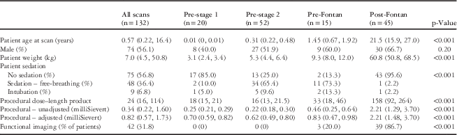
In the whole group, 76 patients were scanned without sedation, 47 were scanned with minimal to moderate sedation, and nine patients were intubated during the scan. Of the nine patients who were intubated during the scan, six were intubated for clinical reasons and three patients specifically for the scan: two for multiple procedures and one for cardiac CT alone.
Overall, 18 patients in the neonatal group were scanned without sedation and two were intubated for clinical management of underlying heart disease. Of the 52 patients scanned between first- and second-stage palliation, 13 patients were scanned without sedation, 34 were freely breathing but were under sedation, and were intubated. Of these five patients, three were intubated for clinical reasons and two patients were intubated with general anaesthesia for multiple concurrent procedures: head CT and cardiac CT on different scanners in a patient with cyanosis; and cardiac CT and gastric tube placement in the other patient. Of the 15 patients scanned between stages 2 and 3 of palliation, two were sedated, 12 were sedated but were freely breathing, and one was intubated for clinical reasons. In the 45 patients undergoing CT scan after Fontan palliation, 43 were scanned without sedation, one patient with developmental delay were sedated for the scan, and one patient with developmental delay and movement disorder was intubated for a breath-holding sequence.
Intravenous-line site and gauge
Information on intravenous-line site and gauge was available for 128 of the 132 scans (97%). Three patients utilised hand-injected of contrast through a central line, one patient utilised power injection through a broviac catheter, one neonate had hand injection through an umbilical venous catheter, and one patient had hand injection through a peripherally inserted central catheter line when other routes were impractical. The remaining 122 patients had contrast injected through a peripheral line. Table 1 lists the peripheral intravenous gauge by stage of palliation for these patients. Peripheral intravenous line location was a right- or left-sided hand vein, right- or left-sided antecubital vein, or a right- or left-sided saphenous vein.
Peripheral IV gauge size by stage of single-ventricle palliationFootnote *.

* Information on IV site and gauge was available for 122 patients
Scan sequences in the patient group
In the neonatal group (n=20), six patients underwent a single anatomical scan and 14 underwent two anatomical scans, making a total of 34 anatomical scans. No patient underwent functional imaging. In all, 27 anatomical scans were performed using a tube potential of 70, and seven anatomical scans were performed using a tube potential of 80.
Among the patients scanned between first- and second-stage palliation, 28 underwent a single anatomical scan and 24 underwent two anatomical scans, making a total of 76 scans. No patient underwent functional imaging. In all, 39 scans were performed using a tube potential of 70, and 37 scans using a tube potential of 80.
Among the patients scanned between the Glenn and Fontan procedures (n=15), six patients underwent a single anatomical scan, six underwent two anatomical scans, and three had underwent an anatomical and a functional scan, making a total of 21 anatomical scans and three functional scans. In all, 13 anatomical scans were performed using a tube potential of 70, and eight anatomical scans using a tube potential of 80. A single functional scan was performed using a tube potential of 70, and two functional scans were performed using a tube potential of 80. After the Fontan procedure (n=45), two patients underwent a single anatomical scan, one patient underwent a single functional scan, four patients underwent two anatomical scans, and 38 patients underwent an anatomical and a functional scan, making a total of 48 anatomical scans and 39 functional scans. Overall, 18 anatomical scans were performed using a tube potential of 70, 27 anatomical scans using a tube potential of 80, and three anatomical scans were performed using a tube potential of 100. In all, four functional scans were performed using a tube potential of 70, 22 functional scans using a tube voltage of 80, 12 functional scans using a tube potential of 100, and one scan was performed using a tube potential of 120.
Estimated radiation dose
Table 2 lists procedural dose–length product and radiation dose estimates for the entire group stratified by stage of palliation. The median procedural dose–length product was 24 mGy-cm (16, 114), and unadjusted and adjusted radiation doses were 0.34 (0.20, 1.80) and 0.82 (0.55, 1.88) mSv, respectively.
Patient demographics and CT scan information.

Results are shown as medians and interquartile ranges
Image quality
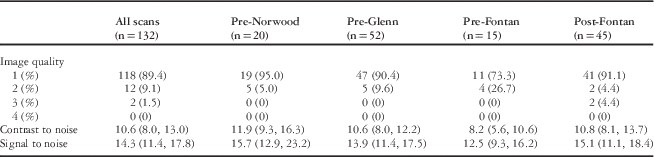
Qualitative image quality for the group showed an image score of 1 for 118 of the 132 scans (89%), an image score of 2 for 12 scans (9%), and an image score of 3 for 2 scans (2%). The two scans that received an image score of 3 were both in the post-Fontan group. Of the 14 scans with image quality score <1, eight had poor contrast opacification in the structures of interest due to scan timing, two had intravenous contrast leak with poor contrast in the structures of interest, two had a contrast scatter artefact, and two had a movement artefact. Table 3 summarises both qualitative and quantitative image quality data.
Scan image quality (qualitative and quantitative).

Adverse events, significant anatomical findings, and correlation with intervention
There was one adverse event of a broviac catheter (placed 2 months before) crack with contrast injection that could be repaired. Multiple attempts at peripheral intravenous placement were unsuccessful in this case. A power injector was used for contrast injection at a rate of 1 ml/s, and the pounds per square inch was acceptable with the saline test injection but the value was over the recommended pounds per square inch for the catheter, with a 50–50% contrast–saline mix at the same injection rate. The catheter crack was repaired and the catheter remained functional through hospital discharge.
In total, 79 patients underwent surgical intervention after CT scan, seven patients underwent catheter-based intervention before surgery, and three patients underwent catheterisation-based intervention alone.
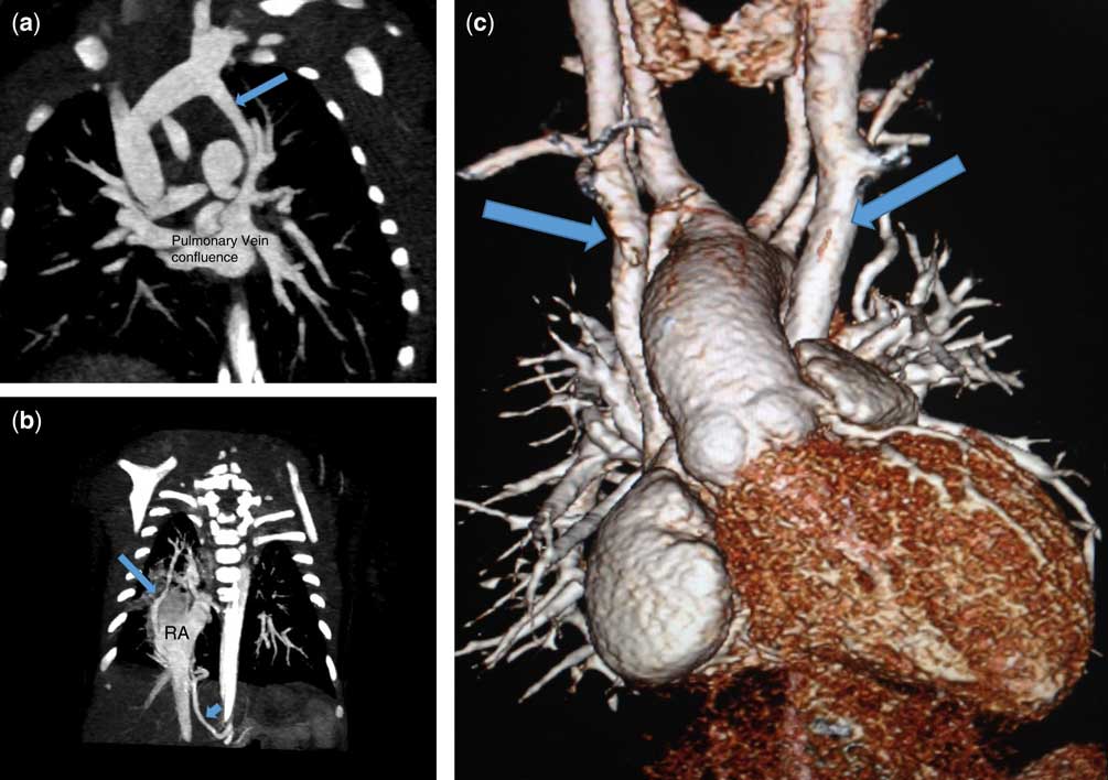
Of the 20 neonates, 18 underwent subsequent staged palliation and two patients had died. No patient had additional advanced diagnostic studies before initial surgical palliation. A minor discrepancy was a difference of opinion regarding the classification of a vessel as an aortopulmonary collateral versus ductus arteriosus. Example neonatal images are shown in Figure 1.
Neonatal evaluation. ( a ) Two-dimensional image of total anomalous pulmonary venous return with a vertical vein (arrow) to the innominate vein in a patient with hypoplastic left heart syndrome. ( b ) Anomalous right pulmonary vein (arrow) and an aortopulmonary collateral (short arrow) in a patient with Scimitar syndrome and hypoplastic left heart syndrome. ( c ) Three-dimensional image of bilateral superior caval (arrows) in a patient with tricuspid atresia.

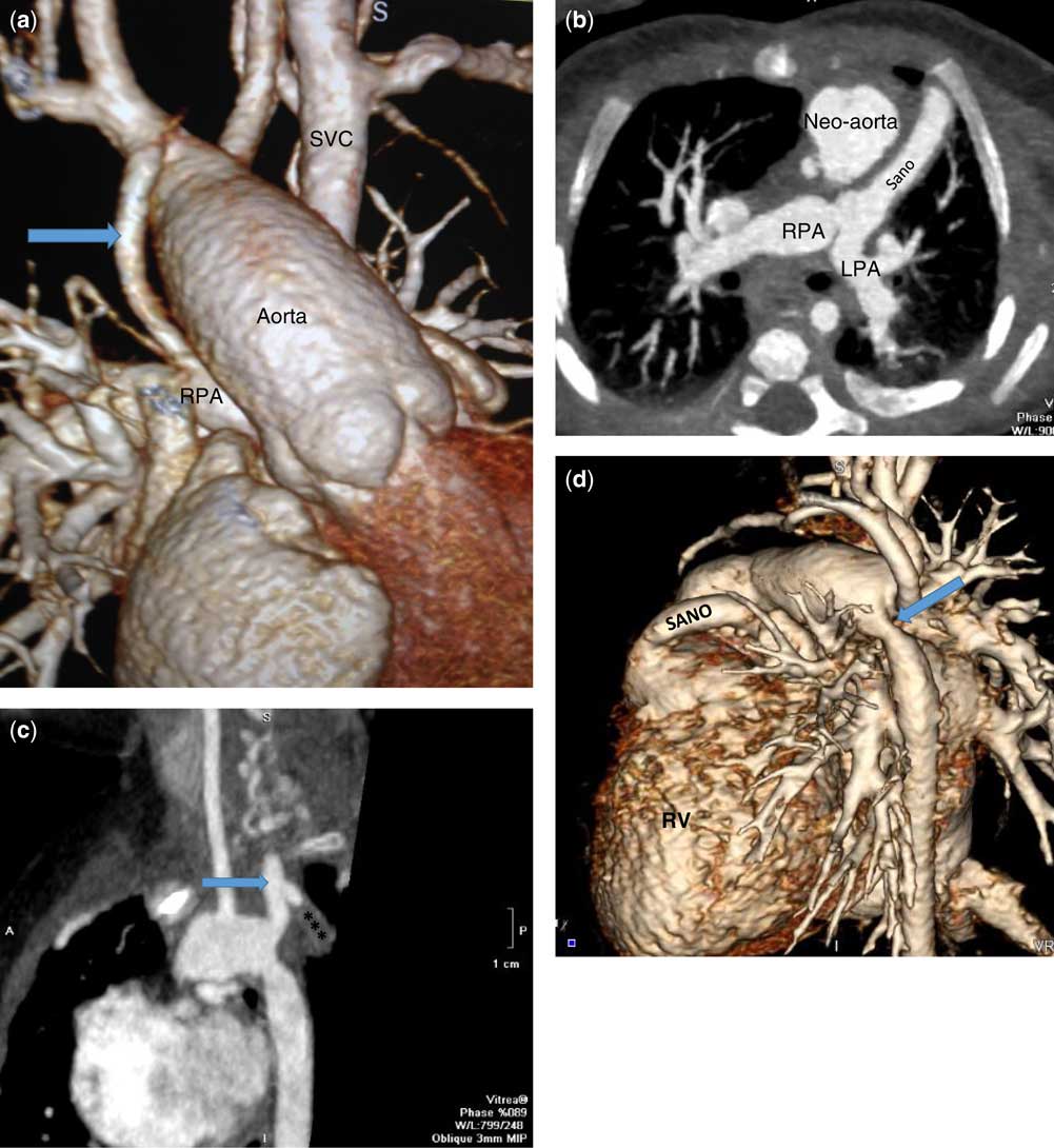
Of the 52 patients who were scanned after first-stage single-ventricle palliation, significant findings other than expected anatomy were seven central venous occlusions, two aortopulmonary shunt or Sano narrowings >50%, 12 significant branch pulmonary artery narrowings, two arch obstructions, and one large pseudoaneurysm at the proximal site of Sano insertion. There were 50 patients who underwent subsequent surgical intervention and two patients who had died: one during anaesthesia induction for planned catheter-based arch angioplasty and one with progressive and severe pulmonary vein stenosis. One surgical intervention was a hybrid procedure with stent placement at the time of Glenn. Four patients underwent catheter-based intervention before surgery, including pulmonary artery balloon angioplasty, pulmonary artery balloon angioplasty with stent placement (n=2), and aortic arch balloon angioplasty. There was one discrepancy compared with interventional findings, which was the presence of right internal jugular vein occlusion in a patient who underwent catheterisation 2 months subsequently. The CT scan in that patient had not been optimised for venous imaging and only a single scan was performed. Examples of pre-Glenn images are shown in Figure 2.
Post-Norwood evaluation. ( a ) Three-dimensional reconstruction of an aortopulmonary shunt (arrow) from the base of the right innominate artery to the right pulmonary artery. Left superior vena cava is noted. ( b ) Axial two-dimensional image of a distal Sano anastomosis to the branch pulmonary arteries. ( c ) Saggital two-dimensional image of an occluded aortopulmonary shunt (***) from the base of the innominate artery (arrow). ( d ) Posterior view of a three-dimensional reconstruction showing aortic coarctation at the distal Norwood anastomosis (arrow) after first-stage Sano palliation.

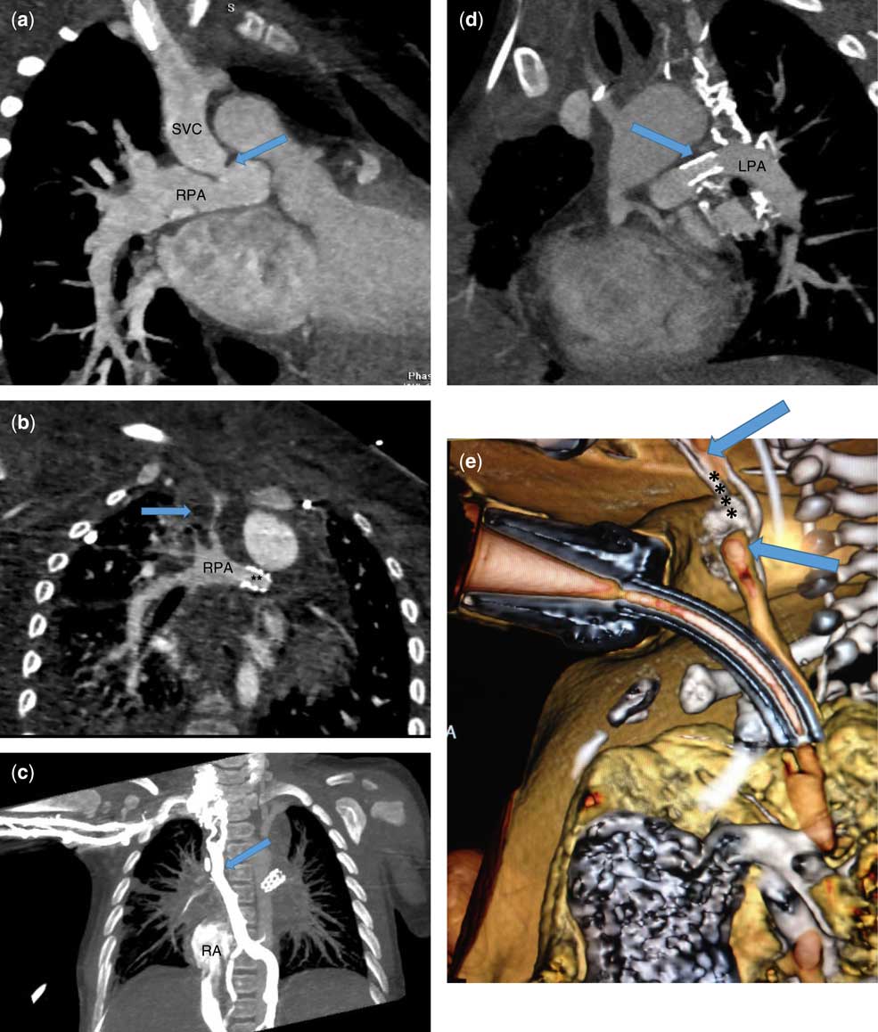
Of the 15 patients imaged between the Glenn and pre-Fontan catheterisation, three instances of central venous occlusion, one of pulmonary artery narrowing, two of arch obstruction, and two instances of large venous collaterals were found. A total of five patients underwent intervention – three catheterisation and two surgical – before third-stage palliation. Interventions included pulmonary arterioplasty, aortic arch angioplasty, aortic arch augmentation, and coiling of large venous collaterals (n=2). There were no discrepancies in this patient subset compared with interventional findings. Examples of images after the Glenn procedure are shown in Figure 3.
Post-Glenn evaluation. ( a ) Coronal two-dimensional image of narrowing (arrow) of the superior vena cava to right pulmonary artery anastomosis after cavopulmonary connection. ( b ) Coronal two-dimensional image showing a clot in the superior vena cava (arrow) and a patent left pulmonary artery stent (**) after Glenn cavopulmonary anastamosis. ( c ) Coronal two-dimensional image showing dense contrast opacification of decompressing venous collaterals (arrow) in a patient with innominate vein and subclavian vein occlusion after glenn cavopulmonartery anastomosis. ( d ) Saggital two-dimensional reconstruction in the same patient showing a patent left pulmonary artery stent (arrow) on a venous phase acquisition. ( e ) Long segment tracheal atresia (****) in a patient after the Glenn procedure. Both the proximal and distal tracheal are visualized (arrows).

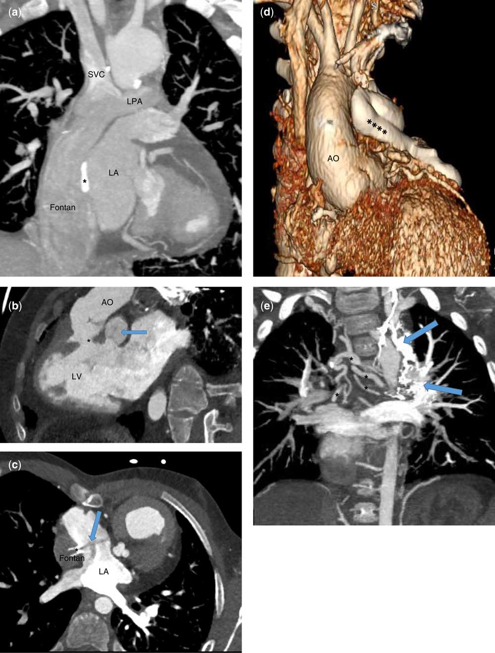
Anatomic findings in 45 post-Fontan patients included six complete central venous occlusions with large decompressing collaterals, one arch obstruction, bulboventricular foramen restriction, an anomalous left coronary artery with interarterial course, and a near-occlusive clot in the superior caval vein. A total of 12 patients underwent intervention: nine surgical and three catheter-based. Surgical interventions included unroofing of a coronary artery and pulmonary valvuloplasty, bulboventricular foramen enlargement, tricuspid valvuloplasty, neo-aortic valve replacement, conversion to extracardiac Fontan, and four pacemaker generator changes. Catheter-based interventions included clot removal, arch angioplasty and stent with vascular plug of collaterals, and vascular plug and coil occlusion of collaterals. There were no discrepancies in this subset of patients compared with interventional findings. Example images after the Fontan procedure are shown in Figure 4.
Post-Fontan evaluation. ( a ) Coronal two-dimensional image from the venous phase scan in a patient after lateral tunnel Fontan. The Fontan fenestration patch is noted (*). ( b ) Two-dimensional image of retained pulmonary valve leaflets (arrow) in a patient after pulmonary artery ligation. The pathway from the systemic left ventricle to the aorta from the systemic ventricle. ( c ) Two-dimensional axial image of a patent Fontan fenestration stent (*) with unopacified flow from the Fontan to the atrium (arrow) after septectomy. ( d ) Three-dimensional reconstruction of a densly opacified large decompressing collateral from the innominate vein after the Fontan procedure (****). ( e ) Two-dimensional coronanal image of both veno-venous (arrows) and arterial collaterals (*) after the Fontan procedure.

Discussion
CT is a robust alternative diagnostic modality across all stages of single-ventricle palliation. For highly select indications, the risk profile may sometimes be in favour of using CT compared with other diagnostic methods when risks from radiation, vascular access, and anaesthesia are considered. Single-ventricle patients are exposed to relatively high cumulative radiation levels during staged palliation.Reference Downing, McDonnell and Zhu 15 , Reference Johnson, Hornik and Li 16 A single institution survey reports a median cumulative effective radiation dose of 25.7 mSv from birth to 33 months of age, of which 78% was from catheterisation, constituting 4% of radiation encounters.Reference Downing, McDonnell and Zhu 15 Another survey of cumulative radiation dose for patients with all forms of CHD showed that 5.3% of patients received over 20 mSv/year with a median follow-up time of 4.3 years.Reference Glatz, Purrington, Klinger, King, Huda and Hlavacek 17 A recent study directly comparing radiation doses from diagnostic catheterisation (n=50 cases) and computed tomography angiography (n=50 cases) in children with CHD has shown 15-fold less radiation from CT angiography, although this was not specific to patients with single-ventricle heart disease.Reference Watson, Mah, Schoepf, King, Huda and Hlavacek 18 Other studies using older CT equipment list doses for CT angiography (n=21) that are twofold higher than those in diagnostic cardiac catheterisation (n=117).Reference Watson, Mah, Schoepf, King, Huda and Hlavacek 18 These results show that the radiation risk from CT varies considerably depending on the scanner platform used and the aggressiveness of dose reduction. The image quality necessary for evaluation of coronary lesions in adult patients is rarely required for congenital applications, and patient-specific dose reduction must be implemented if a diagnostic strategy utilising CT is to be implemented. The standard diagnostic protocol at a majority of centres remains invasive catheterisation before both second- and third-stage palliation, despite data showing a favourable risk profile for non-invasive evaluation.Reference Warnes, Williams and Bashore 3
Non-invasive assessment before second-stage palliation has shown similar operative outcomes compared with invasive catheterisation involving lower risk as measured by radiation exposure, vascular access complications, length of anaesthesia, and adverse events.Reference Brown, Gauvreau and Powell 19 – Reference Han, Vezmar and Lesser 21 Longer-term follow-up, up to 8 years after the Fontan procedure, shows no difference in outcomes between invasive and non-invasive evaluation before stage 2. Our practice now uses CT preferentially for evaluation of anatomy before second-stage palliation. Catheterisation is reserved for patients in whom intervention is likely considered on the basis of echocardiography or clinical examination, and for patients with poor ventricular function and severe valve regurgitation in whom haemodynamics are considered relevant to clinical management. Some experts now propose a non-invasive algorithm for evaluation before both second- and third-stage palliation in patients with single-ventricle heart disease considered to be at a low risk for requiring intervention.Reference Fogel 22 – Reference Prakash, Khan, Hardy, Torres, Chen and Gersony 24
Cardiovascular MRI is the most commonly used non-invasive advanced imaging modality in CHD, but deep sedation or general anaesthesia is required in young children, scan times are relatively long, and gadolinium is used in many patients for angiography. Anaesthesia poses increased risk for patients with complex CHD undergoing MRI evaluation, and there is concern that repeated anaesthesia exposure of young patients may have adverse neurological effects.Reference Ramamoorthy, Haberkern and Bhananker 25 – Reference Odegard, DiNardo and Kussman 34 Gadolinium deposits have been found in brain tissue after repeated dosing in both children and adult patients, the significance of which is not yet known.Reference Miller, Hu, Pokorney, Cornejo and Towbin 35 – Reference Kanda, Fukusato and Matsuda 38 Risk assessment of non-invasive modalities should include assessment of risk from anaesthesia and iodinated or gadolinium-based contrast exposure in addition to radiation exposure and vascular-access requirements.
Cardiovascular CT is an alternative imaging modality that can be used as part of the non-invasive pathway when cardiac MRI is considered to pose a high risk or when there is an imaging artefact.Reference Khairy, Van Hare and Balaji 40 When anaesthesia is needed, a single breath-hold is required for data acquisition and the length of anaesthesia will be relatively short. Functional analysis for both right and left ventricles has been shown to be comparable to cardiac MRI when using CT scanners with acceptable temporal and spatial resolution.Reference Guo, Gao, Zhang, Wang, Yang and Ma 41 – Reference Plumhans, Muhlenbruch and Rapaee 44
Limitations
The findings from this study are retrospective and limited to a single institution. Compared with interventional findings, accuracy is limited to structures present within the surgical field or those imaged using invasive angiography. In addition, these findings may not be generalisable to institutions with different professional expertise in CT operation and interpretation or different CT hardware.
Conclusion
Cardiovascular CT serves as a non-invasive imaging alternative for patients with single-ventricle heart disease for whom advanced imaging is preferred but cardiac MRI poses high risk, has poor image quality, or is contraindicated. Image quality remains good at low radiation exposure, and accuracy is excellent when compared with interventional findings. CT may reduce radiation risk compared with catheterisation and decreases risks from anaesthesia and gadolinium compared with MRI. It should have a role in select patients with single-ventricle heart disease when appropriate staff and CT equipment are available.
Acknowledgements
Financial Support
This research received no specific grant from any funding agency, commercial or not for profit sectors. The Minneapolis Heart Institute Foundation receives research grant support for advanced congenital imaging from Siemens Medical.
Conflicts of Interest
B.K. Han receives institutional grant support from Siemens Healthineers.