Introduction
The visual-evoked potential (VEP) represents the overall changes in the potential of the visual pathway from retinal photoreceptors to the occipital visual area. Abnormal VEP responses have been reported not only in disorders at the optic nerve level, such as optic neuritis (Reference Halliday, McDonald and Mushin1), chiasmal lesions (Reference Halliday, Halliday, Kriss, McDonald and Mushin2) and glaucoma (Reference Atkin, Bodis-Wollner, Podos, Wolkstein, Mylin and Nitzberg3,Reference Towle, Moskowitz, Sokol and Schwartz4), but also in mental disorders, such as schizophrenia (Reference Jibiki, Takizawa and Yamaguchi5), depression (Reference Fotiou, Fountoulakis, Iacovides and Kaprinis6) and dementia (Reference Moore7). Sutter et al. (Reference Sutter8,Reference Sutter and Tran9) reported a method to simultaneously extract many local electroretinograms of the retina through a single contact lens electrode applied to the cornea using multiple random stimulations and a special calculation method. Applying these methods to VEP, Baseler et al. simultaneously measured VEP at many local sites in 1994 (Reference Baseler, Sutter, Klein and Carney10). The standard VEP mainly represents macular responses, whereas the multifocal visual-evoked potentials (mfVEPs) can be simultaneously extracted as responses of the retinal centre over the about 20° peripheral retina. A prolonged peak latency of the standard VEP in the recovery phase of optic neuritis has been reported (Reference Halliday, McDonald and Mushin1), but the severity of the effects of optic neuropathy of the peripheral visual field on mfVEPs measurement was not necessarily consistent (Reference Hood, Odel and Zhang11), and the possibility of a new method to evaluate visual function in intracranial diseases using mfVEPs has also been reported (Reference Watanabe, Shinoda, Kimura, Mashima, Oguchi and Ohde12,Reference Yukawa, Kim, Ueda and Hara13). Furthermore, Yukawa et al. (Reference Yukawa, Kim, Kawasaki, Taketani and Hara14,Reference Yukawa, Matsuura, Kim, Nitta, Taketani and Hara15) compared the mfVEPs responses in children and mental disorder patients with intracranial diseases suspected of having visual field impairment, in whom application of the conventional perimetry is difficult, with those in normal subjects, and identified the usefulness of mfVEPs as an objective visual field evaluation. However, abnormal VEP responses have been reported in mental disorder patients, as described above, suggesting that their mfVEPs responses are also different from those in normal persons. We measured mfVEPs in schizophrenic patients under treatment in whom no abnormality was detected on dynamic perimetry and compared the findings with those in normal subjects to investigate the differences in responses of the peripheral retina.
Materials and methods
The subjects were 31 schizophrenic patients (31 eyes) under treatment at the Psychiatry Department of Hannan Hospital (13 males and 18 females aged 25–70 years, with a mean age of 45.4 years) and 30 age-matched normal subjects (30 eyes) (14 males and 16 females aged 23–70 years, with a mean age of 45.8 years). The subjects underwent a visual acuity test, fundus photography and dynamic perimetry before mfVEPs measurement. Their corrected visual acuity was 20/20 or higher, no ophthalmological disease, including glaucoma, was detected by fundus photography and normal visual field was confirmed by an ophthalmologist. Normal subjects were selected from volunteer staff of the Hannan Hospital and Nara Medical University Hospital.
This study followed the tenets of the Declaration of Helsinki, and signed informed consent was obtained from all subjects before testing began.
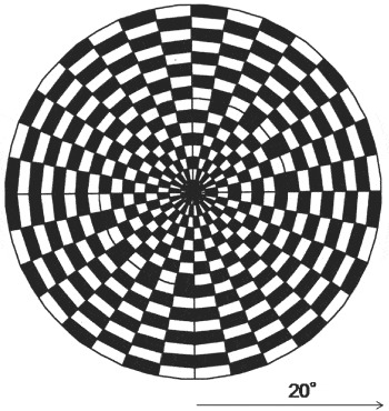
Recording of mfVEPs was performed using a VERIS Junior Science recording apparatus (Mayo, Aichi, Japan). One eye was randomly shielded with an eye patch. A corrective lens was placed 12 mm in front of the eye for optimal focus, 13 cm from the stimulation monitor. The active electrode was placed 4 cm above the inion, the reference electrode at the inion and the ground electrode at the right earlobe. Signals were amplified and bandpass filtered from 1 to 100 Hz. As a stimulation pattern, reversed stimulation with a dart board pattern was used; this consisted of eight elements composed of 64 checks each (Fig. 1). The mean luminance of stimulation was 103 cd/m2 and the contrast was 95%. The stimulus area subtended approximately 20° and the frame rate was 75 Hz. The pseudo-random stimulus presentation, the so-called M-sequence, was 214− 1, and each run was divided into eight equal segments with a total recording time of about 4 min. Responses from the eight sites in each subject were divided into four quadrants (superior and interior temporal quadrants, and superior and inferior nasal quadrants). In each quadrant, two response waves were grouped and averaged, and the height from the peak of the wave at about 70 ms to the peak latency of the main wave at about 100 ms was defined as the amplitude and used for assessment. For the measurement of mfVEPs, the subjects were asked to relax and stare at the centre of the stimulation pattern during examination. For statistical analysis, p < 0.05 was regarded as significant.
Stimulation pattern. A dart board pattern consisting of eight elements composed of 64 checks each was used. The mean luminance of stimulation was 102.5 cd/m2 and the contrast was 95%.

Results
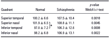
The major oral tranquilizers administered were haloperidol alone in six patients, risperidone alone in seven patients and several combinations of haloperidol, risperidone, sulpiride, carbamazepine, clonazepam, levomepromazine and mosapramine in 18 patients. The means and standard deviations of the peak latency and amplitude in the individual quadrants in the schizophrenic patient and normal subject groups are shown in Table 1 and Table 2. There was a significant difference in the latency between the superior nasal and inferior temporal quadrants in the normal subject group. The amplitude was significantly different between the superior and inferior temporal quadrants, between the superior temporal and inferior nasal quadrants, between the superior nasal and inferior temporal quadrants, and between the superior and inferior nasal quadrants, in both the schizophrenic patient and normal subject groups. The peak latency was increased about 7 and 9 ms in the upper and lower half visual fields, respectively, in the schizophrenic patient group compared to the normal subject group, the amplitude was reduced by about 2 and 5 nV/deg2 in the upper and lower half visual fields, respectively, and significant differences were noted in all quadrants in both parameters between the two groups.
Latencies (ms) on multifocal visual-evoked potentials
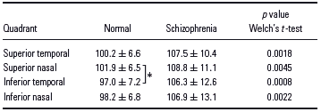
All results given as means ± standard deviations.
Tukey method: *p < 0.05.
Amplitudes (nV/deg2) on multifocal visual-evoked potentials

All results given as means ± standard deviations.
Tukey method: **p < 0.01.
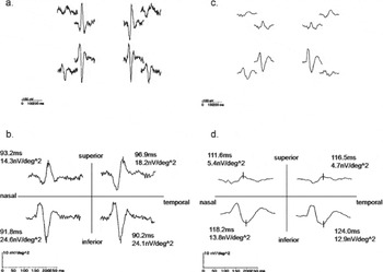
Typical mfVEPs of the schizophrenic patients and normal subjects are shown in Fig. 2.
(a) Original waves obtained from eight sites in a normal subject (right eye of a 47-year-old male). (b) Grouped waves in the four quadrants. The waveforms in the nasal and temporal quadrants were very similar, but the waveforms in the superior and inferior quadrants were mirror images. (c) Original waves obtained from eight sites in a schizophrenic patient (right eye of a 41-year-old male). This patient was under treatment with oral haloperidol, levomepromazine and carbamazepine. (d) Grouped waves in the four quadrants. The latency was apparently prolonged, and the amplitude was reduced in all quadrants, compared with those in normal subjects.

Discussion
Abnormal VEP responses measured in schizophrenic patients have been reported (Reference Jibiki, Takizawa and Yamaguchi5). These patients were under treatment with psychotropics, including haloperidol, and possible influences of these drugs on the VEP waveform have been suggested. Straumanis et al. (Reference Straumanis, Shagass and Roemer16) showed that the amplitude of the transient VEP was reduced in mental disorder patients chronically medicated with psychotropics, and Jibiki et al. (Reference Jibiki, Takizawa and Yamaguchi5) reported that the amplitude of the steady-state VEP waveforms was not affected by changing the check-size of the stimulation optotype in schizophrenic patients, compared with that in normal subjects, suggesting that VEPs were more markedly affected by drugs than by the pathology of schizophrenia. However, to our knowledge, there has been no report on the effects of mental disorders on mfVEPs. Abnormal latency and amplitude of mfVEP waveforms were simultaneously noted in all quadrants in schizophrenic patients in comparison with those in normal subjects, and the characteristic change in normal subjects, namely, greater amplitude in the inferior half of the visual field than in the superior half, was maintained in the schizophrenic patients. By contrast, the latency was not significantly different among the quadrants, but the difference between those in the superior and inferior halves of the visual fields tended to be smaller in schizophrenic patients than in normal subjects. However, since all schizophrenic patients were being treated with psychotropics, it was not clear whether the waveform changes were because of schizophrenia or the drugs, or both. It is necessary to confirm mfVEP waveforms in schizophrenic patients in the absence of treatment to investigate the degree of influence of psychotropics on the mfVEP waveforms.
Regarding the clinical application of mfVEPs, several studies have reported the possibility of objective visual field evaluation (Reference Yukawa, Kim, Kawasaki, Taketani and Hara14,Reference Yukawa, Matsuura, Kim, Nitta, Taketani and Hara15,Reference Klistorner and Graham17–Reference Yukawa, Matsuura, Kim, Taketani and Hara20). In the current perimetry method, the examinee presses the button when he/she senses a light somewhere in the periphery while maintaining sufficient fixation. In mfVEP measurement, reliable waveform responses can be obtained only by staring at the centre of the stimulation optotype in a relaxed state. Using this technique, Yukawa et al. (Reference Yukawa, Matsuura, Kim, Nitta, Taketani and Hara15) measured mfVEPs in patients with mental disorders complicated by intracranial disease, in whom no reliable findings could be obtained by current perimetry, and found that objective evaluation of the visual field was possible in some cases. The subjects were patients in whom reliable perimetry could be performed and no abnormality was detected in the visual field, but their responses on mfVEPs were different from those in the normal subjects, showing deviation between the visual field on perimetry and mfVEPs. The findings also suggested that the prolongation of the latency and amplitude reduction compared to those in normal individuals should be taken into consideration in objective visual filed evaluation using mfVEPs in schizophrenic patients suspected as having optic neuritis or hemianopia associated with intracranial disease.