I. INTRODUCTION
Sol–gel processing techniques have emerged as biocompatible methods to directly interface live cells and tissues with a siliceous mineral phase, conferring novel or enhanced material properties to biological composites. The mechanical strength, porosity, and optical clarity achievable in synthetic silica are advantageous partners for biological functions such as enzymatic activity, bioactivity, and physiologically designed microstructure. Mechanical strength and porosity are especially desirable properties for cell coating materials, with the potential to protect vulnerable cells and regulate transport across the cell surface by size. Reference Jaroch, McLamore, Zhang, Shi, Garland, Banks, Porterfield and Rickus1–Reference Losic, Mitchell and Voelcker4 While traditional established methods for porous silica formation (e.g., block copolymer templating) result in highly regular nano- and microstructural organization and narrow pore size distributions to facilitate precision size exclusion, their synthesis is not compatible with living cells. Reference Zhao, Jianglin, Huo, Melosh, Fredrickson, Chmelka and Stucky5,Reference Beck, Vartuli, Roth, Leonowicz, Kresge, Schmitt, Chu, Olson, Sheppard, McCullen, Higgins and Schlenker6 Adapting biocompatible sol–gel silica hybrid materials to have more precise microstructural properties requires deeper understanding of silica formation kinetics at the organic–inorganic interface.
Formation of porous silica via a two-step sol–gel method involves first hydrolysis and then condensation. Reference Ellerby, Nishida, Nishida, Yamanaka, Dunn, Valentine and Zink7 Hydrolysis of silicon alkoxides in water by either acid or base catalysis produces monosilicic acids and alcohol by-products. Individual monosilicic acids then condense to form siloxane bonds, a process referred to in bulk as polycondensation or gelation. Polycondensation may be described in terms of three interrelated steps: (i) nucleation, the process of individual molecules condensing together to form small particles; (ii) particle growth, the Ostwald ripening Reference Brinker and Scherer8 process in which smaller particles undergo reverse reactions to donate groups to larger particles; and (iii) gel formation, the process of particles condensing together into a porous matrix that spans the volume of the container. Reference Brinker and Scherer8,Reference Iler9 Gels continue to cure over time until all water has been eliminated. Polycondensation is a stochastic process, resulting in porous materials that are structurally amorphous in nature. Reference Brinker and Scherer8 The probability of condensation events, and therefore the overall rate of polycondensation, directly affects the porosity and mechanical compliancy of the resulting material. Reference Brinker, Sehgal, Hietala, Deshpande, Smith, Loy and Ashley10
The kinetics of each stage of silica formation is governed by reaction conditions, including temperature, pH, choice of silicon alkoxide precursor and hydrolysis catalyst, removal of the alcoholic hydrolysis byproduct, and introduction of chemical groups that initiate silica condensation. Reaction kinetics of the particle growth and gelation is often described by complex kinetic models due to the vast number of possible condensation events, but initiation of condensation is a comparatively simpler process. Although polycondensation has been described by rate equations of varying orders depending on reaction conditions and assay method, Reference Tobler, Shaw and Benning11 measurement of mono- and disilicic acid depletion provides a simplified method to characterize condensation in terms of an apparent third-order kinetics; under the assumption that disilicic acids are short-lived intermediates, formation of trimeric species in the bulk solution is approximated as a reaction between three distinct monosilicic acids, so condensation rates measured in this way are dependent only on monosilicic acid concentration. Reference Belton, Deschaume, Patwardhan and Perry12,Reference Patwardhan, Emami, Berry, Jones, Naik, Deschaume, Heinz and Perry13
Introduction of an organic template rich in chemical groups that initiate silica condensation is an effective strategy to increase gelation rates in the bulk solution while also providing a physical scaffold for deposition of the mineral phase. This approach has traditionally been explained by attractive charge interactions between surface groups and condensing silicic acids; Reference Coradin, Nassif and Livage14 however, organic templating is now understood to depend more upon kinetic factors at play in the exchange between silicic acids and cooperating template groups of opposing charge, Reference Wallace, DeYoreo and Dove15 which can bring silicic acid molecules in proximity for condensation. Reference Coradin and Livage16 Proteins with high hydroxyl content in their primary structures, including those known to facilitate silicification in nature (e.g., silicateins and silaffins), Reference Hecky, Mopper, Kilham and Degens17–Reference Müller, Schröder, Burghard, Pisignano and Wang20 can participate in hydrogen bonding, Reference Shimizu, Cha, Stucky and Morse18,Reference Birchall21,Reference Ehrlich, Deutzmann, Brunner, Cappellini, Koon, Solazzo, Yang, Ashford, Thomas-Oates, Lubeck, Baessmann, Langrock, Hoffmann, Wörheide, Reitner, Simon, Tsurkan, Ereskovsky, Kurek, Bazhenov, Hunoldt, Mertig, Vyalikh, Molodtsov, Kummer, Worch, Smetacek and Collins22 electrostatic interactions, Reference Ono, Kanekiyo, Inoue, Hojo, Nango and Shinkai23,Reference Heinemann, Ehrlich, Knieb and Hanke24 and even condensation reactions with silicic acids. Reference Hecky, Mopper, Kilham and Degens17,Reference Ehrlich, Deutzmann, Brunner, Cappellini, Koon, Solazzo, Yang, Ashford, Thomas-Oates, Lubeck, Baessmann, Langrock, Hoffmann, Wörheide, Reitner, Simon, Tsurkan, Ereskovsky, Kurek, Bazhenov, Hunoldt, Mertig, Vyalikh, Molodtsov, Kummer, Worch, Smetacek and Collins22 The involvement of positively charged moieties, especially amines, is also an important attribute of the organic template, as cooperative interactions between differentially ionizable groups, such as hydroxyl and amine partners in close proximity, have demonstrated a greater effect on condensation rates than either hydroxyls or amines alone. Reference Wallace, DeYoreo and Dove15
The natural polymer collagen has been shown to template and direct silica condensation. Reference Kahn, Eren, Campanella, Voytik-Harbin and Rickus25,Reference Niu, Jiao, Qi, Yiu, Ryou, Arola, Chen, Breschi, Pashley and Tay26 Collagen is an excellent candidate protein to study silica templating kinetics, owing to its natural abundance, regular structure, and relevance to human physiology. The primary structure of collagen is high in repetitive (Gly–Xaa–Yaa) n triplets with hydroxylation at the Yaa position, often (Gly–Pro–Hyp) n . Reference Shoulders and Raines27 Multiple hydroxylation sites in close proximity are uniquely capable of interacting with silicic acids, as evidenced by molecular modeling and increased condensation rates for specialized sponge collagen motifs where Xaa is hydroxylated in addition to Yaa. Reference Ehrlich, Deutzmann, Brunner, Cappellini, Koon, Solazzo, Yang, Ashford, Thomas-Oates, Lubeck, Baessmann, Langrock, Hoffmann, Wörheide, Reitner, Simon, Tsurkan, Ereskovsky, Kurek, Bazhenov, Hunoldt, Mertig, Vyalikh, Molodtsov, Kummer, Worch, Smetacek and Collins22 Amines present in abundant Arg, Lys, and Gln residues Reference Shoulders and Raines27,Reference Ramshaw, Shah and Brodsky28 can take part in cooperative interactions with hydroxyls to affect kinetic drivers for silica condensation. Reference Wallace, DeYoreo and Dove15
Our strategy for studying interactions between the condensing silicic acid mineral phase and the collagen-fibril template organic phase employs an uncommon formulation of soluble type I collagen oligomers. Oligomers, which represent soluble aggregates of tropocollagen molecules (e.g., trimers) that retain their natural intermolecular crosslinks, Reference Bailey, Critser, Whittington, Kuske, Yoder and Voytik-Harbin29 exhibit not only fibrillar but also suprafibrillar assembly. Unlike conventional collagen monomers (atelocollagen and telocollagen), which form entangled networks of long individual fibrils, oligomers yield highly interconnected collagen-fibril matrices with dramatically improved mechanical integrity and handling as well as reduced proteolytic degradation. Reference Kreger, Bell, Bailey, Stites, Kuske, Waisner and Voytik-Harbin30 The hierarchical self-assembly of soluble collagen oligomers to form fibrils and fibril networks is spontaneous under neutral conditions at 37 °C and stabilized by inter- and intramolecular crosslinking. Reference Kadler, Holmes, Trotter and Chapman31 Furthermore, oligomers are standardized based on their polymerization or matrix-forming capacity, thereby yielding highly reproducible relationships between oligomer concentration of the polymerization reaction, fibril density, and shear storage modulus (stiffness) of formed matrices. Reference Bailey, Critser, Whittington, Kuske, Yoder and Voytik-Harbin29,Reference Kreger, Bell, Bailey, Stites, Kuske, Waisner and Voytik-Harbin30 We recently demonstrated that the tunability of fibril microstructure afforded by this collagen system is preserved in the templated silica layer, implying a relationship between collagen fibril density and silica condensation potential. Reference Kahn, Eren, Campanella, Voytik-Harbin and Rickus25
The benefit of controlling silica condensation rates at the surface of collagen-fibril templates extends across several active areas of research, including cellular microencapsulation, Reference Jaroch, Lu, Madangopal, Stull, Stensberg, Shi, Kahn, Herrera-Perez, Zeitchek, Sturgis, Robinson, Yoder, Porterfield, Mirmira and Rickus2,Reference Coradin, Nassif and Livage14 bone and tissue engineering, Reference Ramadass, Perumal, Gopinath, Nisal, Subramanian and Madhan32–Reference Heinemann, Heinemann, Wenisch, Alt, Worch and Hanke34 and biomineralization. Reference Wang, Schloßmacher, Schröder and Müller35,Reference Niu, Jiao, Ryou, Diogenes, Yiu, Mazzoni, Chen, Arola, Hargreaves, Pashley and Tay36 In theory, bulk solution and surface gelation rates should have a synergistic influence on hybrid material properties like mineral layer thickness and depth of penetration, while pore size distribution and porosity are controlled both at the level of the collagen-fibril template and silica formation. Reference Kahn, Eren, Campanella, Voytik-Harbin and Rickus25 This report presents the study of sol–gel silica condensation kinetics during interaction with collagen-fibril templates with systematically varied fibril densities. Chemical and rheological analyses provide insight into reaction rates at the surface of the template and in the surrounding bulk solution, leading to a proposed two-phase gelation process describing high initial rates of gelation at the surface that decrease over time in contrast with low initial rates of bulk gelation that increase over time.
II. MATERIALS & METHODS
A. Preparation of collagen-fibril matrices
Type I collagen oligomers were acid solubilized from the dermis of market weight pigs and lyophilized for storage as described previously. Reference Kreger, Bell, Bailey, Stites, Kuske, Waisner and Voytik-Harbin30 Prior to use, lyophilized oligomer was dissolved in 0.01 N hydrochloric acid (HCl) and rendered aseptic by chloroform exposure at 4 °C. Oligomer concentration was determined using a Sirius Red (Direct Red 80) assay. Reference Brightman, Rajwa, Sturgis, McCallister, Robinson and Voytik-Harbin37,Reference Marotta and Martino38 The oligomer formulation was standardized based on molecular composition as well as polymerization capacity according to ASTM standard F3089-14. 39 Here, polymerization capacity is defined by matrix shear storage modulus G′ as a function of oligomer concentration of the polymerization reaction. Collagen-fibril matrices of four different fibril densities (corresponding to shear storage modulus G′ values of 200, 600, 1000, and 1500 Pa) were prepared by dilution in 0.01 N HCl and neutralization with 10× phosphate-buffered saline (PBS, 1× PBS had 0.17 M total ionic strength and pH 7.4) and 0.1 N sodium hydroxide (NaOH) to achieve neutral pH (7.4). Neutralized collagen solutions were kept on ice prior to induction of polymerization at 37 °C. Individual collagen-fibril matrices were drop cast and polymerized in 500 μL volumes in 24-well plates for rheological analysis and in 58 μL volumes in 96-well plates for all other analyses.
B. Preparation of silicifying solution (SS)
To produce concentrated silicic acid, tetramethoxysilane (tetramethyl orthosilicate or TMOS, from Sigma, USA) was hydrolyzed under acidic conditions in nanopure water adjusted to approximately pH 3 with HCl. Acidified water was added to TMOS such that the molar ratio of components (water:TMOS:HCl) was 16:1:0.007. The mixture was vortexed for 5–10 min. Methanol byproduct of TMOS hydrolysis was removed under vacuum by rotary evaporation at 46 °C to reduce the total volume by 40%, forming a concentrated (approximately 3.84 M) solution of silicic acids. The silicic acid solution was placed on ice and used immediately for individual experiments in the form of SS, 3 vol% concentrated silicic acid in 1× PBS, approximately 115 mM.
C. Scanning electron microscopy (SEM)
Collagen-fibril matrices (200 Pa) were exposed to SS (“silicified”) for 0, 15, and 30 min. Following exposure, matrices were washed thrice with 1× PBS to halt silicification. Silicified matrices were prepared for SEM and energy-dispersive x-ray spectroscopy (EDX) by ethanol dehydration (10, 20, 30, 40, 80, 90, and 100% in water), liquid CO2 exchange using a critical point dryer (LADD Research Industries, Williston, VT, USA), and sputter coating with platinum.
High-resolution SEM images were obtained in high vacuum mode with an acceleration voltage of 5 kV using the NOVA nanoSEM FESEM (FEI Company, Hillsboro, OR, USA) equipped with Everhart Thornley Detector and Through-the-Lens Detector.
D. Measurement of monomeric and dimeric silicic acid depletion
Interactions between SS and collagen-fibril matrices were monitored by the depletion of monomeric and dimeric silicic acid species using the blue silicomolybdic acid (SMA) spectrophotometric method, as described in detail by Coradin et al. Reference Coradin, Eglin and Livage40 Briefly, collagen-fibril matrices were immersed in 300 μL SS contained within 48-well plates. Solution samples (100 μL) from independent specimens were removed and diluted to obtain concentrations within the assay linear range. Visible absorbance was recorded at 810 nm and converted to concentration based on a linear calibration from fully concentrated silicic acid solution. The full assay, including calibration, was performed for three independent replicates (n = 3).
E. Kinetic analysis of condensation
Condensation rates were analyzed in terms of an apparent third-order reaction model for silicic acid trimer formation from silicic acid monomers with the assumption that dimers were present only as short-lived intermediates. Reference Harrison and Loton41 The third-order rate coefficient, k 3, was computed from the measured concentration of silicic acid monomers and dimers at five time points (0, 60, 120, 180, and 300 s) for four different collagen templates (200, 600, 1000, and 1500 Pa) and one control without collagen template exposure. Equations (4)–(6) governing reaction rates are found in Sec. III.
F. Real-time viscoelastic analysis of silica gelation
All rheological analyses were performed using a stress-controlled rotational DHR-3 rheometer with Smart Swap™ geometry (TA Instruments, New Castle, Delaware), equipped with parallel plate upper geometry (8 mm diameter) and standard Peltier plate temperature-controlled stationary lower plate. Viscoelastic properties were measured at a constant frequency (0.5 Hz, 3.142 rad/s), constant strain (1%), determined to be within the linear viscoelastic behavior of the sample, and constant temperature (25 °C) during a sampling period of 300–1500 s.
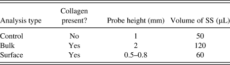
Real-time rheological analysis of gelation can be categorized into three distinct types of analyses: control, bulk, and surface. For control analyses, 50 μL of SS was pipetted directly onto the lower plate of the parallel plate geometry, and the rheometer upper plate was lowered to make contact with SS; for bulk analyses, single 500 μL collagen matrix samples were placed directly onto the lower plate, 120 μL of SS was pipetted on top of the collagen matrix, and the rheometer upper plate was lowered to make contact with SS; for surface analyses, single 500 μL collagen matrix samples were placed directly onto the lower plate, 60 μL of SS was pipetted onto the very surface of the collagen matrix, and the rheometer upper plate was lowered to make contact with the collagen matrix surface, determined by monitoring the axial force read in the instrument when the upper plate was touching the sample. Independent samples for each analytical condition were tested in triplicate (n = 3).
III. RESULTS AND DISCUSSION
A. The oligomeric collagen-fibril template
The collagen-fibril templates used in these studies are polymerized from a set of collagen oligomer building blocks, which are isolated using a unique process that maintains more of collagen's native features including telopeptide sequences and intermolecular crosslinks. Reference Bailey, Critser, Whittington, Kuske, Yoder and Voytik-Harbin29,Reference Kreger, Bell, Bailey, Stites, Kuske, Waisner and Voytik-Harbin30,Reference Whittington, Brandner, Teo, Han, Nauman and Voytik-Harbin42 The oligomer building blocks are quality controlled to such a degree that the matrices' final properties after gelation can be predicted and therefore tuned by the starting oligomer. This feature allowed us to systematically vary the properties of the collagen as a silica template. The relationships between collagen oligomer concentration and matrix shear storage modulus after gelation are known to be highly reproducible and detailed only briefly here. Reference Bailey, Critser, Whittington, Kuske, Yoder and Voytik-Harbin29
Individual extraction/solubilization preparations are characterized by fitting a quadratic function [Eq. (1)] to shear storage modulus G′ measurements (G) for a range of fibril volume fractions (V) of solubilized oligomeric collagen. Collagen fibril volume fraction is defined as the volume of collagen oligomers in solution divided by the total solution volume. Parameters a, b, and c are determined empirically from a quadratic fit to the measured data. Parameters for the two collagen oligomer preparations used in this study are provided in Supplementary Material Table S1.
 $$G = a{V^2} + bV - c\quad .$$
Additionally, solubilized collagen oligomer concentration (C) can be related to measured shear storage modulus G′ via a similar quadratic function [Eq. (2)]. Solubilized collagen oligomer concentration is defined as the concentration of solubilized collagen oligomers (mg/mL) in solution. Constant parameters a′, b′, and c′ are determined empirically from a quadratic fit to the measured data. Parameters for the two collagen oligomer preparations used in this study are also provided in Supplementary Material Table S1.
 $$G = {a\prime }{C^2} + {b{\prime }}C - {c{\prime }}\quad .$$
Solubilized collagen fibril volume fraction and solubilized collagen oligomer concentration can be related proportionally to each other for each collagen oligomer preparation by Eq. (3). The proportionality constant (X) varies slightly for each preparation, and constants for the two collagen oligomer preparations used in this study are provided in Supplementary Material Table S1.
 $$C = XV\quad .$$
For our studies, shear storage modulus G′ was chosen as the main parameter to control and vary, specifically 200, 600, 1000, and 1500 Pa G′ templates. All relevant solubilized fibril collagen volume fraction and concentration values reported have been extracted from their mathematical relationships with measured shear storage modulus G′, as calibrated for each individual collagen oligomer preparation.
B. Collagen-fibril matrices template and direct silica polycondensation
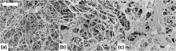
Although silica–collagen interactions are often studied relative to silica penetration of collagen fibrils Reference Niu, Jiao, Ryou, Diogenes, Yiu, Mazzoni, Chen, Arola, Hargreaves, Pashley and Tay36,Reference Nudelman, Pieterse, George, Bomans, Friedrich, Brylka, Hilbers, de With and Sommerdijk43 or silica interaction with soluble collagens in a copolymerization process, Reference Heinemann, Ehrlich, Knieb and Hanke24,Reference Ramadass, Perumal, Gopinath, Nisal, Subramanian and Madhan32,Reference Eglin, Shafran, Livage, Coradin and Perry44 the silica sol–gel system described here primarily serves to encapsulate, not infiltrate, collagen fibrils, penetrating the matrix of fibrils but coating the fibrils themselves. Silica condensation conditions in the present study are characterized by lack of precursor stabilization, removal of methanol hydrolysis byproduct, relatively short gelation time-scale, and high concentration of silicic acid. SEM micrographs in Fig. 1 demonstrate the development of the siliceous mineral phase on a 200 Pa collagen-fibril matrix exposed to SS for 0, 15, and 30 min. The porous silica coating begins with initiation of condensation at the surface of individual collagen fibrils, later condensing together further in web-like structures of silica particles. Images indicate that during the coating development process, silica can penetrate the matrix beyond externally located fibrils to condense upon fibrils that are located deeper in the collagen matrix. As time increases, the coating limits access to the internal matrix fibrils, and porous silica continues to expand on top of previously deposited layers. Macroscopically, we observe that exposure of collagen templates to SS may be allowed to continue for up to 60 min; after 60 min, the gelation of surrounding bulk SS prohibits effective removal and washing of composite materials.
Silica coating develops on the surface of collagen fibrils with increased exposure time. SEM images (20,000×, scale bar 4 μm) of 200 Pa collagen fibril matrices that have been exposed to SS for (a) 0 min, (b) 15 min, and (c) 30 min demonstrate the evolution of silicification over time.

C. Collagen fibril density controls silica condensation rates
To evaluate the effect of collagen fibril density on the kinetics of silica formation, collagen-fibril templates of varying fibril volume fraction corresponding to shear storage modulus G′ values of 200, 600, 1000, and 1500 Pa were exposed to SS for 0–300 s. SS samples were removed from individual sacrificial reactions and assayed for quantification of monomeric and dimeric silicic acid species at 60, 120, 180, and 300 s. Raw data for silicic acid depletion over time are provided in Supplementary Material Figure S1. Based on the high water to silicon molar ratio, the hydrolysis of tetramethoxysilane (tetramethyl orthosilicate, TMOS) via hydrochloric acid catalysis in water was assumed to be complete or nearly so, producing a solution of concentrated monosilicic acids. Reference Iler9 Blue silicomolybdous acid compounds, quantified by their optical absorbance at λ = 810 nm, are formed via complexation and subsequent reduction of silicic acids with ammonium heptamolybdate under acidic conditions. Because this method has been adjusted to detect only monomeric and dimeric silicic acid species, Reference Coradin, Eglin and Livage40 depletion of these species can be modeled as the formation of silicic acid trimers. Reference Belton, Deschaume, Patwardhan and Perry12,Reference Patwardhan, Emami, Berry, Jones, Naik, Deschaume, Heinz and Perry13,Reference Harrison and Loton41 Under the assumption that dimeric silicic acids are short-lived reaction intermediates, Reference Belton, Deschaume, Patwardhan and Perry12 trimer formation is approximated as a reaction between three individual monomeric species. The formation of trimers is therefore only dependent on monomer concentration, a third-order kinetic process presented in Eq. (4).
 $$3{\rm{Si}}{\left( {{\rm{OH}}} \right)_4}\buildrel {{k_3}} \over \longrightarrow {\rm{S}}{{\rm{i}}_3}{{\rm{O}}_2}{\left( {{\rm{OH}}} \right)_8} + 2{{\rm{H}}_2}{\rm{O}}\quad {\rm{.}}$$
The third-order rate coefficient k 3 can be further described in terms of the depletion rate, or the change in monomer concentration over time [Eq. (5)].
 $${{ - {\rm{d}}\left[ {{\rm{monomer}}} \right]} \over {{\rm{d}}t}} = {k_3}{\left[ {{\rm{monomer}}} \right]^3}\quad .$$
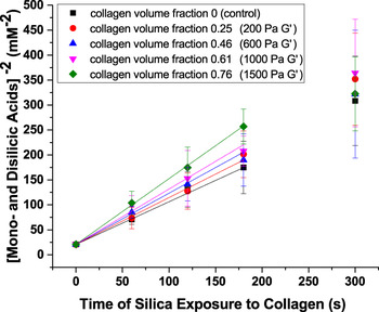
Integrating Eq. (5) reveals a relationship between the inverse square of the monomer concentration and time (t) [Eq. (6)] that enables determination of k 3 from experimental measurements of monomer concentration over time. Figure 2 illustrates relationships between [monomer]−2 (mM−2) and t (s) for four different collagen templates (200, 600, 1000, and 1500 Pa) and a control without collagen, including fits to the linear region of each sample. Beyond this linear region, the apparent third-order kinetics assumption is no longer valid, as the dominant process of trimer formation begins to transition to particle growth through the reaction of monomers with trimers or larger oligomers. Reference Harrison and Loton41 Third-order rate coefficients (k 3) for gelation time t = 0–180 s were determined as half of the slope for each line (all R 2 ≥ 0.9824) and appear to increase with increasing collagen fibril volume fraction.
 $${1 \mathord{\left/ {\vphantom {1 {\left[ {{\rm{monomer}}} \right]}}} \right. \kern-\nulldelimiterspace} {\left[ {{\rm{monomer}}} \right]}}_{\tau = t}^2 = {1 \mathord{\left/ {\vphantom {1 {\left[ {{\rm{monomer}}} \right]}}} \right. \kern-\nulldelimiterspace} {\left[ {{\rm{monomer}}} \right]}}_{\tau = 0}^2 + 2{k_3}t\quad .$$
Determination of the third-order rate coefficient k 3. Concentration of mono- and disilicic acids over time (0–300 s) is plotted in terms of [concentration]−2 as determined by absorbance of SMA chemical assay analysis for 4 different collagen fibril volume fraction levels and a negative control containing silicic acids only with no collagen exposure. With respect to Eq. (6), the slope of each line corresponds to 2k 3, demonstrating an increasing trend with increasing collagen fibril volume fraction.

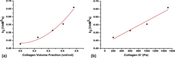
Figure 3 shows that k 3 increases as collagen fibril volume fraction (V) and storage modulus (G′) increase with a quadratic dependence on collagen fibril volume fraction (R 2 = 0.9599) and a linear dependence on shear storage modulus G′ (R 2 = 0.9473). These results are consistent with polymeric dependence on absolute collagen concentration, suggesting a relationship based on the spatial density of cooperatively interacting groups, which should increase due to their proximity to one another, not simply their relative abundance in solution. Availability of sites where cooperative interactions occur between ionizable groups on the fibril surface Reference Wallace, DeYoreo and Dove15 and spatial orientation of silicic acid in relation to the collagen fibril Reference Patwardhan, Emami, Berry, Jones, Naik, Deschaume, Heinz and Perry13,Reference Ehrlich, Deutzmann, Brunner, Cappellini, Koon, Solazzo, Yang, Ashford, Thomas-Oates, Lubeck, Baessmann, Langrock, Hoffmann, Wörheide, Reitner, Simon, Tsurkan, Ereskovsky, Kurek, Bazhenov, Hunoldt, Mertig, Vyalikh, Molodtsov, Kummer, Worch, Smetacek and Collins22 are important kinetic considerations that will not increase linearly with concentration. Although the (Gly–Pro–Hyp) triplet motif is most abundant in type I collagen primary sequences, other triplets containing Arg, Lys, and Gln occur in relatively high frequencies. Reference Ramshaw, Shah and Brodsky28 At silicification pH (∼7.4), deprotonated collagen fibril hydroxyprolines (pKa ∼6) and protonated collagen fibril amines (pKa ∼9–10) are capable of participating in cooperative interactions with each other and also with phosphate salts in the SS. These interactions effectively contribute to increasing silica condensation rates, especially when increasing collagen concentrations decrease the spacing of primary sequence triplets among adjacent collagen helices and fibrils. Reference Wallace, DeYoreo and Dove15 In contrast to our results, Elgin, et al. found that the presence of collagen did not increase the condensation rates of either sodium silicate or silicon catecholate complexes during collagen fibrillogenesis. Reference Eglin, Shafran, Livage, Coradin and Perry44 It is important to consider the difference in silicon sources as well as the 20-fold or greater initial silica concentration in our system, both of which lead to greater reactivity. Additionally, the surface chemistry of collagen proteins during fibrillogenesis, when compared to the surface chemistry of fully formed collagen fibrils within a matrix, may present a different organization of hydroxyl and amine moieties that is less amenable to the formation of sites for initiation of silica condensation.
Silica condensation rate constant increases with collagen-fibril volume fraction and storage modulus. Systematic variation of collagen fibril volume fraction (a), also directly related to quality-controlled collagen shear storage modulus G′ (b), demonstrates control over the apparent third-order reaction rate coefficient for silica condensation such that k 3 increases via second-order polynomial function with increasing collagen fibril volume and linearly with increasing shear storage modulus G′.

D. Bulk SS gelation rates are controlled by collagen fibril density
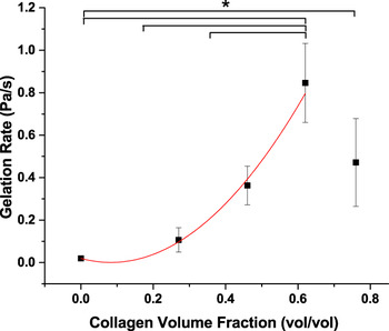
Viscoelastic analysis of condensing SS in the presence of collagen-fibril templates was performed to probe the physical interactions occurring during the early stages of gelation. Initial gelation rates for collagen-fibril templates of varying fibril volume fraction are provided in Fig. 4. Gelation rates were calculated as the slope of the linear region for increasing shear storage modulus G′ data during the first 300 s of gelation for three independent measurements of each sample. Raw data for measurement of shear storage modulus G′ and linear fit for first 300 s of each sample are provided in Supplementary Material Figure S2. Note induction time for all samples that are not silica controls; this represents the lag time between silicic acid application to the collagen surface and data recording by the instrument. In contrast with prior quantitative spectrophotometric analysis, SS gelation rates do not exhibit a strong dependence on the number of collagen fibrils present. Collagen fibril volume fractions up to 0.62 correlate positively with gelation rate, but the gelation rate appears to decrease for collagen volume fraction 0.76. However, comparison of gelation rates for each collagen volume fraction using the Bonferroni Test (α = 0.05) indicates that the gelation rate for collagen volume fraction 0.76 is only statistically different from the silica-only control, suggesting that the decreased gelation at this value may by spurious. The increasing trend for gelation rate between collagen volume fraction 0 and 0.62 exhibits quadratic dependence upon collagen volume fraction (R 2 = 0.9827).
Collagen-fibril template provides control over silica gelation rate. Systematic variation of collagen volume fraction demonstrates control over the rate of bulk silica gelation, as determined by real-time rheological analysis using a rotational rheometer. The gelation rate exhibits quadratic dependence upon collagen volume fraction between 0 and 0.62 (R 2 = 0.9827). Error bars show standard deviation for three independent rheometer tests. * indicates significance determined from Bonferroni Test (α = 0.05) of one-way ANOVA.

Condensation rates that increase with a quadratic dependence on fibril density (measured mono- and disilicic acid depletion) and gelation rates that initially increase but then appear to decrease (measured shear storage modulus G′ over time) support two potential hypotheses. The first is that increased fibril densities, while increasing the total number of silica condensation initiating sites, decrease the depth of silica penetration, which leads to a limit for the actual number of accessible silica condensation initiating sites. The second is that condensation initiating sites induce localized gelation at the fibril surface, leading to increased condensation overall with little occurring in bulk from 0-300 s. Although void spaces between individual collagen fibrils do narrow with increasing fibril density, limitation of silicic acid access to fibril surfaces based on increasing fibril density is unlikely based on previously reported diffusivity values for a comparable range of collagen-fibril matrices. Reference Whittington, Brandner, Teo, Han, Nauman and Voytik-Harbin42 The known templating and directing functions of collagen-fibril matrices support the hypothesis that depletion of silicic acid monomers and dimers is highly influenced by surface-localized condensation upon the organic template. It is also important to note that rheological methods used here only capture viscoelastic properties of the solution to support understanding of chemical phenomena; chemical reactions that do not contribute to creating a significant structured cannot be resolved by this rheological approach.
E. Early stages of silica gelation are localized at the template fibril surface
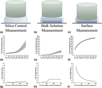
Real-time viscoelastic analysis, focused at the surface of the collagen-fibril template, was performed to understand critical differences in gelation at the surface of the template and the surrounding bulk solution. These experiments require careful placement of the rheometer probe, defined for three types of analyses given in Table I. For the lowest collagen fibril density examined previously (fibril volume fraction 0.25, 200 Pa G′), shear storage modulus G′ was measured in real-time at the surface of the collagen-fibril template and in the surrounding bulk solution for 1500 s. Figure 5 provides shear storage modulus G′ measurements with corresponding rate curves for each analysis and includes cross-sectional illustrations of rheometer probe height and position relative to the collagen template surface and bulk solution. Amplitude sweeps were performed at 0.5 Hz to confirm measurement within the linear viscoelastic region throughout development of the gel during the measurement period and determined to fall within the linear range for 0.001–10% strain. Measurements were recorded at 0.5 Hz and 1% strain. In comparison to SS bulk control without collagen (max 0.2294 Pa/s), the maximum gelation rate from 0–1500 s increases by 206% (max 0.7018 Pa/s) for the bulk SS with collagen and 1754% (max 4.2527 Pa/s) for the collagen surface. Further, the change in rate over time from 0–1500 s indicates a relatively low rate of gelation that increases over time in the bulk but a high rate of gelation that decreases over time for the surface. As gelation rates eventually become limited by availability of condensation sites over time, silica control and bulk gelation rates will begin to decrease as well until the gel has fully cured. Reference Brinker and Scherer8 Surface measurement of collagen-only control (no silicic acid exposure) was also performed and confirmed lack of characteristic rapid gelation (data not shown). Measurement of surface gelation phenomena was also attempted for higher fibril densities, corresponding to shear storage modulus G′ of 600, 1000, and 1500 Pa, but the higher elasticity of these sample surfaces violated the assumptions of our rheological technique.
Experimental parameters for control, bulk, and surface analyses.

Viscoelastic study of silica gelation in the presence of fibrillar collagen template suggests two-phase polycondensation process. Real-time measurement of shear storage modulus G′ was acquired by varying the rheometer upper plate positioning with respect to collagen template and SS (a–c). Error bar represents standard error of the mean for n = 3. Shear storage modulus G′ change during SS interaction with 200 Pa collagen template (0–1500 s) is compared here for (d) SS gelation in the absence of collagen, (e) surrounding SS gelation in the presence of collagen, and (f) gelation at the collagen surface during SS exposure. Change of gelation rate over time (g–i) indicates increased gelation rate overall in the presence of the template. Measurement of (g) silica control and (h) bulk solution measurement demonstrate a point at which gelation rate increases sharply, while (i) surface measurement begins with a very high gelation rate that decreases over time. Note the difference in G′ scale for silica control for ease of visualization (d–f).

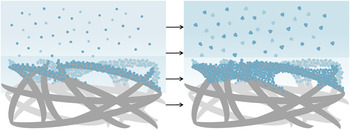
The contrasting gelation rate profiles for surface and bulk support the hypothesis that mono- and disilicic acid depletion, interpreted as silicic acid condensation, is related to surface-localized silica condensation during early gelation. The overall process can be described in terms of two phases, depicted in Fig. 6. During Phase 1, collagen fibrils primarily contribute to localized gelation at the template surface by initiating condensation directly, while simultaneously increasing gelation rates in the bulk through more transient interactions with the surface. During Phase 2, collagen fibril surfaces become increasingly saturated with partially condensed silicate species, so preferential condensation initiation sites on the fibril surface become shielded from reactive silicic acid species. Additionally, the now silicified collagen fibril surface presents silicate groups to the surrounding solution, increasing the overall rate of bulk gelation as greater numbers of condensation events decrease pKa values for available hydroxyls attached to silicon atoms, resulting in deprotonation at the peak gelation condition of neutral pH. Reference Brinker and Scherer8,Reference Iler9 It is important to recognize that surface and bulk gelation occur simultaneously during both Phase 1 and Phase 2. In terms of classical gelation theory, Reference Iler9 the distinction in these phases is whether heterogeneous condensation (i.e., interaction of silicic acids with collagen) or homogeneous condensation (i.e., interaction of silicic acids with themselves) events are kinetically favored. This separation in time of fibril surface-localized silica formation and bulk gelation provides the opportunity to produce different types of collagen-silica composites by controlling the rates of condensation in addition to the exposure time of the collagen to SS.
Proposed two-phase polycondensation process. Our data indicate a two-phase silica polycondensation process, with the collagen-fibril template surface chemistry inducing increased gelation rates both at the surface and in the bulk. Phase 1 is defined by rapid surface-localized gelation (shaded) with slight increases in the bulk compared to control. Phase 2 is defined by limited accessibility to the collagen surface chemistry and high amounts of surface-localized silicate groups, leading to decreasing surface-localized gelation rates with increasing bulk gelation rates (shaded).

IV. CONCLUSIONS
We have demonstrated control over surface-localized silica condensation rates through interaction with collagen-fibril matrices that template and direct silica mineralization. Collagen-fibril templates that are tunable with respect to their microstructure and fibril density enable informed design of the absolute amount of synergistic charge–charge interactions between amine- and hydroxyl-containing collagen side chains with the ability to increase initiation rates of condensing silicic acid species. Our analyses suggest a two-phase process for silica mineralization of the collagen template. During Phase 1, the collagen template primarily increases surface gelation rates through direct surface-localized interactions; during Phase 2, a relatively high quantity of condensed silica contributes to increased rates of bulk gelation.
The key implication of such a two-phase process is that, for any TMOS-derived and collagen-templated silica mineralization process, the initial rate of Phase 1 surface condensation must exceed the rate of Phase 1 bulk condensation to form a continuous mineral layer at the organic template surface and facilitate removal of the bulk prior to complete gelation. Additionally, the rate of Phase 1 surface condensation relative to the rate of Phase 2 bulk condensation should determine the thickness and penetration of the mineral layer. For such diverse applications as cellular microencapsulation, bone engineering scaffolds, and biomimetic organic-inorganic materials, Phase 1 and Phase 2 kinetics are important experimental design factors to control. Study of additional systematic variations to the collagen template (e.g., fibril diameter, interfibril branching, and incorporation of monomeric collagen units) will lead to greater understanding of collagen-silica interactions and advanced optimization of materials for target applications. In the future, exploration of alternate collagen sources, or chemical and genetic tools to introduce additional amines to the protein primary structure should be considered as promising methods to further tune kinetics of silica interactions with the collagen template.
Supplementary Material
To view supplementary material for this article, please visit http://dx.doi.org/jmr.2016.5.
ACKNOWLEDGMENTS
Financial support for this research was provided by the Purdue Research Foundation and Purdue Department of Agricultural & Biological Engineering. The authors would also like to thank Rucha Joshi, Kevin Buno, and Nimisha Bajaj of the Voytik-Harbin lab for their isolation of collagen oligomers and the Purdue Life Science Microscopy Facility, particularly Christopher Gilpin, for their critical sample imaging support.