The ventricular septal defect in tetralogy of Fallot is almost always large and non-restrictive, resulting in equal pressure in the two ventricles. The direction and magnitude of flow across the defect are primarily determined by the relative vascular resistance and by the degree of infundibular obstruction. Typically, if the degree of right ventricular outflow tract obstruction is severe, a right-to-left shunt will yield low pulmonary blood flow and cyanosis, requiring early intervention.Reference Apitz, Webb and Redington 1 , Reference Sommer, Hijazi and Rhodes 2 Here we present a unique case of an infant with tetralogy of Fallot and severe pulmonary valve stenosis, complicated by systemic hypertension, the presence of which altered flow dynamics and possibly prevented cyanosis.
Case
A term male infant antenatally diagnosed with tetralogy of Fallot was assessed at 5 weeks of age in our paediatric cardiology clinic in preparation for elective surgical repair. He had been well with no parental concerns and no noted cyanosis. No medical interventions had been required. Specifically, there was no umbilical artery catheter placement. His weight and height were at the 75th and 40th percentile, respectively.
Four-limb blood pressure measurement in the clinic yielded a right arm pressure of 140/95, left arm of 137/80, right leg of 140/80, and left leg of 137/89. On the basis of age, gender, and size, the definition of hypertension in this patient was ∼104 mmHg systolic. 3 His heart rate was 162 beats/min, pulse oxygenation was 100% on room air, and the child was afebrile. Auscultation revealed a harsh systolic ejection murmur (III/VI) at the left upper sternal border radiating to both lungs. Electrocardiogram showed normal sinus rhythm and right ventricular hypertrophy with an axis at −18°.
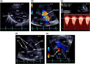
Echocardiogram showed an anterior-malaligned ventricular septal defect that was unrestrictive in nature, an overriding aorta, severe deviated conal septum-type infundibular stenosis, severe pulmonary valve stenosis, mild right ventricular hypertrophy, and normal left ventricular function with no coarctation of the aorta. The pulmonary valve was bicuspid, the annulus was 5 mm (Z score −4.5)Reference Pettersen, Du, Skeens and Humes 4 in diameter, and the right ventricular outflow tract gradient was 147 mmHg. There was a patent foramen ovale with left-to-right shunting. The right pulmonary artery measured 4 mm (Z score −3),Reference Pettersen, Du, Skeens and Humes 4 and the left pulmonary artery measured 3 mm (Z score −4.6)Reference Pettersen, Du, Skeens and Humes 4 (Fig 1). The apical five-chamber view showed left to right flow across the ventricular septal defect. The patient was hypertensive during the echocardiogram, with four-limb blood pressures similar to those noted above. Of note, the right ventricular outflow tract gradient at a community cardiologist's clinic about a week prior was ∼70 mmHg; however, no blood pressure measurement was obtained at the time.
( a ) Parasternal long-axis view in diastole demonstrates the ascending aorta (AAo) overriding the crest of the interventricular septum (*), a classic depiction of tetralogy of Fallot. ( b and c ) Aliasing colour Doppler flow is seen across the right ventricular outflow tract (RVOT) with a peak systolic velocity over 5 m/s (peak systolic gradient of over 130 mmHg). ( d ) On this parasternal short-axis view, the conal septum is displaced anterior causing constriction across the RVOT (*) with a large ventricular septal defect (VSD) noted more posteriorly. The pulmonary valve (PV) appears significantly smaller than the aortic valve (AoV). ( e ) This apical five-chamber view shows unobstructed left to right flow across the VSD and the left ventricular outflow tract into the AAo. The low velocity red colour flow across the large VSD along with the very elevated RVOT gradient depict elevated left ventricular (LV) pressure, consistent with systemic hypertension. RV = right ventricle; LA = left atrium; RA = right atrium; TV = tricuspid valve; PV = pulmonary valve; AoV = aortic valve.

The infant was admitted for pre-operative evaluation of his systemic hypertension. Anti-hypertensives were not initiated to prevent a sudden decrease in pulmonary blood flow secondary to a decrease in systemic vascular resistance. Pre-operatively, the patient had a number of episodes of desaturation, typically occurring when the systolic blood pressure would drop to 80–100 mmHg (30–50 mmHg lower than his typical mean systolic blood pressure levels).
As part of the evaluation for the patient's hypertension, the following tests measured in the normal range: creatinine, blood urea nitrogen, sodium, potassium, aspartate and amino transaminase, thyroid-stimulating hormone, free T4 levels, serum aldosterone, and renin. Urine studies for pheochromocytoma were also normal. Renal Doppler ultrasound revealed no evidence of renal artery stenosis, and both kidneys were normal in cortical echogenicity and corticomedullary differentiation. The right adrenal gland was visualised and was grossly normal. The left adrenal was not visualised. Head ultrasound was normal and did not reveal any intracranial masses. Fluorescence in situ hybridisation was negative for 22q11 microdeletion. Although advanced imaging techniques may have revealed more subtle renovascular abnormalities, these imaging modalities were not considered to be part of the standard of care in the immediate evaluation of neonatal hypertension. In addition, owing to the fragile nature of the patient pre-operatively, such advanced imaging modalities were deferred in favour of maintaining the patient's clinical stability and arranging urgent surgical repair.
The patient was taken to the operating room for a complete tetralogy of Fallot repair, involving patch closure of the ventricular septal defect, trans-annular right ventricular outflow tract patch augmentation, creation of a mono-cusp pulmonary valve, infundibulum muscle resection, and ligation and division of the ductus arteriosus. The patient tolerated the procedure well without residual defects. Post-operatively, the patient continued to have hypertension that was gradually controlled with captopril with resultant systolic blood pressures of 80–90 mmHg and diastolics of 40–60 mmHg. The systolic blood pressure while asleep decreased to 70 mmHg consistently such that captopril could not be titrated to above 0.5 mg/kg per day, and it was eventually discontinued. Therefore, further evaluation of the pre-operative hypertension was not pursued. The patient's desaturation episodes ceased post-operatively.
Discussion
To the best of the authors’ knowledge, this is the first reported case of systemic hypertension in a young infant with unrepaired tetralogy of Fallot. Systemic hypertension could not be adequately managed in this setting given the possibility of a hypercyanotic spell.
Much like infants and older children, the causes of hypertension in neonates are vast,Reference Flynn 5 with the two most common aetiologies being renovascular and renal parenchymal diseases. Other possible causes of neonatal hypertension include aortic coarctation, idiopathic arterial calcification, subdural haematomas, hyperaldosteronism, and adverse events related to umbilical artery catheter insertion.Reference Flynn 5 We were unable to find an aetiology for our patient's pre-operative hypertension despite extensive investigations.
An increase in systemic blood pressure has previously been reported to improve arterial oxygenation and recovery from anoxic spells in tetralogy of Fallot.Reference Oshita, Uchimoto, Oka, Saka, Takeshita and Funatsu 6 – Reference Nudel, Berman and Talner 8 Intravenous phynylephrine is a well-documented therapeutic intervention to raise systemic vascular resistance and consequently increase flow through the right ventricular outflow tract and improve arterial oxygenation.Reference Tanaka, Kitahata, Kawahito, Nozaki, Tomiyama and Oshita 9
This case carries with it a number of clinical considerations for the physician to be aware of. Primarily, the absence of cyanosis does not exclude the presence of severe right ventricular outflow tract obstruction. These patients should be considered for early surgical repair. Secondarily, the clinician should be cautious with respect to the management of systemic hypertension in a patient with tetralogy of Fallot. Decreasing the systemic vascular resistance may decrease pulmonary blood flow, inducing hypoxia.
Conclusion
We report the first case of an infant with unrepaired tetralogy of Fallot and systemic hypertension of undetermined aetiology. The elevated systemic vascular resistance presumably allowed adequate pulmonary blood flow despite a severe right ventricular outflow tract obstruction. This case shows that the clinician should carefully consider the role that systemic blood pressure plays in tetralogy of Fallot physiology.