Introduction
The properties of materials are generally determined by their chemical composition as well as by their internal structure. The chemical nature of most biological materials is limited to polymers, usually composites based on proteins and polysaccharides, which are sometimes reinforced by minerals. Many technically important material classes, such as metal alloys or inorganic semiconductors, do not occur naturally. This relative paucity in the chemical composition of biological materials may be the reason why complex and multiscalar structures appeared in the course of evolution, and they cover an amazing range of physical properties. The colors of beetles or butterflies are often due to photonic crystals based on a transparent base material; the brittleness of silica in the skeleton of sponges is reduced by multilayering of the glass; the magnetic response of some bacteria is tuned by the structure and size of magnetite particles. Reference Fratzl, Dunlop and Weinkamer1
The structure of most biological materials is hierarchical, probably as a consequence of the growth process that leads to assembly of building blocks at various scales. More importantly, this hierarchical structure provides the opportunity for adapting to various functions at the different levels of the hierarchical structure. Evolution has led to a large diversity of interesting and sometimes unusual design principles, which can be extracted and harnessed to develop new types of engineering materials. Reference Fratzl2–Reference Bhushan6 This article extends an in-depth review on natural hierarchical materials published about a decade ago Reference Fratzl and Weinkamer7 and focuses on a few crucial aspects of hierarchical structure, such as the importance of length scales and its potential for multifunctionality.
Basic constituents
The vast variability in organisms stands in striking contrast to the rather parsimonious use of chemical constituents, which are based mostly on light chemical elements, such as H, O, C, and N, and constitute most organic molecules as well as some minerals such as oxides, carbonates, or phosphates of Si, Ca, Mg, and a few other cations. Most other elements only occur as traces in millimolar or even nanomolar concentrations. Reference Spaargaren8
A limiting factor in the choice of chemical elements is that biological materials are formed at moderate ambient temperatures. In contrast to metallurgy involving high temperatures, nature makes use of metals primarily in their ionic form. Metal ions in coordinated interactions with proteins can act as cross-linkers and, consequently, increase the stiffness of biological materials. Reference Degtyar, Harrington, Politi and Fratzl9 Co, Fe, and Zn ions were found to harden the jaws of a marine worm, Reference Lichtenegger, Schoberl, Ruokolainen, Cross, Heald, Birkedal, Waite and Stucky10 the coating of byssal threads used by mussels to anchor to rocks, Reference Harrington, Masic, Holten-Andersen, Waite and Fratzl11 or the venom fang of a spider, Reference Politi, Priewasser, Pippel, Zaslansky, Hartmann, Siegel, Li, Barth and Fratzl12 respectively.
Parsimony is again observed when it comes to the chemical nature of the main components of extracellular matrices, which encompass essentially two main types of macromolecules: proteins and polysaccharides. Proteins include collagen (as a major organic constituent in bone, tooth, skin, blood vessel, muscle, tusk, byssal threads, fish scales), Reference Fratzl13 keratin (hair, nail, horn, claw, beak, feather), Reference Wang, Yang, McKittrick and Meyers14 and fibroin or spidroin as an important component of silk (different varieties of insect and spider silk). Reference Heim, Keerl and Scheibel15,Reference Keten, Xu, Ihle and Buehler16 The most important polysaccharides in biological materials are cellulose (plant cell wall, algae, biofilms) Reference Fratzl and Weinkamer7 and chitin (cell walls in fungi, exoskeletons of crustaceans and insects, the tongue-like feeding apparatus [radula] of mollusks, beaks of squid, or octopuses). Reference Fabritius, Sachs, Triguero and Roobe17 To understand the functionality and, in particular, the formation of tissues, it is not sufficient to only consider the main components of the extracellular matrix. While 95% of the organic matrix of bone consists of type I collagen, noncollagenous proteins play fundamental roles in bone formation, mineralization, and remodeling. Reference Roach18
Fibrils and fibers
Starting from these molecular building blocks, the problem at hand is how to climb the ladder of the different length scales to finally arrive at macroscopic spatial dimensions. An important first step “on the way up” is the formation of small fibrils or larger fibers. Molecular self-assembly plays a crucial role in this.
Fibers as a structural motif are encountered in many, if not all, biological materials. Collagen is a typical example and consists of a staggered arrangement of tropocollagen molecules. Reference Shoulders and Raines19 Similarly, both α- and β-keratin are organized in a filament-matrix structure. Reference Wang, Yang, McKittrick and Meyers14 Fibrils in spider silk consist of protein chains assembling into stiff β-sheet nanocrystals embedded in a softer semi-amorphous phase. Reference Keten, Xu, Ihle and Buehler16 Finally, cellulose and chitin are also organized in partially crystalline nanofibrils, whereby the chitin fibrils are usually wrapped by proteins. Reference Fabritius, Sachs, Triguero and Roobe17
The diameter of all of these fibrils is typically a few nanometers, with the exception of collagen fibrils that may reach diameters of up to several hundred nanometers. The mechanical properties of such fibrils vary widely, depending on their crystalline character and the presence or absence of molecular intrafibrillar cross-links. In some cases, parallel fibrils assemble into wider fibers, sometimes by interfibrillar cross-links. Reference Knott and Bailey20 However, this conserves the high-aspect ratio of the structural motif and, therefore, its principal mechanical weakness: the unsatisfactory performance under compressive load due to fiber buckling. Mineralization is a first strategy to improve the compressive behavior.
Plywood-like structures
A possible way to eliminate the functional anisotropy of a fibrous structure, frequently seen in biological materials, is to stack sheets of parallel fibers under different orientations in a plywood-like manner. This is done by introducing a fixed and rather small twist angle between adjacent fiber sheets. Bouligand pointed out the similarity of the resulting twisted plywood structure with cholesteric liquid crystals; he also lists biological materials wherein this structure is found. Reference Bouligan21 Examples include insect cuticles, bone, and blood vessel walls. Since then, this list has been significantly extended by structural analyses with modern imaging technology. Reference Fabritius, Sachs, Triguero and Roobe17,Reference Zimmermann, Gludovatz, Schaible, Dave, Yang, Meyers and Ritchie22,Reference Langer, Pacureanu, Suhonen, Grimal, Cloetens and Peyrin23 The twisted plywood structure naturally gives rise to a lamellar structure, where one 180° rotation of the fiber sheet corresponds to one lamella.
Size matters
Living organisms are subjected to many different challenges and, consequently, it is likely that most biological materials have to fulfill a variety of roles, and they should be considered as (at least potentially) multifunctional. Hierarchical structure presents unique opportunities for this, since it puts at their disposal several structural levels that may be separately adapted to address different functions. Examples of biological materials where this strategy seems to be followed are presented in the next sections. Prior to this, it is useful to consider some fundamental constraints of this implementation, due to the fact that physical effects often have a specific length scale. “Size matters” in defining magnetic, optical, and mechanical, as well as surface properties.
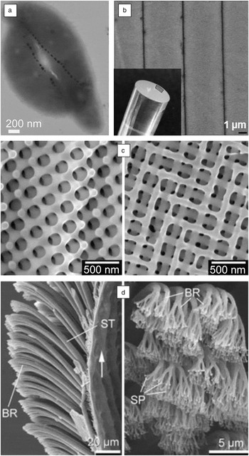
Magnetotactic bacteria contain chains of magnetic particles that induce a passive alignment along the Earth’s magnetic field that helps them efficiently navigate along oxygen gradients ( Figure 1a). Reference Lefevre, Bennet, Landau, Vach, Pignol, Bazylinski, Frankel, Klumpp and Faivre24 For magnetite (Fe3O4), stable single-domain ferrimagnets are only obtained in a size range between 20 nm to 80 nm at room temperature. For larger sizes, the particles split up magnetically into multiple domains thereby reducing their coercivity (i.e., resistance against demagnetization). For particles below 20 nm, thermal fluctuations cause continuous magnetic spin flipping preventing a stable magnetic dipole. Reference Faivre25 This then defines a typical size for such particles and the size distribution in the bacteria is centered at 50 nm, well within the range of a single magnetic domain. Reference Chen, Berounsky, Chan, Blackford, Cady, Moskowitz, Kraal, Lima, Kopp, Lumpkin, Weiss, Hesse and Vella26
(a) Two magnetotactic bacteria (Magnetospirillum magneticum) with linearly aligned magnetic particles of roughly 50 nm in size. Courtesy of Damien Faivre. (b) The structure of the deep-sea sponge Monorhaphis chuni: the inset shows the whole spicule (with the position of the organic layers drawn schematically); scanning electron microscope (SEM) micrograph of the spicule cross section shows alternating biosilica–organic layers (thin dark layers). Reference Zlotnikov, Shilo, Dauphin, Blumtritt, Werner, Zolotoyabko and Fratzl29 (c) SEM micrographs showing the microstructure of photonic crystals in the small scales covering the beetle Entimus imperialis. Reprinted with permission from Reference Reference Wu, Erbe, Raabe and Fabritius30. © 2013 Wiley. (d) Toe of a gecko shown by SEM micrographs of different magnifications: setae (ST) split up into branches (BR) to end in spatula (SP), the finest terminal branches of seta. Reference Fratzl, Kolednik, Fischer and Dean40

Fracture properties of biological materials can be strongly influenced by appropriate dimensioning of their components. Mineral crystal sizes of just a few nanometers, as found in composites like bone or teeth, result in a fracture strength that is not lowered by the presence of flaws in the crystals. Reference Gao, Ji, Jager, Arzt and Fratzl27 On a much larger length scale, strong spatial variation in the elastic modulus, can reduce the driving force for the propagation of a crack. The ideal length scale for this phenomenon depends on the magnitude of the modulus variation. Reference Kolednik, Predan, Fischer and Fratzl28 It is in the micrometer range, for example, in the spicules that provide structural support in deep-sea glass sponges such as Monorhaphis chuni (Figure 1b), where the elastic modulus varies by a factor of about 50 between silica and the protein interlayers. Reference Zlotnikov, Shilo, Dauphin, Blumtritt, Werner, Zolotoyabko and Fratzl29
Another example is optical properties that can be most effectively manipulated on a length scale close to the wavelength of visible light, that is, in the submicron range. This is mirrored in the structure of biological materials that fulfills functions like transparency, camouflage, or coloring. Structural colors are obtained in a photonic structure by a combination of two materials with different refractive indices in a periodic arrangement (Figure 1c). Reference Wu, Erbe, Raabe and Fabritius30 Depending on the wavelength, light reflection from the different layers of the photonic structure leads to constructive or destructive interference. Reference Vukusic and Sambles31,Reference Parker32 The resulting colors often depend on the angle of observation. Such iridescent structures are observed in plants Reference Glover and Whitney33 and diverse animal taxa with functions going beyond communication and avoidance of predation (e.g., thermoregulation and water repellency). Reference Doucet and Meadows34
In a photonic structure, colors can also be tuned by a change in the periodicity of the structure. In longhorn beetles, this occurs by swelling from water infiltration and the resulting change from a dry to a wet state, Reference Liu, Dong, Liu, Zheng and Zi35 and in the freshwater neon tetra fish by a change in the tilt angle of stacks of intracellular guanine crystals. Reference Gur, Palmer, Leshem, Oron, Fratzl, Weiner and Addadi36
The size of surface patterns modifies adhesion and interaction with liquids. Reference Su, Ji, Huang and Hwang37 In the case of dry adhesion, the contact splitting model predicts that the scale of the surface structuring is mainly controlled by the required adhesion strength, which depends on the size of the animal from the fruit fly to the gecko. Reference Arzt, Gorb and Spolenak38 Splitting the contact area into n smaller elements of the same shape as the original contact results in an increase in the adhesion force by √n, as seen in the gecko toe (Figure 1d). Reference Arzt39,Reference Fratzl, Kolednik, Fischer and Dean40
Accommodating conflicting properties
If one considers that specific functions require a “correct length scale” to adapt the material’s properties, structural hierarchy can be utilized to address different properties at different hierarchical levels. While, for example, the laws of optics fix the length scale to produce colors in the (sub)micron range, mechanical reinforcement can be tackled on lower or higher hierarchical levels. A new challenge emerges when considering conflicting properties to be accommodated in the hierarchical structure. A classical pair of conflicting mechanical functions is stiffness and toughness—a high resistance against deformation is usually accompanied by poorer fracture properties for any given material. This function tradeoff can be newly negotiated using structural hierarchy as discussed in the following few examples.
Subdivision of space into a tessellation with large tiles or bricks separated by small gap regions introduces an additional level of structural hierarchy defined by the size of the bricks. Reference Gao, Wang, Yao, Gorb and Arzt41 In the case where the tiles are stiff and the gaps are small and filled with a compliant matrix, the resulting composite can almost retain the elastic modulus of the tiles, while crucially improving the toughness. Well-known examples of such brick-and-mortar structures are nacre, Reference Sun and Bhushan42 enamel, and the mineralized collagen fibril, which is a basic building block of bone and dentine. Reference Fratzl and Weinkamer7
Tessellation is used by many organisms to introduce an extra length scale to manipulate mechanical behavior, such as in the scales of fish, Reference Bruet, Song, Boyce and Ortiz43 the carapace of turtles, Reference Gao, Wang, Yao, Gorb and Arzt41,Reference Krauss, Monsonego-Ornan, Zelzer, Fratzl and Shahar44 the osteoderm of the armadillo (used as defensive armor), Reference Meyers, Chen, Lopez, Seki and Lin45 or the human skull. The interdigitated structures of sutures in the carapace and in the skull allow unconstrained movement as long as deformations are small, but provide interlocking strength for larger deformations. The tiles in the armadillo and the cranial bones are held together by Sharpey’s fibers, Reference Meyers, Chen, Lopez, Seki and Lin45 which are partly nonmineralized, and therefore, compliant collagen fibers. But tessellation serves not just for improving mechanical properties, as demonstrated by the lyriform slit organ found in spiders. Roughly parallel slits in the spider’s exoskeleton function as a mechanical sensor, where strains (i.e., displacement changes) between the slits as small as 10–6 can trigger a nerve impulse in surrounding sensory cells. Reference Fratzl and Barth46
An attractive example of multifunctionality is provided by the peacock feather. The tail feathers of the Indian peacock—known as its train—display a variety of colors with conspicuous eyespots. Both the brightness and iridescent properties of the plumage correlate positively with male mating success. Reference Loyau, Gomez, Moureau, Thery, Hart, Saint Jalme, Bennett and Sorci47 From the central shaft (rachis) of the peacock tail feather, so-called barbs branch off, which are themselves branched to form flat barbules. The outer cortex of barbules was shown to contain a two-dimensional photonic-crystal structure made up of melanin rods connected by keratin. The different colors are produced by simply varying the lattice constant and the number of periods of the photonic crystal, Reference Zi, Yu, Li, Hu, Xu, Wang, Liu and Fu48 which correspond to the melanin rod spacing and the number of melanin rod layers, respectively. The extreme lengths of these feathers of up to 1 m, together with their slenderness, challenges mechanical stability under its own weight. Structurally, the load-bearing rachis of the feather consists of a closed foam in the center that is sheathed by a dense cortex. Along most of the rachis, not only the diameter, but also the thickness of the cortex decreases linearly giving rise to a self-similar conical geometry, where the cortex provides bending rigidity. Reference Weiss and Kirchner49 The keratin nanostructure of the fibrous cortex does not change along the rachis. Reference Pabisch, Puchegger, Kirchner, Weiss and Peterlik50
A last example of multifunctionality realized on different hierarchical levels is provided by the tracheid cells found in vascular plants. On the one hand, these elongated cells use their inner hollow space for transport of water and mineral salts. On the other hand, the cell wall contributes to structural support of the plant. The mechanical properties of the cell wall depend crucially on the spatial arrangement of cellulose microfibrils, in particular, the orientation angle of the fibrils with respect to the tracheid long axis known as the microfibril angle (MFA). A small MFA results in a stiff material with low fracture strain. With an increase in the MFA to about 50°, the fracture strain can be increased by an order of magnitude at the expense of a decrease in stiffness of a similar amount. Reference Fratzl and Weinkamer7 Spatial variations in the MFA and the resulting different swelling behaviors are also employed by plant tissues to generate internal stresses (e.g., to counteract the bending forces of tree branches). Reference Fratzl and Barth46
Transport in biological organisms
For solving problems of transport, one again faces a pair of conflicting properties. The transport should provide efficient accessibility to all places. Conversely, transport paths such as streets and tracks are seen as a “necessary evil” that should be kept to the unavoidable minimum. Again, a compromise is made possible by structural hierarchy. In the human body, tree-like networks of hierarchical character serve transport functions. A well-known example is the lower respiratory tract, where air is transported in a hierarchical succession of ducts of decreasing diameter from the trachea to the bronchi, bronchioles, and alveolar ducts. Another tree-like network is seen in the vascular system, with diameters ranging over almost four orders of magnitude, from the aorta (2–3 cm) to the capillaries (5–10 µm).
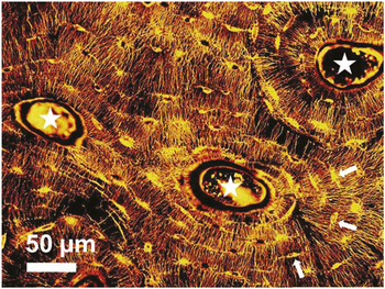
In the human body, nutrients have to reach cells that are sometimes difficult to access, such as osteocytes. These most-abundant bone cells are walled in the mineralized bone matrix Reference Bonewald51 and are only accessible by a network-like system of canals called canaliculi with a diameter of roughly 300 nm ( Figure 2 ). This porosity-permeating bone is also used by the osteocytes to connect to each other via their long cytoplasmic extensions (i.e., dendritic cell processes). Although our understanding of the canalicular porosity and the osteocyte network is still limited, it is clear that its function extends beyond that of a transport network. A prominent theory to explain the mechanosensitivity of bone is that loading due to physical activity causes fluid flow through the canalicular network. The resulting shear forces on their surfaces are then sensed by the osteocytes. This mechanical information, when transmitted to bone-forming and resorbing cells, enables adaption of the bone structure to changing mechanical needs. Reference Fratzl and Weinkamer7 The vast surface provided by the canalicular porosity, for instance, with 80% of the bone materials closer than 1.4 µm to the next canaliculus in mouse bone, Reference Kerschnitzki, Kollmannsberger, Burghammer, Duda, Weinkamer, Wagermaier and Fratzl52 is further used to influence the surrounding bone material. In a process called osteocytic osteolysis, the osteocytes can dissolve bone mineral in their vicinity, making readily available elements like calcium essential for cell physiology. Reference Bonewald51
The network-like microporosity of equine cortical bone imaged by a confocal laser scanning microscope after staining the bone sample. The central canal (marked by white stars) of the roughly circular structures called osteons house a blood vessel. Some positions where the osteocytes are located are marked by white arrows. The small canals called canaliculi connect the cell lacunae and have a diameter of about 300 nm. Figure courtesy of Michael Kerschnitzki.

Conclusions
The goal of this article was to demonstrate that structural hierarchy opens the possibility of addressing different functionalities at different hierarchical levels. When these functionalities are conflicting, biological materials show how hierarchical structuring helps to circumvent such dilemmas. Hierarchy allows, for example, mechanical properties like stiffness and toughness to be partly conciliated. The key is that the various laws of physics define a specific length scale where a given property can be adapted, so that hierarchical structuring is a promising strategy wherever different scales need to be addressed at the same time.
Acknowledgments
P.F. acknowledges support by the German Science Foundation Leibniz Prize. R.W. and P.F. acknowledge support by the German Science Foundation and Cluster of Excellence “Image Knowledge Gestaltung.”