Introduction
The initial development of fish comprises a series of morphological events with a duration that varies among species, depending on the egg size and reproductive strategy (Kolm & Ahnesjö, Reference Kolm and Ahnesjö2005), especially in neotropical species (Borçato et al., Reference Borçato, Bazzoli and Sato2004; Gomes et al, Reference Gomes, Scarpelli, Arantes, Sato, Bazzoli and Rizzo2007, Reference Gomes, Sato, Rizzo and Bazzoli2013; Amorim et al., Reference Amorim, Gomes, Gomes, Sato, Gomes, Martins, Sato, Rizzo and Bazzoli2009; Perini et al., Reference Perini, Sato, Rizzo and Bazzoli2010, Reference Perini, Sato, Rizzo and Bazzoli2013; Weber et al., Reference Weber, Sato, Santos, Rizzo and Bazzoli2012). Due to the scarcity of knowledge and technology regarding the culture of native fish species, Brazilian fish culture has mainly developed with exotic species. In order to exploit native fish species for aquaculture, basic reproductive parameters of native species have to be described, analysed, and incorporated into practice. In this context, artificial reproduction, embryogenesis, and larval ontogeny are of fundamental importance for the culture and conservation of the Prochilodus hartii.
Among the teleosts of the Jequitinhonha River basin, the ‘curimba’ P. hartii belongs to the Prochilodontidae family, one of the most important for commercial and sport freshwater fishing in the South American continent (Castro & Vari, Reference Castro, Vari, Reis, Kullander and Ferraris2003). Species of the genus Prochilodus perform long reproductive migrations (Godinho & Kynard, Reference Godinho and Kynard2006; Arantes et al., Reference Arantes, Santos, Rizzo, Sato and Bazzoli2011) and are considered to be key species on the structuring of aquatic ecosystems due to their detritivorous feeding habits (Flecker, Reference Flecker1996).
Prochilodontidae prepare for captive breeding, completing vitellogenesis, but ovulation and spawning only occur after hormonal induction (Sato et al., Reference Sato, Cardoso, Godinho and Godinho1996). Induced reproduction is appropriate for species that do not naturally breed in captivity or for those whose stock is declining. In such cases, hormonal therapies are used to obtain quality gametes (Zaniboni-Filho et al., Reference Zaniboni-Filho, Reynalte-Tataje and Weingartner2006; Arantes et al., Reference Arantes, Sato, Sampaio, Rizzo and Bazzoli2013). Several hormonal therapies are used to induce spawning in fish, including: gonadotropin-releasing hormone (GnRH) (Crim & Bettles, Reference Crim, Bettles, Fingerman, Nagabhushanam and Thompson1997; Mylonas & Zohar, Reference Mylonas and Zohar2001); treatment with synthetic hormone (Sugumar & Munuswamy, Reference Sugumar and Munuswamy2006); and hypophysation with crude common carp pituitary extract (Woynarovich & Horváth, Reference Woynarovich and Horváth1980; Sato et al., Reference Sato, Cardoso, Godinho and Godinho1996; Arantes et al., Reference Arantes, Santos, Rizzo, Sato and Bazzoli2011).
Along with the reduction of stocks of natural fisheries, there is an increasing need to develop artificial reproduction technology for species that are of high commercial value in order to improve the production of fingerlings for fish culture and reintroduction (Arantes et al., Reference Arantes, Sato, Sampaio, Rizzo and Bazzoli2013). As Prochilodontidae are large, their meat has desirable organoleptic characteristics combined with a good carcass income, making them good candidates for aquaculture.
To date there have been no detailed studies about the early development of P. hartii, therefore this study analysed the embryogenesis and larval ontogenesis of this species, aiming to contribute to the understanding of its conservation, taxonomy, larviculture, and breeding, as well as improving induced reproduction techniques.
Materials and methods
Fish
Specimens of P. hartii were captured in the Jequitinhonha River and transferred to the Machado Mineiro Pisciculture Station (15°31′64″S and 41°30′ 18″W), Minas Gerais, Brazil. The fish were kept in 200 m2 tanks with an average depth of 1.8 m at a density of 0.1–0.2 kg of fish/m2 and were fed daily with a commercial pelleted diet containing 37% crude protein at a ratio of 1.5–2.0% of body weight per day. At the time of reproduction, the female showed a flaccid, swollen abdomen and reddish and edematous genital papilla. The males snored and oozed semen through the genital papilla when handled.
Induced reproduction, eggs, and embryogenesis
For the induced reproduction process performed in January 2013, 10 females with a total length of 46 ± 1.28 cm and body weight of 1.3 ± 0.08 kg and 10 males with a total length of 41 ± 3.7 cm and body weight of 1.0 ± 0.33 kg were transferred to hypophysation tanks (dimensions: 3.0 × 1.0 × 0.8 m) with constant water circulation passing through filtering, aeration, and heating systems that had the following characteristics: constant water flow at 26°C; dissolved oxygen, 5.5–6.0 mg/l; pH, 6.0–7.0; and electric conductivity, 50–75 μS/cm2. During the induced reproduction process, the light cycle (photoperiod) was 12 h. Reproduction was induced by hypophysation, injecting commercial pituitary extract of common carp (Cyprinus carpio) from the United Agro Fisheries laboratory, using a 0.9% saline solution as solvent. This was injected into the coelomic cavity in two doses (2.5 and 5.0 mg/kg) for males and three doses (2.5, 5.0, and 2.5 mg/kg) for females. The doses were given with a 24-h period between the first and second and a 12-h interval between the second and third dose. The extrusion of oocytes and sperm was performed manually and fertilization was performed immediately by the dry method using individual plastic buckets for each female and male. From each female that underwent hypophysation, the extruded oocytes (ova) were weighed and samples were taken for counting and estimating the number of oocytes released. Then, water was added to the plastic buckets for oocyte hydration. The eggs were kept in funnel-type incubators with a 20-l capacity and constant water flow at 23°C; 200 g of ova/incubator were placed in 20-l incubators where the larvae were kept after hatching, according to Woynarovich & Horváth (Reference Woynarovich and Horváth1980). The diameters of the freshly spawned non-hydrated and hydrated eggs were measured under a stereomicroscope. Fertilized eggs and embryos were collected every 10 min (20 samples from each embryonic development stage) and were immediately photographed under a stereomicroscope for morphological analyses and determination of the duration of the main events of embryonic development. The fertilization rate was calculated as follows: from each female (n = 10) three samples of ova were collected, from which 100 oocytes were randomly counted, and the viable ones were separated from the non-viable ones, totaling 3000 oocytes. Finally, using a simple rule of three, the fertilization rate was determined (% viable oocytes) following Woynarovich & Horváth (Reference Woynarovich and Horváth1980).
Histology, larval ontogenesis and biometrics
After hatching, 10 larvae were collected daily for 7 days. Larvae were anesthetized with MS222, fixed in Bouin's liquid for 8–12 h, and embedded in historesin. Histological sections of 3 μm thickness were stained with toluidine blue-sodium borate (AT) and analysed by light microscope in order to monitor larval development.
For biometric analysis, 30 larvae were collected daily, anesthetized with MS222, fixed in 4% formaldehyde, and analyzed under a stereomicroscope. Based on the methodology proposed by Guimarães-Cruz et al. (Reference Guimarães-Cruz, Santos, Sato and Veloso-Júnior2009), the following parameters were measured: Total length (TL), standard length (SL), snout length (SNL), eye diameter (ED), head length (HL), head height (HH), body height (BH) pre-anal distance (PAD), length of the yolk sac (LSV), and height of the yolk sac (YSH).
Results
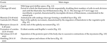
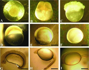
Recently extruded non-hydrated P. hartii oocytes re spherical, and non-adhesive with a diameter of 1480 ± 39 μm and after hydration, the diameter, including the perivitelline space and chorion, was 2860 ± 120 μm. The positive response to hypophysation was 100% for females and 80% for males. Spawning occurred 7 h after the third hormonal dose. In this experiment, 1.103 ± 98 oocytes/g of ova were obtained, totaling 343,000 ± 181,000 oocyte. The fertilization rate was 77% at 23°C. Non-viable or faulty oocytes represented 23% of the total number. The blastopore closed at 6 h 45 min and the embryonic development was completed 36 h 10 min after fertilization. The main events of the embryogenesis and their respective times are found in Table 1. In this study, the embryonic stage was considered valid when the frequency of each event was higher than 70%.
Main events of the embryonic development of P. hartii and their respective h after fertilization (hpf) at 23°C

Main stages of the embryo development of P. hartii. (A) Zygote; (B) two blastomeres; (C) 16 blastomeres; (D) blastula; (E) gastrula; (F) blastopore closure; (G) somites differentiation (arrow = Kupffer's vesicle); (H) tail release; and (I) hatching. Scale bar = 400 μm.

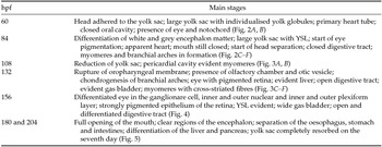
The main morphological characteristics of the larval ontogeny, determine the time required in hours post fertilization (hpf) are found in Table 2.
Morphological characteristics during the development of P. hartii larvae in hours post fertilization (hpf)

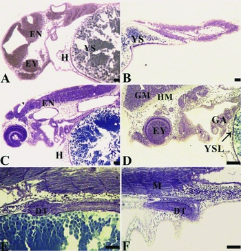
Sagittal histological sections of the ontogenesis stages of P. hartii larvae, stained with toluidine blue-sodium borate (AT). (A) 60 hpf post hatching: cranial portion, yolk sac (YS), eye (EY), encephalon (EN), (H) heart. (B) 60 hpf post hatching: caudal portion, yolk sac (YS). (C) 84 hpf: encephalon (EN) and heart (H). (D) 84 hpf: eye (EY), gill arches (GA), head mesenchyme (HM) and grey matter (GM) and yolk syncytial layer (YSL). (E) 84 hpf: digestive tract (DT). (F) 84 hpf: myomeres (M), digestive tract (DT). Scale bar = 100 μm.

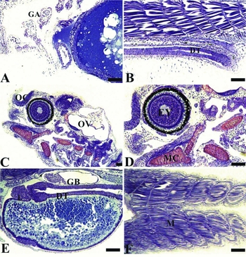
Sagittal histological sections of the ontogenesis stages of P. hartii larvae, stained with toluidine blue-sodium borate (AT). (A) 108 hpf: gill arches (GA). (B) 108 hpf: digestive tract (DT). (C) 132 hpf: otic vesicle (OV), olfactory chamber (OC). (D) 132 hpf: eye (EY), Meckel's cartilage (MC). (E) 132 hpf: gas bladder (GB), liver (L), digestive tract (DT). (F) 132 hpf: myomeres (M). Scale bar = 100 μm.

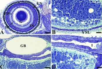
Sagittal histological sections of the ontogenesis stages of P. hartii larvae, stained with toluidine blue-sodium borate (AT). (A) 156 hpf: eye with ganglion cells (G), inner plexiform layer (EP), outer plexiform layer (IP), inner nuclear layer (IN), outer nuclear layer (EN). (B) 156 hpf: yolk syncytial layer (YSL). (C) 156 hpf: gas bladder (GB). (D) 156 hpf: intestine (I). Scale bar = 100 μm.

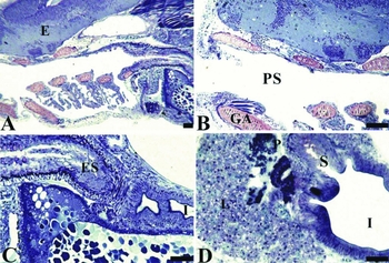
Sagittal histological sections of the ontogenesis stages of P. hartii larvae, stained with toluidine blue-sodium borate (AT). (A) 180 hpf: encephalon (E). (B) 180 hpf: gill arches (GA), pharyngeal space (PS). (C) 180 hpf: esophagus (ES), intestine (I). (D) 204 hpf: liver (L), intestine (I), pancreas (P), stomach (S). Scale bar = 100 μm.

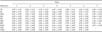
Considering the larvae, the biometric parameters presented a gradual increase from the first to the seventh day after hatching in all measured structures, except for length and height of the yolk sac, which showed a gradual decrease from the first to the seventh day, when the structure was completely resorbed (Table 3).
Body measurement (in mm) of P. hartii larvae from the first day to the seventh day after hatching

Total length (TL), standard length (SL), snout length (SNL), eye diameter (ED), head length (HL), head height (HH), body height (BH) pre-anal distance (PAD), length of the yolk sac (LSV) and (YSH) height of the yolk sac.
Discussion
Most neotropical reproductive migratory teleost species do not naturally reproduce in captivity, thus induced spawning is necessary. During the induced reproduction procedures, water parameters limit the success of final oocyte maturation, ovulation, and spawning (Mylonas & Zohar, Reference Mylonas and Zohar2001). This technique is widely used due to its low operational cost, high production control and a well defined and largely known protocol. Furthermore, fertilization by extrusion reduces oocyte handling, an important factor for the embryo development, as it decreases occurrence of fungus, increases the fertilization rate and increases the quality of the larvae produced (Reynalte-Tataje et al., Reference Reynalte-Tataje, Lopes, Ávila-Simas, Garcia and Zaniboni-Filho2013). The injection dosage and time for oocyte extrusion have similar values for different species of the same family, as noted by Sato et al. (Reference Sato, Fenerich-Verani, Verani, Vieira and Godinho2000) and Arantes et al. (Reference Arantes, Sato, Sampaio, Rizzo and Bazzoli2013). The fertilization rate is the percentage of viable oocytes per spawning and it depends on several factors such as oocyte quality, hormonal treatment, and chemical and physical characteristics of the water, all of which are important parameters for pisciculture stations (Sato et al., Reference Sato, Fenerich-Verani, Verani, Vieira and Godinho2000).
In the present study, the positive response to hypophysation by P. hartii females at 26°C was 100% using three doses, which is a better percentage than that reported for other Prochilodontidae at the same temperature and when using only two doses, e.g. for P. argenteus (75%) and P. costatus (80%) (Arantes et al., Reference Arantes, Sato, Sampaio, Rizzo and Bazzoli2013).
Recently extruded oocytes were non-adhesive and were telolecitic, similar to P. argenteus (Sato et al. Reference Sato, Cardoso, Godinho and Godinho1996) and P. lineatus (Ninhaus-Silveira et al., Reference Ninhaus-Silveira, Foresti and De Azevedo2006).
Egg diameter can influence the development and survival of the species as larger eggs produce larger larvae with a greater amount of yolk and slower embryogenesis (Kolm & Ahnesjö, Reference Kolm and Ahnesjö2005). Recently extruded P. hartii oocytes presented a diameter of 1480 ± 39 μm, a value close to that observed in P. argenteus 1583 ± 38 μm and P. costatus 1511 ± 46 μm (Sato et al., Reference Sato, Fenerich-Verani, Nuñer, Godinho, Verani, Godinho and Godinho2003a). The hydrated eggs of P. hartii presented a diameter approximately two times larger than that observed in the non-hydrated eggs, a pattern similar to other neotropical teleosts (Gomes et al., Reference Gomes, Sato, Rizzo and Bazzoli2013). Hydration of eggs is accomplished via incorporation of water through the membrane protein, aquaporin forming the perivitelline space, which protects the embryo against mechanical shocks (Andrade-Talmelli et al., Reference Andrade-Talmelli, Kavamoto, Romagosa and Fenerich-Verani2001; Lubzens et al., Reference Lubzens, Young, Bobe and Cerdà2010).
Knowledge of the initial development of a species provides information on environmental conditions, reproductive strategy, life cycle, and taxonomy (Gomes et al, Reference Gomes, Scarpelli, Arantes, Sato, Bazzoli and Rizzo2007; Marques et al., Reference Marques, Nakaghi, Faustino, Ganeco and Senhorini2008). Furthermore, knowledge of a species’ embryonic development has many applications, including biotechnology experiments, genetic engineering, and evaluation of the effect of toxic substances on ichthyofauna (Ninhaus-Silveira et al., Reference Ninhaus-Silveira, Foresti and De Azevedo2006; Marques et al., Reference Marques, Nakaghi, Faustino, Ganeco and Senhorini2008). In this sense, our study analysed for the first time the embryogenesis and larval ontogeny of P. hartii submitted to induced spawning.
Blastopore closure is important in aquaculture as a sign of successful fertilization, and is therefore the precise moment to estimate the fertilization rate, as unfertilized oocytes can parthenogenetically develop up to blastopore closure (Gomes et al., Reference Gomes, Sato, Rizzo and Bazzoli2013). In P. hartii, blastopore closure occurred at 6 h 45 min hpf.
The fertilization rate of oocytes varies among species, depending on oocyte quality and the hormone protocol used for inducing final oocyte maturation and ovulation (Mylonas et al., Reference Mylonas, Fostier and Zanuy2010). In this study, fertilization rate was 77%, slightly higher than that obtained for other fish of the Prochilodus genus, such as P. costatus (71%) and P. argenteus (69%) (Sato et al., Reference Sato, Fenerich-Verani, Godinho, Godinho and Godinho2003b).
After spawning, cytoplasmic movements separate the vegetative pole, formed by the yolk cells, from the animal pole, where the blastodisc formed (Langeland & Kimmel, Reference Langeland, Kimmel, Gilbert and Ramio1997). In this study, the first cleavages formed blastomeres similar to those in several neotropical teleost species (Ninhaus-Silveira et al., Reference Ninhaus-Silveira, Foresti and De Azevedo2006; Gomes et al., Reference Gomes, Sato, Rizzo and Bazzoli2013). Multiple layers of cells are formed by successive cleavage of the blastomeres, culminating in the flattening of the blastomeres in order to form the common standard blastocyst in the embryonic development of teleosts (Langeland & Kimmel, Reference Langeland, Kimmel, Gilbert and Ramio1997). In the gastrula stage, the cells of the blastoderm migrate along the surface of the yolk cells over the yolk syncytial layer (YSL). This movement is due to the expansion of the microtubule network of the YSL as it moves towards the vegetative pole, culminating in blastopore closure (Langeland & Kimmel, Reference Langeland, Kimmel, Gilbert and Ramio1997).
Migratory species present fast embryogenesis, whereas sedentary species have a long embryogenesis (Godinho et al., Reference Godinho, Lamas and Godinho2010). In the present study, embryonic development within 36 h 10 min hpf at 23ºC was longer than that found for other migratory fish with total spawning and non-parental care, e.g. P. costatus, 21 h 10 min hpf at 24°C (Sato et al., Reference Sato, Fenerich-Verani, Godinho, Godinho and Godinho2003b); P. argenteus, 19 h 00 min hpf at 24°C (Sato et al., Reference Sato, Fenerich-Verani, Godinho, Godinho and Godinho2003b); and P. lineatus, 22 h 00 min hpf at 25°C (Ninhaus-Silveira et al., Reference Ninhaus-Silveira, Foresti and De Azevedo2006). These findings show the influence of temperature on the embryonic development of P. hartii. In fact, high temperatures, between 24°C and 26°C (Sato et al., Reference Sato, Cardoso, Godinho and Godinho1996), accelerate embryogenesis, chorion rupture, and hatching of neotropical fish that usually spawn during the warmer seasons (Ninhaus-Silveira et al., Reference Ninhaus-Silveira, Foresti and De Azevedo2006). Conversely, lower temperatures, within certain limits, increase hatching and larval development time (Woynarovich & Horváth, Reference Woynarovich and Horváth1980), as also observed in this study.
After hatching, there are important morphometric changes that are responsible for the larvae's body shape change towards the acquisition of juvenile characteristics in a short period of time (Osse et al., Reference Osse, Van Den Boogaart, Van Snik and Vander Sluys1997). Recently hatched larvae of P. hartii are transparent with a TL of 5.0 ± 0.11 mm, larger values than those observed in other Prochilodontidae, such as P. argenteus, 3.20 ± 0.04 mm (Sato et al. Reference Sato, Fenerich-Verani, Godinho, Godinho and Godinho2003b). In this study, the biometric parameters presented a gradual increase between the different developmental stages, with the exception of those variables related to the yolk sac, similar to that noted by Guimarães-Cruz et al. (Reference Guimarães-Cruz, Santos, Sato and Veloso-Júnior2009). Analyses of morphometric measurements allow comparisons of different developmental stages between species and, together with other morphologic characteristics, are useful in species identification (Sanches et al., Reference Sanches, Nakatani and Bialetzki1999).
The YSL was observed in embryos of P. hartii from 84 hpf. This layer, located between the blastodisc and the yolk, is the result of incomplete blastomere division. (Ninhaus-Silveira et al., Reference Ninhaus-Silveira, Foresti, De Azevedo, Agostinho and Veríssimo-Silveira2007).
After 180 hpf, the larvae reached a mean SL of 6.56 ± 0.14 mm with a completely resorbed yolk sac, similar in length to P. costatus (6.10 ± 0.29 mm) and P. argenteus (6.0 ± 0.18 mm) (Godinho et al. Reference Godinho, Santos, Sato, Godinho and Godinho2003). The transition between endogenous and exogenous feeding is critical for the survival of larvae, and is a limiting factor for successful pisciculture (Baras et al., Reference Baras, Ndao, Maxi, Jeandrain, Thome, Vandewalle and Melard2000; Zaniboni-Filho et al., Reference Zaniboni-Filho, Reynalte-Tataje and Weingartner2006). The moment the rupture of the oropharyngeal membrane varies among teleost species (Ninhaus-Silveira et al, Reference Ninhaus-Silveira, Foresti and De Azevedo2006; Oka & Higashiji, Reference Oka and Higashiji2012). In P. hartii, rupture of oropharyngeal membrane happened within 132 hpf, while in P. costatus and P. argenteus it occurred within 72 h hpf (Godinho et al., Reference Godinho, Santos, Sato, Godinho and Godinho2003). In this study, larvae 180 hpf were already ready for exogenous feeding. In the present study, exogenous feeding was not offered to the larvae before the collection of biological material. After this procedure the larvae were fed ad libitum.
The results of this study provide information to better understand the induced reproduction, breeding, and management in captivity of P. hartii, one of the main species for fisheries in the Jequitinhonha River Basin, Brazil.
Acknowledgements
The authors thank the Hydrobiology and Hatchery Station of Machado Mineiro-CEMIG for technical support, FAPEMIG, CNPq and CEMIG for financial support.