Somatic milk cells [polymorphonuclear neutrophils (PMN), macrophages (Mφ), lymphocytes (L), and some epithelial cells] are crucial for an effective immune defence in the mammary gland. ‘Resident’ cells which are already in the milk without a preceding immune stimulation are the first cells that come into contact with invading pathogens. Shortly after pathogens are recognized, additional leucocytes are recruited into the milk through the blood-milk barrier and thus somatic cell count (SCC) is increased. This is initiated by inflammatory mediators. Therefore, SCC is used worldwide as an indirect indicator to diagnose intramammary infections and to control bacterial contamination of the udder and milk (Brolund, Reference Brolund1985; Harmon, Reference Harmon1994).
In healthy cows, macrophages are the predominant cell population particularly in the cisternal milk fraction which is close to the entrance port of the pathogenic agents (Sarikaya et al. Reference Sarikaya, Schlamberger, Meyer and Bruckmaier2006). They are able to actively recognize and engulf pathogens, to release chemotactic and inflammatory mediators, and to initiate the inflammatory response (Rainard & Riollet, Reference Rainard and Riollet2006). PMN are the predominant cells recruited in large amounts from the blood into the milk during the acute phase of inflammation to fight the causative organism (Harmon & Heald, Reference Harmon and Heald1982; Kehrli & Schuster, Reference Kehrli and Shuster1994; Sordillo & Streicher, Reference Sordillo and Streicher2002; Mehrzad et al. Reference Mehrzad, Duchateau and Burvenich2004). The active stage of immune defence requires viable and immune competent somatic cells for a rapid and efficient elimination of the pathogen. After diapedesis into the mammary gland and participation in the immune response the cells die via apoptosis or necrosis pathways (Sládek & Ryšánek, Reference Sládek and Ryšánek1999, Reference Sládek and Ryšánek2000) and their remains are removed by phagocytosis. Apoptosis is the result of interactions between death modulators and death receptors (e.g.: Fas/APO-1 (CD95) (Menaker & Jones, Reference Menaker and Jones2003) and Caspases [e.g. caspase3 (Casp3); Robertson et al. Reference Robertson, Orrenius and Zhivotovsky2000].
The immune response is initiated by cell wall components of invading pathogens such as lipopolysaccharide (LPS) (Burvenich et al. Reference Burvenich, Paape, Hill, Guidry, Miller, Heyneman, Kremer and Brand1994; Boudjellab et al. Reference Boudjellab, Chan-Tang, Li and Zhao1998; Schmitz et al. Reference Schmitz, Pfaffl, Meyer and Bruckmaier2004). Therefore, LPS is often used in experiments in vivo (Bannerman et al. Reference Bannerman, Paape, Lee, Zhao, Hope and Rainard2004; Didier & Bruckmaier, Reference Didier and Bruckmaier2004; Mehrzad et al. Reference Mehrzad, Dosogne, Meyer and Burvenich2001a) or in vitro (Boudjellab et al. Reference Boudjellab, Chan-Tang, Li and Zhao1998; Wellnitz & Kerr, Reference Wellnitz and Kerr2004) to stimulate somatic cells and to mimic a bacterial infection. LPS is recognized by the toll like receptor-4 (TLR4) (Takeuchi et al. Reference Takeuchi, Hoshino, Kawai, Sanjo, Takada, Ogawa, Takeda and Akira1999). The different mechanisms of the immune response are regulated by immunomodulators (Rainard & Riollet, Reference Rainard and Riollet2006) such as cytokines e.g. tumour necrosis factor α (TNF-α) or interleukin 8 (IL-8), antibacterial proteins e.g. lactoferrin (Lf), and lysozyme (Lz), lipid mediators e.g. cyclooxygenase-2 (COX-2), or other factors e.g. granulocyte macrophage colony stimulating factor (GM-CSF). Their expression is unregulated in mammary tissue and milk cells during an immune response (Pfaffl et al. Reference Pfaffl, Wittmann, Meyer and Bruckmaier2003).
The differential count of leucocytes can be influenced by milk fraction and SCC (Sarikaya et al. Reference Sarikaya, Schlamberger, Meyer and Bruckmaier2006). In addition, the mRNA expression of inflammatory factors is lower in cells of low-SCC milk. High SCC levels >100×103cells/ml are highly correlated with mastitis (Kehrli & Shuster, Reference Kehrli and Shuster1994). In addition, elevated SCC can reduce the susceptibility to mastitis (Suriyasathaporn et al. Reference Suriyasathaporn, Schukken, Nielen and Brand2000). Therefore it is possible that the immunological activity of resident cells is different in high- and low-SCC milk. The milk fraction can also have an influence on the SCC and the cell viability. Sarikaya et al. (Reference Sarikaya, Werner-Misof, Atzkern and Bruckmaier2005) found a higher SCC and cell viability in the foremilk fraction compared with the alveolar fraction.
This study was performed to investigate the importance, for the immunity of the mammary gland, of cells already resident in milk before an infection occurs. The ex-vivo condition of the study allowed us to investigate these cells after removal from the udder in their natural environment, the milk, without the influence of new cells recruited from the blood or contribution of the mammary tissue. The viability, cell differentiation and mRNA expression of several immunomodulating factors of these cells were tested. A special focus was the comparison of viability and cell differentiation between quarters with high and low SCC and of foremilk and alveolar milk fractions.
Materials and Methods
Experiment 1
Ten lactating dairy cows were chosen at random (5 Red Holstein, 5 Holstein). Cows were in months 2 to 9 of their 2nd to 7th lactation with a SCC of 140±57×103 cells/ml. Whole milk was harvested by machine milking in the morning. Five-hundred ml was filtrated (pore size 100 μm) and incubated at 37°C. After 0, 1·5, 3, 4·5 and 6 h of incubation, SCC was measured with an automated milk cell counter (DCC; DeLaval, Tumba, Sweden) to evaluate changes of SCC during incubation.
Cell isolation was performed according to Sarikaya et al. (Reference Sarikaya, Prgomet, Pfaffl and Bruckmaier2004) with modifications as follows: after each incubation period, 30 ml of the milk samples were diluted 1:1 with cold (4°C) sterile phosphate-buffered saline (PBS; pH=7·4) and centrifuged at 1000 g at 4°C for 15 min. The fat layer and supernatant were removed and the cell pellet resuspended and centrifuged twice in 30 ml PBS (400 g and 300 g at 4°C for 10 min). The final pellet was resuspended in cold PBS (depending on the original total cell concentration in 50 μl to 1 ml to obtain an optimal cell concentration for differentiation). Twenty-five μl of the cell suspension was used for determination of cell count and viability in a Neubauer's counting chamber (Brand, Wertheim, Germany) with direct light microscopy using a Trypan blue staining (1:1 dilution). Viable cells appeared shining white under the microscope while dead cells appeared blue.
Experiment 2
Eleven lactating dairy cows (4 Red Holstein, 4 Holstein, 3 Brown Swiss) were selected based on milk SCC <150×103 cells/ml (60±10×103 cells/ml) determined by the DCC. The cows were in months 2 to 8 of their 1st to 6th lactation. They were clinically healthy and had no signs of inflamed udders such as redness, swelling or heat. Whole udder milk was collected during morning machine milking and kept at 4°C for approximately 1 h until further preparation. The milk was filtrated (pore size, 100 μm) and separated into aliquots of 500 ml. One sample from each cow was treated with 0·5 μg/ml LPS from Escherichia coli (O111:B4; SIGMA, Buchs, Switzerland) another sample from each cow was not treated (controls). All samples were incubated at 37°C for 1, 3 or 6 h. Then samples were centrifuged and the cells washed as for Experiment 1. Final pellets were resuspended in 2 ml of cold PBS.
Cell count and cell viability was determined microscopically (see Experiment 1). Cell differentiation was performed using a panoptic staining method according to Pappenheim modified for milk cells (Sarikaya et al. Reference Sarikaya, Prgomet, Pfaffl and Bruckmaier2004).
After the last step, remaining milk cells from 6 of these cows (SCC=51±7×10³ cells/ml) were centrifuged at 300 g at 4°C for 5 min and pellets stored at −80°C until RNA extraction. Total RNA was extracted using RNeasy Mini Kit (Quiagen, Hilden, Germany) according to the manufacturer's instructions. Final RNA concentration was quantified by spectrophotometry (Biophotometer, Vaudaux-Eppendorf, Basel, Switzerland) from the optical density at 260 nm. One microgram of RNA was reverse transcribed with 200 U Moleney Murine Lekaemia Virus Reverse Transcriptase RNase H minus, Point Mutant (MMLV-RT, Promega, Madison WI, USA) using 100 pmol random hexamer primers (Invitrogen, Leek, The Netherlands).
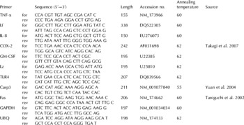
Quantitative RT-PCR analysis was carried out on a Rotor Gene 6000 (Corbett Research, Sydney, Australia) using the Sensimix DNA Kit (Quantace, Biolabo, Châtel St Denis, Switzerland). Primers for the housekeeping [GAPDH, ubiquitin (UBQ)] and target genes (TNF-α, IL-8, Lf, Lz, GM-CSF, COX-2, TLR4, Casp3, and Fas) were synthesized commercially (Microsynth, Balgach, Switzerland) using previously published or newly designed primer sequences (Table 1). The following 3-step programme was used: 10 min at 95°C, 40 cycles of 95°C for 15 s, primer specific annealing temperature for 30 s, and 20 s 72°C, ending with a melting curve programme (60–99°C, heating rate of 0·1°C/s, continuous measurement). Take-off values (second derivative maximum) were achieved by Rotor Gene software version 1.7.40. Target gene expressions were normalized to housekeeping genes.
Sequence of PCR Primers forward (for) and reverse (rev), PCR product length (bp), accession number, Annealing temperature (°C), and reference of primers. The primers without a given reference were designed in our laboratory by using the sequence found with the accession numbers. The product of all primers gave good efficiency, and they were suitable for qPCR in this study

Experiment 3
Milk from 12 quarters of 6 randomly chosen dairy cows (3 Red Holstein, 1 Holstein, 2 Brown Swiss) was used. The cows were in months 3 to 10 of their 5th to 7th lactation and were clinically healthy. Six quarters with higher SCC (HQ) (253±169×103 cells/ml) and six quarters with lower SCC (LQ) (17±11×103 cells/ml) were used. The milk was taken during morning milking in two fractions: foremilk (FM) (defined as <1·5 min after teat cup attachment) and alveolar milk (AM) (>1·5 min after teat cup attachment) using a quarter milking device for separate quarter milk collection (Sarikaya et al. Reference Sarikaya, Werner-Misof, Atzkern and Bruckmaier2005). To obtain a FM fraction free of alveolar milk, milking was performed without any udder preparation to avoid milk ejection (Bruckmaier & Blum, Reference Bruckmaier and Blum1996). Milk was filtrated (pore size, 100 μm) and incubated at 37°C in 50-ml aliquots with or without 0·5 μl/ml LPS from Esch. coli (O111:B4; SIGMA, Buchs, Switzerland) for 0, 0·5, 1, 1·5 and 2 h. Thereafter, samples were centrifuged and pellets were washed as for Experiment 1. Final cell pellets were resuspended in cold PBS (depending on the original total cell concentration between 100 μl and 1·5 ml). Cell count, cell viability and differential cell count was determined (see Experiments 1 and 2).
Statistical analysis
Results are presented as means±sem. SCC is presented in percentages wherein SCC at time 0 h is 100%. Differences in cell count, cell viability, cell differentiation and mRNA expression between control samples and LPS-treated samples, as well as between FM and AM or between different incubation times were tested for significance by paired t test using Sigma Plot 10.0 (Systat Software Inc., San Jose CA, USA). P<0·05 was considered as significant.
Results
Experiment 1
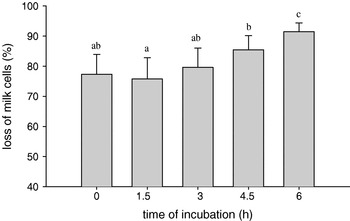
SCC measured by the DCC before centrifugation of the milk was largely higher than the microscopic cell count after centrifugation calculated to the total cell count of 30 ml milk. The proportion of cell loss (in %) over the time and by cell isolation increased significantly (Fig. 1). Viability of the somatic cells at 0 h was 75·3±5·6% and a significant decrease to 65·4±5·3% between 0 h and 6 h was observed.
Decrease of milk cell count (%) by centrifugation at 0, 1·5, 3, 4·5, and 6 h post incubation. Values are means with sem for n=10. a–c: Means without common letters differ significantly (P<0·05). The cell loss by centrifugation increased with incubation time.

Experiment 2
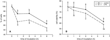
Mean cell counts (Fig. 2) derived from control and LPS-treated milk decreased from 0 to 1 h, remained stable from 1 to 3 h, and decreased from 3 to 6 h. Compared with controls, LPS-treated samples contained a lower number of cells at 1, 3 and 6 h. Cell viability (Fig. 2) at 0 h was 74·7±3·5% and decreased between 3 and 6 h in control samples. In LPS-treated samples cell viability decreased between 3 and 6 h.
Milk cell count (A) and cell viability (B) of control (•) and LPS-treated samples (○) at 0, 1, 3 and 6 h. Values are means with sem for n=11. a–d: means without common letters differ significantly (P<0·05).

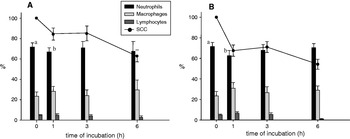
At time 0 h, the cell population contained 71±3·9% PMN, 23·5±4·3% Mφ, and 5±0·8% L. Mean PMN concentration decreased between 0 and 1 h in control and in LPS-treated samples (Fig. 3). Percentages of L and Mφ did not change significantly with time.
Decrease of milk cell count in relation to time (line) and cell distribution (bars) at 0, 1, 3 and 6 h: Neutrophils (black), Macrophages (grey), and Lymphocytes (dark grey), of control (A) and LPS samples (B). Values are means with sem for n=11. a,b: different letters represent significant (P<0·05) differences between time points within cell populations: PMN decreased between 0 h and 1 h in A and B.

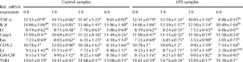
mRNA expression (Table 2) of Casp3, TLR4, and GM-CSF did not change within incubation time. Fas, TNF-α, IL-8, and Lf mRNA expression was decreased after 6 h. COX-2 expression was decreased in LPS-treated samples at 6 h. Lz expression was decreased at 3 and 6 h.
Relative mRNA expression (±sem for n=6) of immunomodulators and apoptosis factors in control and LPS-treated samples at 0, 1, 3 and 6 h. Casp3, TLR4, and GM-CSF mRNA expression did not change within incubation time. Fas, TNF-α, IL-8, and Lf mRNA expression was decreased after 6 h. COX-2 expression was decreased in LPS-treated samples after 6 h. Lz expression decreased after 3 h

a–e : Means within a row with different superscript letters differ (P<0·05)
Experiment 3
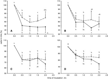
At 0 h the number of cells in the 50-ml milk samples after centrifugation was 142±40×103 in LQ_FM, 136±38×103 in LQ_AM, 5016±1067×103 in HQ_FM, and 2587±1071×103 in HQ_AM. In LQ milk no significant difference between FM and AM within cow was detected. In HQ milk within cow there was a decrease of the number of cells between FM and AM in four, an increase in one, and no variation in one sample. In each group at each time point the decrease of the number of cells in foremilk was numerically, but not significantly, larger than in alveolar milk (Fig. 4). There was no significant difference between control and LPS-treated samples.
Milk cell count (%) of LQ_control (A), LQ_LPS treated (B), HQ_control (C) and HQ_LPS treated samples (D) at 0, 0·5, 1, 1·5 and 2 h; foremilk (•) and alveolar milk (○). Values are means with sem for n=4–6. a–c: means within one group without common letters tended to differ (P<0·1). Cell counts decreased within one hour of incubation with no significant differences between foremilk and alveolar milk samples and between control and LPS treatment.

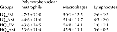
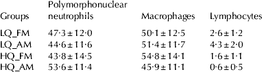
At 0 h the proportion of viable cells ranged in LQ_FM from 23 to 61%, in LQ_AM from 28 to 60%, in HQ_FM from 22 to 82% and in HQ_AM from 15 to 70%. In LQ milk the viability in AM was lower in three, elevated in two, and the same in one quarter compared with FM. In HQ milk the viability of cells in AM was lower in four, elevated in one and the same in one quarter compared with FM. No significant change of the viability was observed within the 2 h of the experiment. The distribution of cell populations is shown in Table 3 and did not change during the experiment.
Cell populations (in %) at time 0 h for each group: low- (LQ) and high-SCC quarter milk (HQ), foremilk (FM) and alveolar milk (AM). Values are means with sem for n=6. No significant changes were observed within the 2 h of the experiment
Discussion
In this ex-vivo model, cells were studied in their natural environment, the milk, compared with in-vitro studies where the cells were cultured in media. The design offers the possibility of analysing the behaviour of resident somatic cells without interaction with mammary tissue or newly migrated leucocytes. To study the immune capacity of mammary glands and milk cells, several studies have been performed in vivo with LPS by intramammary challenges (Mehrzad et al. Reference Mehrzad, Dosogne, Meyer and Burvenich2001a; Didier & Bruckmaier, Reference Didier and Bruckmaier2004; Schmitz et al. Reference Schmitz, Pfaffl, Meyer and Bruckmaier2004) or in vitro (Boudjellab et al. Reference Boudjellab, Chan-Tang, Li and Zhao1998; Prgomet et al. Reference Prgomet, Sarikaya, Bruckmaier and Pfaffl2005). In vivo, besides resident milk cells mammary epithelial cells of the tissue as well as newly infiltrating immune cells can react. For this reason, with experiments in vivo it is difficult to isolate the response to LPS stimulation of the resident somatic cells, which are already in the milk, from other cells. Milk cells were studied in vitro in culture media, which provide an optimal environment; however, this does not correspond to the conditions of somatic cells in vivo. Therefore in the present study the ex-vivo model was used to study the competence of the somatic milk cells to immunologically respond in their natural environment, i.e. milk, without any addition of substitutes or interaction with other cells.
The first experiment was performed to investigate the stability of the cells in the model. The selection criterion of cows was the clinical status (health) of the cow and the udder, but not the SCC or the breed. A lower cell number in the microscopic count (after centrifugation) showed that, obviously, the cell preparation is responsible for the destruction of the cells. The centrifugation forces may have a stronger destructive influence on the milk cells, which are already extenuated. Therefore, the increase of cell loss after 6 h post incubation most likely reflects the fact that cells are less resistant after a long presence in the milk. These findings explain the rapid decrease of the number of cells in the milk with incubation time in Experiments 2 and 3 independently of SCC and milk fraction. During the migration from blood into milk PMN utilize energy reserves needed for efficient phagocytosis and killing of invading pathogens (Newbould, Reference Newbould1973). In the milk, striking changes occur in the morphology of PMN owing to the ingestion of fat globules and casein (Paape et al. Reference Paape, Guidry, Kirk and Bolt1975; Paape & Guidry, Reference Paape and Guidry1977) by endocytosis. From this internalization of cell membrane result intracellular membrane bound vacuoles, a loss of pseudopodia, and cell rounding (Paape et al. Reference Paape, Bannerman, Zhao and Lee2003; Rainard & Riollet, Reference Rainard and Riollet2006; Burvenich et al. Reference Burvenich, Bannerman, Lippolis, Peelman, Nonnecke, Kehrli and Paape2007). These morphological and physiological changes consume a lot of energy which induces a decrease in phagocytic and bactericidal activity and, therefore, an impairment of the mammary gland defence (Paape et al. Reference Paape, Bannerman, Zhao and Lee2003; Rainard & Riollet, Reference Rainard and Riollet2006; Burvenich et al. Reference Burvenich, Bannerman, Lippolis, Peelman, Nonnecke, Kehrli and Paape2007). Blood PMN have a short half-life (Carlson & Kaneko, Reference Carlson and Kaneko1975) and they spontaneously undergo apoptosis (Fadeel et al. Reference Fadeel, Ahlin, Henter, Orrenius and Hampton1998; Smits et al. Reference Smits, Burvenich, Guidry, Heyneman and Massart-Leen1999). According to Paape et al. (Reference Paape, Mehrzad, Zhao, Detilleux and Burvenich2002) PMN function as phagocytes persists for 1–2 d after leaving the blood circulation. Furthermore, migration of PMN across collagen-coated membranes induces an apoptotic response (Van Oostveldt et al. Reference Van Oostveldt, Paape and Burvenich2002b). In addition, milk, rich in casein and fat globules, is not an optimal culture medium (Paape et al. Reference Paape, Bannerman, Zhao and Lee2003; Burvenich et al. Reference Burvenich, Bannerman, Lippolis, Peelman, Nonnecke, Kehrli and Paape2007). Comparable mechanisms are probably involved in the disappearance of leucocytes other than PMN in milk. Therefore, the cells were expected to have a short life in the removed milk and the rapid disappearance of leucocytes incubated in milk was not surprising.
In Experiment 2 (SCC of 60±10×103 cells/ml) LPS treatment reduced the number of cells compared with control samples. However, no differences between controls and LPS treatment could be seen in the Experiment 3 with high (>200×103) and low (<50×103) cell counts between cisternal and alveolar milk. Van Oostveldt et al. (Reference Van Oostveldt, Tomita, Paape, Capuco and Burvenich2002a) showed that apoptosis of blood PMN after an intramammary challenge with Esch. coli was accelerated. On the other hand, LPS stimulation of blood leucocytes in vitro caused spontaneous inhibition of PMN apoptosis (Mangan et al. Reference Mangan, Welch and Wahl1991; Colotta et al. Reference Colotta, Re, Polentarutti, Sozzani and Mantovani1992; Hachiya et al. Reference Hachiya, Takeda, Miyata, Watanabe, Yamashita and Sendo1995). In addition, after LPS intramammary challenge, programmed cell death of milk PMN is decreased (Ryśanek et al. Reference Rysánek, Sládek, Vasícková and Faldyna2005). It remains unclear why LPS had different influences on the cells in the different experiments of this study.
In the present study the viability of the cells differed widely in all experiments. However, in all experiments the viability was stable within the first 3 h of incubation. Nevertheless, the viability (Mehrzad et al. Reference Mehrzad, Dosogne, Meyer, Heyneman and Burvenich2001b, Reference Mehrzad, Duchateau and Burvenich2004) of cells in our study was lower than in other studies where the viability was above 88% (Sarikaya et al. Reference Sarikaya, Werner-Misof, Atzkern and Bruckmaier2005) and was still 88% after 8 h incubation at 37°C (Prgomet et al. Reference Prgomet, Sarikaya, Bruckmaier and Pfaffl2005). It is most likely that in these studies dead cells were already cracked and washed away by centrifugation and washing steps according to Experiment 1. Previous studies compared cells from different milk fractions and described an increase in SCC and in cell viability from FM to AM (Sarikaya et al. Reference Sarikaya, Werner-Misof, Atzkern and Bruckmaier2005). Surprisingly, in our investigation, the variation of cell viability between both fractions did not follow a definite pattern and differed in each quarter. The relatively high proportion of PMN can indicate a previous or incipient inflammation (Persson-Waller, Reference Persson-Waller2000; Jacobsen et al. Reference Jacobsen, Toelboell and Andersen2005) and the infection status has an influence on PMN viability (Mehrzad et al. Reference Mehrzad, Dosogne, Meyer and Burvenich2001a). However, at the time point of sampling the SCC of the sampled quarter was low with no signs of detectable inflammation. This shows that the definition of the milk samples through only the SCC is not sufficient and other parameters such as previous infections, lactation stage (Mehrzad et al. Reference Mehrzad, Dosogne, Meyer, Heyneman and Burvenich2001b), milk composition, and surely other parameters like the lactation number (Mehrzad et al. Reference Mehrzad, Duchateau, Pyörälä and Burvenich2002) have to be considered to explain the differences in cell populations.
The differential cell count with the microscope was performed by one examiner, because there is a subjective influence (Schröder & Hamann, Reference Schröder and Hamann2005). The distribution of cell populations at 0 h showed a difference in Experiments 2 and 3. In Experiment 2 (mean SCC of 60±10×103 cells/ml) PMN represented the biggest fraction of somatic cells. In Experiment 3 [FM and AM fractions, low (<50×103 cells/ml) and high (>200×103 cells/ml) SCC] the distribution of cells was almost 50% PMN and 50% Mφ in all groups. Sarikaya et al. (Reference Sarikaya, Schlamberger, Meyer and Bruckmaier2006) showed the proportion of Mφ to be the highest in milk with a SCC of (12–100)×103 cells/ml, almost equal with PMN in milk with a SCC of (100–350)×103 cells/ml, and the proportion of PMN the highest in milk with a SCC>350×103 cells/ml. On the other hand, Olde Riekerink et al. (Reference Olde Riekerink, Barkema, Veenstra, Berg, Stryhn and Zadoks2007) found Mφ to be always the predominant fraction of milk cells independently of the SCC. The reason for the differences in our study is, again, most likely due to the definition of the milk samples through only the SCC.
LPS stimulation increases mRNA expression of immunomodulators in mammary tissue (Schmitz et al. Reference Schmitz, Pfaffl, Meyer and Bruckmaier2004). In milk cells Prgomet et al. (Reference Prgomet, Sarikaya, Bruckmaier and Pfaffl2005) found increased mRNA expression of TNF-α, IL-6, and IL-1β in response to LPS with a peak after 1–3 h wherein the cells were incubated in RPMI 1640 medium, supplemented with 10% inactivated FCS. The decrease of mRNA expression of immune factors and of TLR4 that recognizes LPS (Werling et al. Reference Werling, Piercy and Coffey2006) in cells incubated in milk is rather a lack of the ability to respond to this stimulation than a regulated mechanism. It shows the degenerative changes of the cells. It reflects a reduced immune competence and confirms that milk is not a good environment to maintain the efficiency and the viability of the cells (Paape et al. Reference Paape, Bannerman, Zhao and Lee2003; Burvenich et al. Reference Burvenich, Bannerman, Lippolis, Peelman, Nonnecke, Kehrli and Paape2007).
In freshly removed milk, Pfaffl et al. (Reference Pfaffl, Wittmann, Meyer and Bruckmaier2003) showed that Lf and COX-2 mRNA expression was higher in cells from high SCC quarters (>150×103 cells/ml) than in cells from low SCC quarters. If we assume that a high SCC is due to pathogen stimulation, we can compare these results of high SCC with our results of LPS stimulation. In addition, Lee et al. (Reference Lee, Bannerman, Paape, Huang and Zhao2006) showed that after intramammary challenge with Esch. coli mRNA expression of TNF-α was elevated at 8 h after infection and the mRNA expression of IL-8 was elevated at 16 h and 24 h after infection. However, the discrepancy with our experiments may be explained by the ex-vivo conditions. It shows that cells newly recruited into the mammary gland after pathogen contact are most likely responsible for an increased expression of immunomodulators in the milk and not the cells that are already resident. Furthermore, under ex-vivo conditions, resident cells have no contact and, therefore, no possible interaction with mammary tissue. Eukaryotic cells generally die either by necrosis or by apoptosis (Duvall & Willie, Reference Duvall and Willie1986). Both forms of extinction exist in the cavity system of the juvenile bovine mammary gland to eliminate PMN (Sládek & Ryšánek, Reference Sládek and Ryšánek2000). Didier & Bruckmaier (Reference Didier and Bruckmaier2004) found an increased mRNA expression of the apoptosis factor Fas in mammary tissue after LPS challenge, but could not detect a change of the mRNA expression of these factors in somatic milk cells. These results show that the milk cells do not respond to LPS challenge with an increased mRNA expression of apoptosis factors and that accords with our ex-vivo study where no changes in Fas or Casp3 mRNA expression in cells incubated in milk and stimulated with LPS could be observed. Milk cells seem to undergo necrosis some hours after the diapedesis from blood into the milk. The cells, obviously, do not undergo apoptosis but necrosis, which explains the decrease in cell count after 3 h of incubation, the loss of cells after centrifugation, and also the percentage of viable cells in relation to intact cells.
In conclusion, this study showed that that resident milk cells were less viable, less able to respond to immunological stimulation, and less immune competent than freshly migrated cells from blood into the milk. Therefore, cells that are already in milk are less important for a good immune competence, and the best protection of the mammary gland against infection is a fast recruitment of new immune cells from the blood which is indicated by a rapid increase of the SCC after pathogen invasion.
This study was supported by the H Wilhelm Schaumann-Stiftung, Hamburg, Germany. We are grateful to C Morel and C Philipona for their excellent technical assistance.