Introduction
Luminescent materials are everywhere in our daily lives, from display devices, to biomedical research, personal safety, and solid lighting. They make our life colorful and exciting. Despite the fact that many luminescent materials are very emissive in dilute solutions, their luminescence is weakened or even completely quenched at higher concentrations, partially due to aggregate formation. As common luminescent materials naturally aggregate in the solid state and in aqueous media, the phenomenon of aggregation-caused quenching (ACQ) is a problem in many applications, especially in light-emitting devices and biomedical research. Over the past few decades, tremendous efforts have been made to solve the ACQ problem, with limited success. Due to their large aromatic rings and planar structures, most fluorophores naturally aggregate in the solid state and in aqueous media, but researchers are trying their best to separate them. Reference Hong, Lam and Tang1
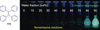
One of the most straightforward solutions to address the ACQ problem was the invention of luminophores with aggregation-induced emission fluorogens (AIEgens). Tang et al. first coined the concept of aggregation-induced emission (AIE) in 2001. Reference Luo, Xie, Lam, Cheng, Chen, Qiu, Kwok, Zhan, Liu, Zhu and Tang2 AIE molecules generally have rotor structures, and one of the most common examples is tetraphenylethylene (TPE [ Figure 1 ]). Reference Feng and Liu3 When it is dissolved in a good solvent such as tetrahydrofuran, there is barely any fluorescence due to the free motions of the molecular rotors, which deactivate the radiative decay channel. Upon aggregate formation caused by the addition of water to the solution, the restriction of the molecular motions lead to bright fluorescence in an aggregate or thin-film state. Detailed studies over the past decade reveal that the mechanism underlying the AIE phenomenon is the restriction of intramolecular motions in the aggregated state. Reference Feng and Liu3 The rotor structures also prevent the molecules from approaching each other closely, which results in fluorescence quenching. Although the restriction of intramolecular motion has been used as a primary mechanism to explain the AIE phenomenon, several complementary theories and mechanisms, such as the suppression of Kasha’s rule, have also been proposed. Reference Mei, Hong, Lam, Qin, Tang and Tang4
The rotor-shaped chemical structure of tetraphenylethylene (TPE) and aggregation-induced emission upon addition of water to its tetrahydrofuran solution. Reprinted with permission from Reference Reference Feng and Liu3. © 2016 Wiley.

Over the past 15 years, we and many other research groups have designed and synthesized a series of AIEgens. Almost all of them show very low luminescence as molecular species, but quantum yields of up to 100% (all absorbed photons emitted as light) have been obtained in the film state. Reference Qian, Cousins, Horak, Wakefield, Liptak and Aprahamian5 Due to their unique features, AIEgens have been employed in a wide range of research applications, including smart materials that are responsive to external stimuli, active materials for energy devices, and tools to monitor biological or physical self-assembly processes. They have proven useful in environmental engineering, such as for water and food quality control, and gas detection. They have been used for biomedical research in areas such as sensing, imaging, and therapy. This article highlights the utilization of special properties of the AIEgens to design a series of AIE bioprobes. Reference Mei, Leung, Kwok, Lam and Tang6,Reference Ding, Li, Liu and Tang7 The extremely low fluorescence as molecular species and the intensified fluorescence as nanoaggregates of AIEgens have allowed us to design and invent a series of probes for use in various biomedical applications.
AIE light-up probes
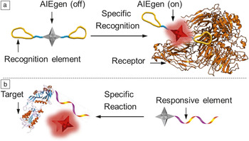
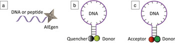
AIE light-up probes were synthesized by attaching water-soluble recognition elements to AIEgens, which are not emissive in aqueous media ( Figure 2 ). Reference Liang, Tang and Liu8 Once the probes interact with a biomolecule, such as a protein, bacteria, or DNA, the restriction of the intermolecular motion turns on the fluorescence (Figure 2a). Reference Shi, Liu, Geng, Tang and Liu9 Some of these probes may also undergo chemical or biological (e.g., enzymatic) reactions to change the solubility of the AIEgens in aqueous media to give intensified fluorescence as the signal output (Figure 2b). Reference Shi, Kwok, Liu, Xing, Tang and Liu10 As compared to the popular fluorescence resonance energy-transfer-based probes ( Figure 3 ), AIE probes have a distinct advantage—they only require the presence of a single molecule to report a biological process. In comparison, a Förster resonance energy transfer (FRET) probe requires at least a pair of molecules—a donor and an acceptor, or a donor and a quencher—to report the presence of the target.
Illustration of design principles for aggregation-induced emission (AIE) light-up probes. Reference Liang, Tang and Liu8 Note: AIEgen, AIE fluorogen.

The representative design of an aggregation-induced emission (AIE) probe and two Förster resonance energy transfer (FRET) probes. (a) AIE probe; (b) FRET probe with a donor and a quencher; and (c) FRET probe with a donor and an acceptor. Note: AIEgen, AIE fluorogen.

The applications of AIE light-up probes can be illustrated using drug screening and prodrug (a compound that, after administration, can be converted into a pharmacologically active drug in the body) tracking as examples. Reference Yuan and Liu11 As many drugs can induce cell apoptosis (cell death), we started by designing a cell apoptosis probe through integration of a caspase substrate peptide to tetraphenylethylene. Reference Shi, Kwok, Liu, Xing, Tang and Liu10 The probe is not emissive in either aqueous media or inside cells. However, when a cell undergoes apoptosis, the caspase enzymes inside the cells are activated, which cleave the hydrophilic peptide from the hydrophobic AIEgen to yield fluorescence. This probe not only offers the chance to realize real-time monitoring of cell apoptosis, but also makes it possible to conduct drug screening for toxicity evaluation. The probe fluorescence responds well to different drugs at various concentrations.
As prodrugs are generally less toxic than real drugs, they are under intensive study. To track prodrug delivery and monitor its activation, we used tetraphenylenethylene as an energy donor and doxorubicin (dox), a red emissive chemical drug, as the energy acceptor. Reference Yuan, Kwok, Zhang, Tang and Liu12 The donor and acceptor were linked using a platinum prodrug, which can be activated inside the cells. Due to energy transfer between the donor and the acceptor, the probe shows red emission for prodrug tracking. Upon cellular uptake and activation of the platinum prodrug inside the cells, separation of the donor and acceptor occurs, with blue emission observed in the cytoplasm and red emission in the nucleus. This is indicative of successful drug activation. Detailed studies revealed that the drug molecules migrate into the cells in 1 h. In 2 h, activation of the drug becomes obvious, reflected by the color change in the cytoplasm. The activated dox takes about 6 h to reach the nucleus, which is similar to that for free dox, indicating that the prodrug probe does not alter the function of the dox drug.
To monitor how effectively a specific drug can kill cancer cells, an AIE light-up apoptosis probe was conjugated to a platinum prodrug, together with a targeting ligand. Due to the cell surface receptor-mediated endocytosis, the prodrug was selectively sent into cancer cells, and we were able to achieve in situ monitoring of the drug’s therapeutic function. Reference Yuan, Kwok, Tang and Liu13 Once the prodrug enters the cancer cells via target-mediated endocytosis, it can automatically undergo reduction to release the actual drug that will migrate to the nucleus to activate apoptosis. Meanwhile, the AIE apoptosis probe remains silent until the caspase-3 is activated by the drug to cleave the peptide from the AIEgen, which yields green fluorescence. Although the process is complicated, this AIE probe has been successfully used as a tool to visualize each step. Effective cell death was observed in <4 h by monitoring the green fluorescence as an indication of the therapeutic function, which is much more efficient than traditional cell liability assays. The probe was also found to be selective to cancer cell lines. Reference Yuan, Kwok, Tang and Liu13
Efforts have also been made to design a molecular probe that can simultaneously visualize drug delivery, drug activation, and the therapeutic function of the drug in a specific cancer cell. For this, we designed a macromolecular probe that incorporates an AIEgen and an ACQ photosensitizer. Reference Yuan, Kwok, Tang and Liu14 A small amount of diisopropylamine molecule was included so that the macromolecule can respond to solution pH, producing nanoparticles under basic conditions and extended polymer chains under acidic conditions. Due to the different nature of the AIE and ACQ molecules, the macromolecular probe is green emissive at pH = 7.4, and red emissive at pH = 5.5. Detailed analysis also revealed that the aggregation of the photosensitizer at high pH not only quenched its fluorescence, but it also reduced its ability to produce singlet oxygen, which motivates us to develop AIE photosensitizers. This will be discussed later.
When the macromolecular probe is used for cancer cell imaging and ablation, it shows green fluorescence in the cytoplasm. Once the probe migrates into the lysosome through the endocytosis pathway, the pH change activates the photosensitizer due to a change in polymer conformation. As a consequence, red emission from the photosensitizer is observed, which can be used to guide the cell ablation. Under white light illumination, the singlet oxygen produced is able to disrupt the lysosome membrane, so that the polymer probe can be released from the lysosome. The change in environmental pH will then cause the macromolecular probe to self-assemble into green emissive nanoparticles. The appearance of green nanoparticles in the cytoplasm indicates the positive therapeutic function. Reference Yuan, Kwok, Tang and Liu14
After designing a series of AIE light-up probes, it occurred to us that if we could integrate therapeutic functions into the AIEgen itself, then our light-up probe could immediately be transformed into a theranostic (therapeutic and diagnostic) probe that can offer sensing, imaging, and therapeutic functions using a simple molecular design ( Figure 4 ). Reference Yuan and Liu11 Based on the observations discussed earlier, traditional photosensitizers such as chlorin e6 and rose bengal are good photosensitizers as molecules. However, once they have aggregated in the culture medium or inside cells, the fluorescence, as well as the ability to produce singlet oxygen, could be completely quenched. To address this issue, we proposed modifying the existing AIEgens by reducing the energy gap between S1 and T1 states to enhance the intersystem crossing. Reference Xu, Yuan, Cai, Zhang, Hu, Liang, Zhang, Zhang and Liu15 A series of AIEgens were designed through the separation of the highest occupied molecular orbital (HOMO) and lowest unoccupied molecular orbital (LUMO) electron distribution to yield an energy difference ranging between 1.2 eV and 0.1 eV. As a consequence of this modification, AIEgens with more efficient singlet oxygen production than rose bengal have been successfully produced.
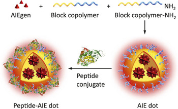
Illustration of aggregation-induced emission (AIE) dot synthesis through the nanoprecipitation method. Reference Yuan, Zhang and Liu18 Note: AIEgen, AIE fluorogen.

Subsequently, our AIE photosensitizers were integrated with a targeting molecule, as well as a short peptide sequence responsive to the enzymes overexpressed in specific cancer cells, to form a theranostic probe. Reference Yuan, Zhang, Gao, Zhang, Tang and Liu16 With the double-targeting effect, the probe shows higher selectivity toward cancer cells versus normal cells. Once the probe enters the cancer cells and localizes into the lysosome, the overexpressed cathepsin B in the cancer cell activates the probe. The activated probe light emission is able to guide further white light irradiation to produce singlet oxygen to kill the cells. This probe shows low toxicity in the dark and has good selectivity toward cancer cells. Based on a similar strategy, once the AIE photosensitizers are conjugated to another recognition element, such as a peptide drug that is specific to bacteria, the new probes can be used for detection and in situ killing of the specific bacteria in just a few minutes. Reference Feng, Yuan, Hu, Zhang, Xing, Zhang, Zhang and Liu17
In addition to the molecular probe, the AIE photosensitizers have been integrated into macromolecules to serve as effective carriers for drug and gene delivery. Reference Yuan and Liu11,Reference Yuan, Zhang and Liu18 In one example, we created a three-dimensional (3D) macromolecule by integrating an AIE molecule with the small oligomer ethylene imine. Reference Yuan, Zhang and Liu18 The obtained macromolecule is highly effective for gene delivery. More importantly, we can control the dose of light irradiation to accelerate the escape of the gene carrier from the lysosome. An even higher light dose can cleave the 3D structure to facilitate gene release. As a result, the new carrier has been demonstrated to show enhanced gene transfection efficiency by nearly 100% using the gold standard PEI 25 K in different cell lines.
AIE dot probes
AIE dots are small organic nanoparticles with AIEgens at the core and block polymers as the shell (Figure 3). Reference Li, Qin, Ding, Tomczak, Geng, Liu, Liu, Zhang, Liu, Liu and Tang19 They can also be designed to have multiple functionalities on their surface, which allows further conjugation with targeting molecules for different applications. The dot synthesis takes only a few minutes, and the dot size can be well controlled in the nanometer range. Most AIE dots showed good photo- and physical stability. They also show good biocompatibility in living systems. More importantly, some of the organic AIE dots were found to be brighter than commercial quantum dots with similar emission wavelengths. In one example, a factor of 10–40 more photons were emitted by AIE dots in 100 s than by quantum dots. Reference Li, Qin, Ding, Tomczak, Geng, Liu, Liu, Zhang, Liu, Liu and Tang19 We also observed that the blinking (on/off emission) phenomenon for quantum dots was absent in the AIE dots, Reference Li, Qin, Ding, Tomczak, Geng, Liu, Liu, Zhang, Liu, Liu and Tang19 which made the AIE dots ideal for continuous tracking at the single nanoparticle level.
To demonstrate potential applications of AIE dots, we studied their suitability for cell labeling and noninvasive long-term cell tracking. Reference Ding, Mao, Li, Wang, Qin, Liu, Chiam, Tomczak, Yang, Tang, Kong and Liu20 Taking the study of stem cell homing and therapeutic function monitoring as an example, AIE dot-labeled stem cells were applied to wounds on mice. Reference Ding, Mao, Li, Wang, Qin, Liu, Chiam, Tomczak, Yang, Tang, Kong and Liu20 Monitoring was carried out over a period of six weeks to study the function of the stem cells. It was observed that the stem cells indeed stayed at the disease site, where they helped to reconstruct blood vessels and promote the growth of regular muscle cells. Clear overlay between the green fluorescence from the fluorescent protein and red emission from the AIE dots at day 30 and day 42 indicated the accuracy of AIE dots in long-term tracing of biological therapeutic cells. Detailed studies also revealed that tracking with AIE dots did not require genetic modification, and that they were superior to commercial quantum dots and organic dye-based tracker products in terms of tracking time. Reference Ding, Mao, Li, Wang, Qin, Liu, Chiam, Tomczak, Yang, Tang, Kong and Liu20
Recently, AIE dots have also been successfully used for multiphoton in vivo imaging of vascular structures in the brain, ear, bone, and muscle. Reference Lou, Zhao and Tang21 Using multiphoton absorption means the excitation wavelengths can be extended from the original 400–600 nm to 1000 or even 2000 nm to realize better penetration depths. Reference Ding, Mao, Li, Wang, Qin, Liu, Chiam, Tomczak, Yang, Tang, Kong and Liu20 Similar nanoparticles have also been developed to visualize the leakage of the blood vessel and even to quantify the leakage. Good correlations between the fluorescence intensity at the leakage site have been observed in different disease models.
Once AIEgens with therapeutic functions are incorporated into nanoparticles, these AIE dots can be immediately utilized for imaging, image-guided surgery, and therapy. Reference Lou, Zhao and Tang21,Reference Liu, Chen, Ji, Liu, Ding, Zhao and Liu22 Taking a tumor-bearing mouse as an example, upon injection of the AIE dots from the tail vein, the dots migrated through the whole body and accumulated at the tumor region. Obviously, the whole tumor is lit up by the fluorescent AIE dots, which is essential for image-guided surgery. Subsequent photodynamic ablation of the residue cells after tumor removal significantly improved the survival rate of the mice. Reference Lou, Zhao and Tang21
Summary
AIE fluorogens are a special class of molecules with rotor structures, molecular vibrators, or a combination of both. They operate primarily on the restriction of intramolecular motion, although other complementary theories have been proposed to explain the AIE phenomenon. Due to their unique low fluorescence as molecular species and intense fluorescence when they form nanodots, they have shown interesting properties as biomolecular probes in sensing, imaging, and therapy. With the development of multifunctional AIEgens, where both imaging and therapy functions are integrated into the same molecules, simple but effective AIEgen-based theranostic platforms have been developed that exhibit promising performance. There has been tremendous recent growth in publications and citations of AIE research, which clearly indicates that this is an interesting field worth pursuing.
Acknowledgments
The Singapore NRF Competitive Research Program (R279-000-483-281), NRF Investigatorship (R279-000-444-281), and the National University of Singapore (R279-000-482-133) are gratefully acknowledged for their financial support.

Bin Liu joined the Chemical and Biomolecular Engineering Department of the National University of Singapore (NUS), in 2005, and was named Dean’s Chair Professor in 2014. She received her BS degree from Nanjing University, China, and her PhD degree from NUS. She completed her postdoctoral training at the University of California, Santa Barbara. Her current research focuses on organic nanomaterials for biomedical and energy applications. She is a Fellow of the Royal Society of Chemistry and serves as the associate editor for the journal Polymer Chemistry. Liu can be reached by phone at 65-65168049 or by email at cheliub@nus.edu.sg.