




No CrossRef data available.
Article contents
Molecular analysis of astacin-like metalloproteases of Ostertagia ostertagi
Published online by Cambridge University Press: 13 December 2004
Abstract
In this study, we describe the molecular analysis of zinc-metalloproteases from the abomasal nematode Ostertagia ostertagi which were exclusively recognized by local antibodies of immune cattle. Full-length or partial coding sequences of 4 different zinc-metalloprotease cDNAs of Ostertagia (met-1, -2, -3 and -4) were amplified using gene-specific primers using the 3′- and 5′-Rapid Amplification of cDNA Ends (RACE) technique. Sequence analysis identified the cDNAs as encoding zinc-metalloproteases, which showed between 62% and 70% homology to a metalloprotease 1 precursor of Ancylostoma caninum. The full-length cDNA of met-1 consists of an open reading frame (ORF) of 586 amino acids which contains 5 potential N-glycosylation sites and a predicted zinc-binding domain (HEBXHXBGFXHEXXRXDRD). The complete coding sequence of met-3 contains an ORF of 508 aa and the same conserved zinc-binding domain. These domains are signature sequences of the astacin family of the superfamily of metzincin metalloproteases. The presence of a threonine amino acid after the third histidine in MET-1 and MET-3, however, may place them in a new family or subfamily. Real-time PCR analysis of L3, exsheathed L3, L4 and adult cDNA identified transcription of the 4 metalloproteases in different life-stages. The protein MET-1 was expressed in insect cells using the baculovirus expression system but the immunization of calves with this molecule did not lead to protection against challenge infection.
Information
- Type
- Research Article
- Information
- Copyright
- © 2004 Cambridge University Press
INTRODUCTION
Proteolytic enzymes have been identified as vaccine candidates against a range of parasites, as they mediate several important physiological processes in the life-cycle (Tort et al. 1999; Knox, 2000). Members of the superfamily of zinc metalloproteases are or are proposed to be involved in numerous processes (Zhan et al. 2002) as diverse as immunomodulation (Culley et al. 2000), embryonic development and organ shape in Caenorhabditis elegans (Blelloch & Kimble, 1999; Moerman, 1999), tissue penetration in Onchocerca volvulus (Haffner et al. 1998), Trichuris suis (Hill et al. 1993) and Ancylostoma caninum (Hotez et al. 1990), activation to parasitism at infection in A. caninum (Hawdon et al. 1995; Hawdon et al. 1996; Jones & Hotez, 2002), exsheathment in Haemonchus contortus (Gamble, Purcell & Fetterer, 1989) and moulting in Brugia pahangi (Hong et al. 1993; Lustigman, 1993). Such functions suggest a critical role in the invasion/infection of the host, making metalloproteases potential targets for the development of a vaccine against gastrointestinal nematodes. In H. contortus, a highly protective gut membrane protein is an aminopeptidase (Munn et al. 1997) and a component of the highly protective gut glycoprotein complex H-gal-GP has been identified as a family of 4 zinc metalloendopeptidases (MEPs 1–4) (Redmond et al. 1997; Smith et al. 2003).
In a previous study (De Maere et al. 2002), several potential vaccine candidate antigens of Ostertagia ostertagi were identified by screening cDNA libraries with local antibodies from the abomasum and the abomasal lymph node lymphocytes from immune cattle. Of these cDNAs, 3 showed homology to metalloproteases (met-1, -2 and -3). While cDNAs met-1 and met-3 were found in the library prepared from the adults of O. ostertagi, met-2 cDNA was found in the library representing the fourth larval stage (L4) (De Maere et al. 2002). All of these cDNAs showed between 62% and 70% homology to a metalloprotease 1 precursor of A. caninum (GenBank Accession no. AAK62032) (Zhan et al. 2002). In the present study, we report the molecular characterization of four metalloproteases, met-1, -2, -3 and -4, of O. ostertagi and their relationship with the astacin family of metalloproteases. The complete coding sequences of the genes met-1 and -3 and their expression profiles during the development of the parasite were compared. The full-length cDNA of met-1 was cloned into a baculovirus expression vector, and a vaccination trial in cattle was conducted using the expressed proteins.
MATERIALS AND METHODS
Parasites
Third-stage larvae (L3), fourth-stage larvae (L4) and adults of O. ostertagi were collected as described by Geldhof et al. (2000). In short, L3 larvae were obtained by culturing the faeces from calves infected with a strain of O. ostertagi maintained in the laboratory by passage through parasite naïve calves. The L4 and adults were collected 10 and 21 days, respectively, after infection of nematode-free calves with L3. Somatic extracts were made by grinding the L3, L4 or adults in a mortar under liquid nitrogen and resuspending them in PBST (0·05 M PBS, 0·05% Tween-20). Insoluble fragments were centrifuged (25000 g, 4 °C) and the clear supernatant fractions were filter-sterilized (0·2 μm, Costar). Excretory/Secretory (ES) products were collected as described by Geldhof et al. (2000).
Isolation and cloning of cDNAs
Based on the sequence of clone met-1 (GenBank Accession no. AJ419180) (De Maere et al. 2002), specific primers were designed for 5′- or 3′-RACE. The cloning strategy is shown in Fig. 1A. The 5′-RACE kit from Invitrogen™ was employed to identify the 5′-end of met-1. First-strand cDNA was produced using the Metal I Rev1 primer on 2 μg of adult total RNA. This cDNA was poly C-tailed at its 3′-end with terminal deoxytransferase and used as a template in the PCR with anchor primer AAP (Abriched Anchor Primer, Invitrogen™) and gene- specific primer Metal I Rev2. The PCR product was cloned, sequenced and designated as Met I 5′-RACE clone. The 3′-RACE kit (Invitrogen™) was employed to identify the 3′-end of the gene met-1. Briefly, first strand cDNA was produced in a reverse transcription reaction using an oligo(dT)-containing Adapter Primer (AP) using 2 μg of adult RNA. This cDNA was used as a template in the PCR with a gene-specific primer Metal I For and the Universal Amplification Primer (UAP, Invitrogen™). The PCR product was cloned, sequenced and designated as Met I 3′-RACE clone (Fig. 1A). Alignment of the Met I 5′-RACE and Met I 3′-RACE sequences made it possible to design new gene-specific primers, respectively FormetalI and RevmetalI, including the start and stop codon. The SUPERSCRIPT™ Pre-amplification System for First Strand cDNA Synthesis (Invitrogen™) was used to produce template for the PCR with these primers to obtain the full-length cDNA (FL clone, Fig. 1A).

Fig. 1. Cloning strategy to obtain full-length cDNAs met-1, met-2, met-3 and met-4 and gene specific primers.
5′- and 3′-RACE were performed as described above using gene-specific primers (Fig. 1B and C), designed to the sequences of the clones Metal II and Metal III (De Maere et al. 2002). A nested PCR was then performed on the Met III 3′-RACE clone with a second internal gene-specific primer.
Sequence analysis and phylogenetic analysis
Alignment and analysis of the partial nucleotide sequences and deduced amino acid sequences of the met clones was conducted using SEQMAN from the DNASTAR software program (DNAstar INC.). Database searches were performed using the servers NCBI (www.ncbi.nlm.nih.gov/blast) and PARASITE-BLAST (www.ebi.ac.uk/blast2/parasites. html). Amino acid sequences were analysed for potential N-glycosylation and phosphorylation sites using Prosite (www.expasy.ch/tools/scanprosite) and for signal peptides using SignalP (www.cbs.dtu.dk/services/SignalP).
For phylogenetic analysis, the full length of met-1 and met-3 Ostertagia metalloproteases and 11 other astacin-like proteins from a variety of other organisms were aligned with the Bionumerics 3.5 (Applied Maths BVBA, Belgium) program. Phylogenetic analyses were carried out with this program using the neighbour-joining method (Saitou & Nei, 1987) and the maximum parsimony method (Felsenstein, 2004). The reliability of the tree was tested by bootstrap analysis with 1000 replications.
RNA extraction
Total RNA was extracted from L3, exsheathed L3, L4 and adult O. ostertagi by grinding previously frozen (−70 °C) parasites in liquid nitrogen, followed by TRIZOL-treatment (Invitrogen™). The RNA pellet was subsequently treated with DNAse I, air-dried and dissolved in DEPC-treated water. The quality of the extracted total RNA was verified on an agarose gel and its concentration measured at 260 nm.
Real-Time PCR
This method was used to measure the expression of met transcripts in different developmental stages of O. ostertagi. Total RNA (3 μg) from L3, exsheathed L3 larvae, L4 or adults was used for the cDNA synthesis using an oligo(dT) primer (Life Technologies). Primers were designed for amplification of cDNAs of ~300–500 bp representing the met-1, met-2, met-3 and met-4 transcripts (Table 1). Actin (Oo-act), described by Vercauteren et al. (2003), was used as a constitutively expressed ‘house-keeping’ gene control to determine the uniformity of the reverse transcription reactions. The cDNAs met-1, -2, -3 and -4 were amplified and quantified using the Light Cycler and the lightcycler-faststart DNA master SYBR green I kit (Roche, Mannheim, Germany). The reaction mixture consisted of a master mix containing Taq DNA polymerase, dNTP mixture and SYBR green I, 2 mM MgCl2, 5 pmol of each primer and 2 μl of cDNA template in a volume of 20 μl. The specificity of the PCR products was confirmed by subjecting these products to a melting curve analysis, subsequent agarose gel electrophoresis and sequencing. The PCR analysis was performed in triplicate and quantification was done using external plasmid standards for met-1, met-2, met-3 and met-4 and Oo-act cDNA. Calculations were performed with the Lightcycler analysis software. The relative amount of met-1, met-2, met-3 and met-4 transcript expression was plotted as a ratio [(=copy numbers of Metal/copy numbers of house keeping gene)×100000].

SDS-PAGE and Western blotting
SDS-PAGE gels and Western blots were carried out as described previously (De Maere et al. 2002). In short, 10 μg of recombinant expressed antigen, somatic extract or ES product were separated under reducing conditions in a 12% polyacrylamide gel (Laemmli, 1970). Proteins were transferred to a polyvinyldine difluoride (PVDF) membrane (Immobilon®, Millipore) by semi-dry electrophoretic transfer at 250 mA for 2 h. Blots were blocked with 10% (v/v) horse serum in PBST for 1 h and then washed with PBST. Blots were incubated overnight at 4 °C with a mouse anti-His antibody (1/3000), pooled pre-immune sera (1/500) or the pooled sera from calves after the second immunization (1/500) and subsequently with conjugated secondary antibodies (1/8000). The detection of antigenic components was carried out using 3′,3′ diaminobenzidine tetrachloride in PBS containing 0·02% (v/v) H2O2.
Expression of the gene met-1 in insect cells and vaccination trial
The full coding sequence (1722 bp) of met-1 was subcloned for expression into the baculovirus pVec 35 vector (Intervet International bv.), in frame with a carboxy-terminal 6xHistidine tag. The recombinant baculovirus was constructed by co-transfection of the purified pVec35-met-1 plasmid and the recipient wild-type virus into SF9 insect cells to promote in vivo recombination. The recombinant was isolated and purified and the virus amplified for subsequent infection of an insect cell culture in order to produce the MET-1 recombinant antigen. Cell supernatant and cell pellet (in PBS) were inactivated with 25 kGray and tested for antigen production by immunoreactivity with conjugated anti-His antibody.
The resultant recombinant MET-1 protein band was analysed by MALDI-TOF mass spectrometry (Ultraflex, Bruker Daltonics, Bremen, Germany) following tryptic digestion (Gevaert et al. 2000). The identity of MET-1 was determined after comparing its experimental peptide mass fingerprint with theoretical masses of peptides, produced by in silico digestion of the protein sequence stored in a public database (GenBank Accession no. AJ439463).
A vaccination trial was designed using the same timetable, infection regime and sampling schedule as described by Geldhof et al. (2002), except that calves received 30000 L3 instead of 25000. Calves were administered 60 μg of baculovirus-expressed MET-1 mixed with 750 μg QuilA (Superfos Biosector, Denmark) at each immunization. Parasitological parameters were determined as described by Geldhof et al. (2002).
RESULTS
Isolation and cloning of cDNAs
The full-length coding sequence of met-1 (GenBank Accession no. AJ439463) was 1722 bp long. The complete amino acid sequence of the inferred protein MET-1 is presented in Fig. 2 in an alignment with other metalloproteases. MET-1 is 586 aa in length, with a predicted molecular weight of 65·6 kDa and a pI 8·40. The N-terminus contains a putative signal sequence which is cleaved between aa 16 and 17 (Glycine-Glycine). The sequence contains 5 N-glycosylation sites (N-X-S/T) at Asn38,134,194,198 and Asn541, a zinc-binding region (cd00203) and a pfam01400 domain which is specific for the family of astacins (Peptidase family M12A) (Bode, Gomis-Rüth & Stöckler, 1993).

Fig. 2. Deduced amino acid sequence of Ostertagia ostertagi metalloproteases aligned with Ancylostomum caninum metalloprotease 1 precursor (Genbank™ Accession No. AAK62032). Two open boxes represent conserved zinc-binding domains (cd00203) and pfam01400 astacin domain with the met-turn, characteristic for the family of astacins. A putative 16 aa hydrophobic N-terminal signal peptide present in met-1 and met-3 is double overlined with predicted cleavage site between aa 16–17 (arrowhead). Nineteen cysteine residues conserved in all 5 sequences are indicated (). Two additional cysteines are present in both met-1 and met-3 but are not conserved (). EGF domains are indicated by grey box. Presumed propeptide cleavage site is single overlined. Presumed cleavage site of truncated baculo-expressed protein is dashed overlined.
The 3′- and 5′-RACE with gene-specific primers from the met-2 clone (De Maere et al. 2002) revealed a 747 bp long sequence which included a poly-A tail and a stop codon (GenBank Accession no. AJ420009). This cDNA did not belong to the same cluster when aligned with the original met-2 clone. The translated protein sequences were homologous to each other and to the A. caninum metalloprotease 1 precursor (89% positives/84% identity). This cDNA was termed met-4 (GenBank Accession no. AY536377) and represented a new member of the metalloprotease family. Attempts to find more 5′-end and 3′-end sequence information for met-2 and more 5′-end information for met-4 were not successful.
The 5′- and 3′-RACE with gene-specific primers designed on the met-3 clone revealed additional sequence information which comprised a start codon and a stop codon, followed by the poly-A tail. The full-length sequence of met-3 contains 1524 bp and represents a protein of 508 aa with an estimated molecular weight 58 kDa and a pI of 7·2 (GenBank Accession no. AJ426428). The N-terminus contains a putative signal sequence which is cleaved between aa 16 and 17 (Alanine-Glycine). It contains 3 N-glycosylation sites at Asn39,187 and Asn492, a cd00203 zinc region and a pfam01400 astacin region (Fig. 2).
Classification of metalloproteases
Sequence information for the 4 cDNAs of Ostertagia revealed a close relationship with other metalloproteases of the superfamily of metzincins. Families belonging to the metzincins are characterized by a short N-terminal signal peptide followed by a pro-peptide that has to be removed enzymatically to activate the metalloprotease. The astacin family is typified by an extended zinc-binding site, HEBXHXBGFXH, where B stands for a bulky apolar residue and X for any amino acid. The 3 histidine residues are directly involved in binding to zinc. Based on the residue present immediately after the third H in the motif, the metalloprotease enzymes can be divided into several families. One of the distinguishing features of the astacin family is the glutamic acid (E) following the third histidine (H) and a second highly conserved region which contains the Met-turn, SBMHY (Bode et al. 1993). In contrast to the astacin protein from Astacus astacus (crayfish) itself, most other members of the family also contain C-terminal domains, including variable numbers of epidermal growth factor (EGF) and CUB domains.
Both MET-1 and MET-3 contain these characteristic features, including the extended zinc binding region (Fig. 2). No sequence information is available for this region in the cDNAs met-2 and met-4 but MET-4 contains the last 3 aa of the extended zinc binding region (DRD). The MET-1 and MET-3 amino acid sequence contains an amino acid N-terminal domain with a predicted four amino acid recognition site (R144-T145-K146-R147) and (M144-K145-R146-Q147) respectively for a trypsin-type processing enzyme (Bond & Beynon, 1995). Proteolysis at this site would activate the MET-1 enzyme to a putative protein of 47·8 kDa and a pI of 8·56, and the MET-3 enzyme to a protein of 40·6 kDa with a pI of 5·2. Although the rest of the conserved zinc-binding sequence is 35–60% similar to those of the astacins and MET-1, -3 and -4 contain the conserved Met-turn located 37 amino acids downstream of this region, the MET-1 and -3 of Ostertagia do not contain glutamic acid but threonine following the third histidine (Fig. 2). MET-1 possesses a domain with homology to EGF, from aa 368 to 379. Amino acids 386 to 509 show weak homology to the CUB domain. MET-1 also possesses an RGD-like sequence (aa 254 to 256) or a disintegrin domain (Ruoslahti & Pierschbacher, 1986) that is involved in cell attachment. MET-2, of which only 108 aa are known to date, contains the pfam01400 astacin region with the typical Met-turn and an EGF-like domain from aa 86–97. The catalytic domain of MET-3 is followed by an EGF-like domain from aa 370 to 381 and a CUB-like domain from aa 388 to 507. Although the sequence information available on MET-4 does not prove the presence of the extended zinc-binding site, the predicted protein contains the characteristic pfam01400 astacin domain with the Met-turn, an EGF-like domain from aa 113 to 124 and a CUB-like domain from aa 161 to 248.
The phylogram (Fig. 3), made by the neighbour-joining method, shows a cluster of nematode astacin-like proteins separated from the mammalian proteins, the Drosophila proteins and the crayfish astacin. The Ostertagia proteins cluster with the Ancylostoma Ac-MPT-1 homologue and the C. elegans nematode astacin-like 31 protein. This protein of C. elegans has an additional 6 cysteine motifs next to the CUB-domains which are also present in the proteins Ac-MPT-1 and MET-1 and partially in MET-3. The homologue of H. contortus does not possess a CUB-domain, which explains the branch separating from the other C. elegans NAS-proteins which all contain CUB-domains (Möhrlen, Hutter & Zwilling, 2003). The robustness of the tree was verified additionally by the maximum parsimony method. The same clustering of the MET-1, MET-3, Ac-MPT-1 and Ce NAS-31 proteins was obtained, with a bootstrap value of 100 (data not shown).

Fig. 3. Phylogenetic relationship of astacin-like proteins, including the full-length met-1 and met-3 O. ostertagi metalloproteases and selected examples from other organisms. The tree was deduced by neighbour-joining analysis based on the alignment of the amino acid sequences. At branching points, bootstrap values greater than 1000 replications are given as indication for the confidence of the tree presented. The scale bar represents % sequence dissimilarity. HS-BMP-1, Homo sapiens bone morphogenetic protein 1 (P13497); AA, Astacus astacus (crayfish) astacin (CAA64981); Ce NAS-31, -25, -16, -17, Caenorhaditis elegans Nematode Astacin like proteins (CAA98503, CAA98501, CAA91045, CAA98497); Ace-mpt-1, Ancylostoma ceylanicum metalloprotease precursor 1 (AAN11401); Ac-mpt-1, Ancylostoma caninum metalloproteases precursor 1 (AAK62032); Hc-mpt, Haemonchus contortus metalloprotease (CAA99352); DM astacin-like, Drosophila melanogaster astacin-like protein (AAF44953); DS astacin-like, Drosophila simulans astacin-like protein (AAG10257).
Developmental expression using RT-PCR
Levels of expression of the 4 metalloproteases of O. ostertagi were distinctly different among the 3 developmental stages. The level of expression of met-1 was highest in the adult stage, which was 12-fold more than in the exsheathed L3 (Fig. 4A). The expression of met-2 was highest in L4, 1·5 times more than in the ensheathed L3 larvae (Fig. 4B). Expression of met-3 was highest in exsheathed L3 (Fig. 4C), 3 times more than in adults. Met-4 was expressed at the highest levels in the L4 which was 33-fold more compared with the adults (Fig. 4D).

Fig. 4. Stage specificity of metalloprotease-1, -2, -3 and -4 by Real-Time PCR. The relative amount of met-1, met-2, met-3 and met-4 expression was plotted as the ratio [(=copy numbers of met/copy numbers of house-keeping gene)×100000]. L3+s: ensheathed L3; L3 ex: exsheathed L3.
Recombinant expression of met-1 and mass spectrometric analysis
When expression was induced in the insect cell line, a band was detected in the cell pellet of the culture by Western blot using anti-His antibody. This protein was 30–35 kDa whereas the predicted molecular weight of MET-1 was ~65·6 kDa. In-gel trypsin digest analysis of the 30–35 kDa band by MALDI-MS peptide mass fingerprinting revealed peptide masses which were detected in an in silico tryptic digest of met-1, thus verifying the nature of the expressed protein. These peptides were all located towards the carboxy-end of the MET-1 mature protein.
Vaccination of calves with baculovirus-expressed MET-1
During the entire course of the trial there was a trend for higher faecal egg counts in the baculo MET-1 vaccinated calves compared with the control QuilA group. The geometric mean-cumulative faecal egg counts for the baculo MET-1 vaccinated calves (29454, range 16612 to 55250) were 17% higher than for the control group (21713, range 10850 to 49275). Vaccinated animals had 25% more adult worms (7231, range 5851 to 9104) compared with the control group (5773, range 2900 to 8360).
Recombinant baculo MET-1 was recognized by sera from all vaccinated animals on Western blot (data not shown). Sera from vaccinated calves were tested on native proteins in L3, L4 and adult extract and ES, but no proteins were recognized.
DISCUSSION
In this study we report the molecular analysis of 4 O. ostertagi zinc metalloproteases belonging to the family of astacins (Dumermuth et al. 1991). The astacin family, typified by astacin, a digestive enzyme from the crayfish A. astacus, consists of several proteins from diverse sources, including mammalian metallo-endoproteases and developmentally regulated proteins from human, fruit fly, frog and sea urchin (Hooper, 1994). The 4 Ostertagia metalloproteases described in this study are members of the astacin family, based on several typical domains and regions such as the zinc-binding region and the Met-turn (Bode et al. 1993). Nevertheless, MET-1 and MET-3 differ in their amino acid sequence after the third histidine. Since members of the metzincin superfamily are divided into distinct families based on this amino acid (Hooper, 1994), one could suggest that these two metalloproteases of O. ostertagi belong to a new family or that the astacin family should be divided further into subfamilies. This amino acid substitution is also found in proteins of the free-living nematode C. elegans (GenBank Accession no. Q20975, O16977, Q21178 and Q20459), the canine hookworm A. caninum (GenBank Accession no. Q962D4) and the fruitfly Drosophila melanogaster (GenBank Accession no. Q9V402 and Q9GTN2). The metalloproteases of O. ostertagi also contain an EGF-like domain and CUB domains (PDOC00021 and PDOC00908 respectively according to ScanProsite). EGF-like domains are sequences of 30 to 40 aa residues and have been found to be relatively conserved for a large number of extracellular matrix proteins and cell surface proteins of animals. The functional significance of EGF domains with respect to their adhesive properties is reflected by the fact that they are present in extracellular domains of membrane-bound proteins or in proteins known to be secreted (Bork et al. 1996). CUB domains are common in astacin metalloproteases and are extracellular domains of ~110 residues which are found in functionally diverse, mostly developmentally regulated proteins (Bork, 1991; Bork & Beckmann, 1993). The 4 metalloproteases of Ostertagia are developmentally regulated. Met-2 is switched on during exsheathment and in the L4 stage, with met-1 being almost adult specific and met-4 being L4 specific. Therefore, these proteins are predicted to have distinct functions in the nematode life-cycle or can co-operate and be complementary to each other. In C. elegans, there are 151 genes which code for metalloproteases (Möhrlen et al. 2003) and 40 astacin-like proteases. Considering that human and mouse have 6 astacin genes, and D. melanogaster has 12, the number of genes in C. elegans is intriguing. For nematodes, it could suggest very specific roles in the life-cycle and may relate to the great diversity of processes discussed above in which metalloproteases can be involved.
Thus far, the only published successful vaccination trials against the abomasal nematode O. ostertagi were with gut membrane glycoproteins (Smith, Smith & Pettit, 2000), an ES-thiol fraction enriched for cysteine proteases (Geldhof et al. 2002), and a partially purified Ostertagia Polyprotein Allergen (OPA) (Vercauteren et al. 2004) using the adjuvant Quil A. Since recombinant expression of the metalloproteases in E. coli failed (data not shown), met-1 was cloned into a baculovirus plasmid to express the proteins in an insect cell line. Although the full-length cDNA was cloned and transferred to the virus, a short version of the protein was detected in the cell pellet of the culture. Since the His-tag is located at the carboxy-terminus of the protein, it may be that the Western blot result is merely a reaction against part of the mature metalloprotease. Based on the molecular weight of the 30–35 kDa molecule detected, the transcription of the recombinant could start near the conserved pfam01400 astacin domain containing the Met-turn (as depicted in Fig. 2) and end at the stop codon.
Vaccination of calves with the truncated baculo-expressed MET-1 antigen did not provide any protection against challenge infection with O. ostertagi. Sera from the vaccinated animals did recognize the truncated baculo-expressed MET-1 antigen but did not recognize the native 65·6 kDa enzyme. This lack of recognition could be explained by the fact that the truncated baculo-expressed antigen did not fully resemble the native protein but comprised the conserved regions and thus did not raise a protective immune response. It is more likely that an immune response is raised against the ‘variable regions’ of a protein and not against the conserved regions, avoiding cross-reaction with host-specific mammalian metalloproteases. Lack of protection of the baculo-expressed MET-1 could also be explained by the fact that the parasite can possibly switch to other metalloproteases which can exercise the same function. However, the protective capacity of the native metalloprotease-1 antigen has not yet been tested in an immunization trial. The possibility cannot be excluded that the native metalloprotease-1 protein cannot elicit a protective immune response. Therefore, polyclonal antiserum raised against synthetic proteins covering a part of the conserved region was used in an attempt to purify the native metalloproteases. This approach has been unsuccessful due to the diversity of proteins recognized by this antiserum (data not shown).
In conclusion, based on the knowledge acquired about the possible functions of metalloproteases in other organisms, metalloproteases are considered a group of very interesting enzymes and could represent potential vaccine targets. However, due to the nature of these proteins and the number of different members within the metalloprotease family, it appears complex to purify enzymatically-active native or recombinant proteins and to direct the immune response to particular members of the family crucial for survival of the parasite.
The authors would like to thank S. Casaert, I. Peelaers and L. Braem for their excellent technical assistance and Dr T. Coenye for his help with the phylogenetic analysis. This research was supported by a grant of the ‘Institute for the Promotion of Innovation by Science and Technology in Flanders (IWT)’, by a grant of ‘Fund for Scientific Research-Flanders’ (F.W.O.-Flanders) (G.0229.02) and by Intervet International. K.G. is a Postdoctoral Fellow of the ‘Fund for Scientific Research-Flanders’ (Belgium) (F.W.O.-Flanders). I.V. is Postdoctoral Fellow of the ‘Institute for the Promotion of Innovation by Science and Technology in Flanders’.
References
REFERENCES
BLELLOCH, R. &
KIMBLE, J. (
1999).
Control of organ shape by a secreted metalloprotease in the nematode Caenorhabditis elegans.
Nature, London
399,
586–590.CrossRefGoogle Scholar
BODE, W.,
GOMIS-RÜTH, F. X. &
STÖCKLER, W. (
1993).
Astacins, serralysins, snake venom and matrix metalloproteinases exhibit identical zinc-binding environments (HEXXHXXGXXH and Met-turn) and topologies and should be grouped into a common family, the ‘metzincins’.
FEBS Letters
331,
134–140.CrossRefGoogle Scholar
BOND, J. S. &
BEYNON, R. J. (
1995).
The astacin family of metalloendopeptidases.
Protein Science
4,
1247–1261.CrossRefGoogle Scholar
BORK, P. (
1991).
Complement components C1r/C1s, bone morphogenic protein 1 and Xenopus laevis developmentally regulated protein UVS.2 share common repeats.
FEBS Letters
282,
9–12.Google Scholar
BORK, P. &
BECKMANN, G. (
1993).
The CUB domain. A widespread module in developmentally regulated proteins.
Journal of Molecular Biology
231,
539–545.CrossRefGoogle Scholar
BORK, P.,
DOWNING, A. K.,
KIEFFER, B. &
CAMPBELL, I. D. (
1996).
Structure and distribution of modules in extracellular proteins.
Quarterly Reviews of Biophysics
29,
119–167.CrossRefGoogle Scholar
CULLEY, F. J.,
BROWN, A.,
CONROY, D. M.,
SABROE, I.,
PRITCHARD, D. I. &
WILLIAMS, T. J. (
2000).
Eotaxin is specifically cleaved by hookworm metalloproteases preventing its action in vitro and in vivo.
Journal of Immunology
165,
6447–6453.CrossRefGoogle Scholar
DE MAERE, V.,
VERCAUTEREN, I.,
SAVERWYNS, H.,
CLAEREBOUT, E.,
BERX, G. &
VERCRUYSSE, J. (
2002).
Identification of potential protective antigens of Ostertagia ostertagi with local antibody probes.
Parasitology
125,
383–391.CrossRefGoogle Scholar
DUMERMUTH, E.,
STRECH, E.,
JIANG, W.,
WOLZ, R.,
BOND, J.,
FLANNERY, A. &
BEYNON, R. (
1991).
The astacin family of metalloendopeptidases.
Journal of Biochemistry
266,
21381–21385.Google Scholar
FELSENSTEIN, J. (
2004).
Inferring phylogenies.
Sinauers Associates Inc,
Sunderland, MA, USA.
GAMBLE, H. R.,
PURCELL, J. P. &
FETTERER, R. H. (
1989).
Purification of a 44 kDa protease which mediates the ecdysis of infective Haemonchus contortus larvae.
Molecular and Biochemical Parasitology
33,
49–58.CrossRefGoogle Scholar
GELDHOF, P.,
CLAEREBOUT, E.,
KNOX, D. P.,
AGNEESSENS, J. &
VERCRUYSSE, J. (
2000).
Proteinases released in vitro by the parasitic stages of the bovine abomasal nematode Ostertagia ostertagi.
Parasitology
121,
639–647.CrossRefGoogle Scholar
GELDHOF, P.,
CLAEREBOUT, E.,
KNOX, D.,
VERCAUTEREN, I.,
LOOSZOVA, A. &
VERCRUYSSE, J. (
2000).
Vaccination of calves against Ostertagia ostertagi with cysteine proteinase enriched protein fractions.
Parasite Immunology
24,
263–270.Google Scholar
GEVAERT, K.,
EGGERMONT, L.,
DEMOL, H. &
VANDEKERCKHOVE, J. (
2000).
A fast and convenient MALDI-MS based proteomic approach: identification of components scaffolded by the actin cytoskeleton of activated human thrombocytes.
Journal of Biotechnology
78,
259–269.CrossRefGoogle Scholar
HAFFNER, A.,
GUILAVOGUI, A. Z.,
TISCHENDORF, F. W. &
BRATTIG, N. W. (
1998).
Onchocerca volvulus: microfilariae secrete elastinolytic and males nonelastinolytic matrix-degrading serine and metalloproteases.
Experimental Parasitology
90,
26–33.CrossRefGoogle Scholar
HAWDON, J. M.,
JONES, B. F.,
PERREGAUX, M. A. &
HOTEZ, P. J. (
1995).
Ancylostoma caninum: metalloprotease release coincides with activation of infective larvae in vitro.
Experimental Parasitology
80,
205–211.CrossRefGoogle Scholar
HAWDON, J. M.,
JONES, B. F.,
HOFFMAN, D. R. &
HOTEZ, P. J. (
1996).
Cloning and characterization of Ancylostoma-secreted protein.
Journal of Biological Chemistry
271,
6672–6678.CrossRefGoogle Scholar
HILL, D. E.,
GAMBLE, H. R.,
RHOADS, M. L.,
FETTERER, R. H. &
URBAN, J. F. (
1993).
Trichuris suis – a zinc metalloprotease from culture fluids of adult parasites.
Experimental Parasitology
77,
170–178.CrossRefGoogle Scholar
HONG, X. Q.,
BOUVIER, J.,
WONG, M. M.,
YAMAGATA, G. Y. L. &
McKERROW, J. H. (
1993).
Brugia pahangi – Identification and characterization of an aminopeptidase associated with larval molting.
Experimental Parasitology
76,
127.CrossRefGoogle Scholar
HOOPER, N. M. (
1994).
Families of zinc metalloproteases.
FEBS Letters
354,
1–6.CrossRefGoogle Scholar
HOTEZ, P.,
HAGGERTY, J.,
HAWDON, J.,
MILSTONE, L.,
GAMBLE, H. R.,
SCHAD, G. &
RICHARDS, F. (
1990).
Metalloproteases of infective Ancylostoma hookworm larvae and their possible functions in tissue invasion and ecdysis.
Infection and Immunity
58,
3883–3892.Google Scholar
JONES, B. F. &
HOTEZ, P. J. (
2002).
Molecular cloning and characterization of Ac-mep-1, a developmentally regulated gut luminal metallopendopeptidase from adult Ancylostoma caninum hookworms.
Molecular and Biochemical Parasitology
119,
107–116.CrossRefGoogle Scholar
KNOX, D. P. (
2000).
Development of vaccines against gastrointestinal nematodes.
Parasitology
120,
S43–S61.CrossRefGoogle Scholar
LAEMMLI, U. K. (
1970).
Cleavage of structural protein during the assembly of the head of bacteriophage T4.
Nature, London
227,
680–685.CrossRefGoogle Scholar
LUSTIGMAN, S. (
1993).
Molting enzymes and new targets for chemotherapy of Onchocerca volvulus.
Parasitology Today
9,
294–297.CrossRefGoogle Scholar
MOERMAN, D. G. (
1999).
Organ morphogenesis: a metalloprotease prepares the way.
Current Biology
9,
R701–R703.CrossRefGoogle Scholar
MÖHRLEN, F.,
HUTTER, H. &
ZWILLING, R. (
2003).
The astacin protein family in Caenorhabditis elegans.
European Journal of Biochemistry
270,
4909–4920.CrossRefGoogle Scholar
MUNN, E. A.,
SMITH, T. S.,
SMITH, H.,
JAMES, F. M.,
SMITH, F. C. &
ANDREWS, S. J. (
1997).
Vaccination against Haemonchus contortus with denatured forms of the protective antigen H11.
Parasite Immunology
19,
243–248.CrossRefGoogle Scholar
REDMOND, D. L.,
KNOX, D. P.,
NEWLANDS, G. &
SMITH, W. D. (
1997).
Molecular cloning and characterization of a developmentally regulated putative metallopeptidase present in a host protective extract of Haemonchus contortus.
Molecular and Biochemical Parasitology
85,
77–87.CrossRefGoogle Scholar
RUOSLAHTI, E. &
PIERSCHBACHER, M. D. (
1986).
ARG-GLY-ASP – a versatile cell recognition signal.
Cell
44,
517–518.CrossRefGoogle Scholar
SAITOU, N. &
NEI, M. (
1987).
The neighbour-joining method: a new method for reconstructing phylogenetic trees.
Molecular and Biological Evolution
4,
406–425.Google Scholar
SMITH, W. D.,
SMITH, S. K. &
PETTIT, D. (
2000).
Evaluation of immunization with gut membrane glycoproteins of Ostertagia ostertagi against homologous challenge in calves and against Haemonchus contortus in sheep.
Parasite Immunology
22,
239–247.CrossRefGoogle Scholar
SMITH, W. D.,
NEWLANDS, G. F. J.,
SMITH, S. K.,
PETTIT, D. &
SKUCE, P. J. (
2003).
Metalloendopeptidases from the intestinal brush border of Haemonchus contortus as protective antigens for sheep.
Parasite Immunology
25,
313–323.CrossRefGoogle Scholar
TORT, J.,
BRINDLEY, P. J.,
KNOX, D.,
WOLFE, K. H. &
DALTON, J. P. (
1999).
Proteinases and associated genes of parasitic helminths.
Advances in Parasitology
43,
161–166.CrossRefGoogle Scholar
VERCAUTEREN, I.,
GELDHOF, P.,
PEELAERS, I.,
CLAEREBOUT, E.,
BERX, G. &
VERCRUYSSE, J. (
2003).
Identification of excretory-secretory products of larval and adult Ostertagia ostertagi by immunoscreening of cDNA libraries.
Molecular and Biochemical Parasitology
126,
201–208.CrossRefGoogle Scholar
VERCAUTEREN, I.,
GELDHOF, P.,
VERCRUYSSE, J.,
PEELAERS, I.,
VANDEN BROECK, W.,
GEVAERT, K. &
CLAEREBOUT, E. (
2004).
Vaccination with an Ostertagia ostertagi polyprotein allergen protects calves against homologous challenge infection.
Infection and Immunity
72,
3002–3010.CrossRefGoogle Scholar
ZHAN, B.,
HOTEZ, P. J.,
WANG, Y. &
HAWDON, J. M. (
2002).
A developmentally regulated metalloprotease secreted by host-stimulated Ancylostoma caninum third-stage infective larvae is a member of the astacin family of proteases.
Molecular and Biochemical Parasitology
120,
291–296.CrossRefGoogle Scholar

Fig. 1. Cloning strategy to obtain full-length cDNAs met-1, met-2, met-3 and met-4 and gene specific primers.

Table 1. Position and sequence information of primers used in Real-Time PCR
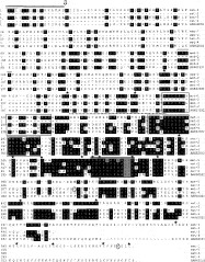
Fig. 2. Deduced amino acid sequence of Ostertagia ostertagi metalloproteases aligned with Ancylostomum caninum metalloprotease 1 precursor (Genbank™ Accession No. AAK62032). Two open boxes represent conserved zinc-binding domains (cd00203) and pfam01400 astacin domain with the met-turn, characteristic for the family of astacins. A putative 16 aa hydrophobic N-terminal signal peptide present in met-1 and met-3 is double overlined with predicted cleavage site between aa 16–17 (arrowhead). Nineteen cysteine residues conserved in all 5 sequences are indicated (). Two additional cysteines are present in both met-1 and met-3 but are not conserved (). EGF domains are indicated by grey box. Presumed propeptide cleavage site is single overlined. Presumed cleavage site of truncated baculo-expressed protein is dashed overlined.
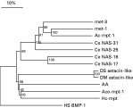
Fig. 3. Phylogenetic relationship of astacin-like proteins, including the full-length met-1 and met-3 O. ostertagi metalloproteases and selected examples from other organisms. The tree was deduced by neighbour-joining analysis based on the alignment of the amino acid sequences. At branching points, bootstrap values greater than 1000 replications are given as indication for the confidence of the tree presented. The scale bar represents % sequence dissimilarity. HS-BMP-1, Homo sapiens bone morphogenetic protein 1 (P13497); AA, Astacus astacus (crayfish) astacin (CAA64981); Ce NAS-31, -25, -16, -17, Caenorhaditis elegans Nematode Astacin like proteins (CAA98503, CAA98501, CAA91045, CAA98497); Ace-mpt-1, Ancylostoma ceylanicum metalloprotease precursor 1 (AAN11401); Ac-mpt-1, Ancylostoma caninum metalloproteases precursor 1 (AAK62032); Hc-mpt, Haemonchus contortus metalloprotease (CAA99352); DM astacin-like, Drosophila melanogaster astacin-like protein (AAF44953); DS astacin-like, Drosophila simulans astacin-like protein (AAG10257).
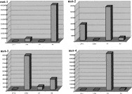
Fig. 4. Stage specificity of metalloprotease-1, -2, -3 and -4 by Real-Time PCR. The relative amount of met-1, met-2, met-3 and met-4 expression was plotted as the ratio [(=copy numbers of met/copy numbers of house-keeping gene)×100000]. L3+s: ensheathed L3; L3 ex: exsheathed L3.
 You have
Access
- 15
- Cited by