

Leukoaraiosis (LA), also named white matter hyperintensities (WMH), involving both periventricular and subcortical area on magnetic resonance imaging (MRI) is related to poor clinical outcomes and increases the risk of stroke, dementia and overall mortality and morbidity (Au et al., Reference Au, Massaro, Wolf, Young, Beiser, Seshadri and DeCarli2006; Kuller et al., Reference Kuller, Longstreth, Arnold, Bernick, Bryan and Beauchamp2004). Age and hypertension are the most importantly established risk factors for WMH though the exact pathogenic mechanism remains unclear (Lin, Huang, & Tzeng, Reference Lin, Huang and Tzeng2015). It is often ascribed to small vessel disease as its association with cerebrovascular risk factors and similarity in location to lacunar infarctions (Del Bene et al., Reference Del Bene, Ciolli, Borgheresi, Poggesi, Inzitari and Pantoni2015; Turk, Zaletel, & Pretnar-Oblak, Reference Turk, Zaletel and Pretnar-Oblak2016). What is more, other vascular risk factors such as diabetes are also found to relate to WMH (Lucatelli et al., Reference Lucatelli, Montisci, Sanfilippo, Sacconi, Suri, Catalano and Saba2016; Park et al., Reference Park, Ryoo, Kim, Kim, Chung, Lee and Bang2012). It seems that WMH share some common risk factors with arthrosclerosis. Metabolic syndrome (MetS) is a commonly used term to describe the clustering of central obesity, hypertension, hyperglycaemia, dyslipidaemia and is commonly thought as an important risk factor of cerebrovascular disease. We hypothesise that MetSmay also be a risk factor of WMH. Previous studies had different findings on whether MetS and its components relate to WMH (Knopman, Mosley, Catellier, & Sharrett, Reference Knopman, Mosley, Catellier and Sharrett2005; Liao et al., Reference Liao, Cooper, Cai, Toole, Bryan, Burke and Heiss1997; Park et al., Reference Park, Yasuda, Toyonaga, Nakabayashi, Nakasato, Nakagomi and Shimizu2007; Tiehuis et al., Reference Tiehuis, van der Graaf, Mali, Vincken, Muller and Geerlings2014). Except for different definition of LA, the subjects of these studies are heterogeneous leading to conflicting conclusions. Tiehuis et al. (Reference Tiehuis, van der Graaf, Mali, Vincken, Muller and Geerlings2014) found that WMH has no relation to MetS in patients manifesting arterial disease while the other studies drew the contrary conclusion in health subjects (Lucatelli et al., Reference Lucatelli, Montisci, Sanfilippo, Sacconi, Suri, Catalano and Saba2016; Park et al., Reference Park, Yasuda, Toyonaga, Nakabayashi, Nakasato, Nakagomi and Shimizu2007). Knopman et al. (Reference Knopman, Mosley, Catellier and Sharrett2005) restricted the subjects to middle-aged while the WMH is more common in elderly. There is no research on the relation between MetS and WMH in stroke patients until now. As to the components of MetS, the conclusions are also not unified either. In addition, studies revealed that there may be common underlying mechanisms of WMH and MetS, such as inflammatory reaction, molecular and genetic basis (Lin et al., Reference Lin, Huang and Tzeng2015; Roriz-Filho et al., Reference Roriz-Filho, Sá-Roriz, Rosset, Camozzato, Santos, Chaves and Roriz-Cruz2009). The aim of this study is to clarify the association of WMH with MetS and the components of MetS in stroke patients.
Material and Methods
Patient Selection
The hospital-based cross-sectional study took place in a teaching hospital, the first affiliated hospital of Wenzhou Medical University, China. We analysed consecutive Chinese patients with ischemic stroke recorded in a prospectively collected stroke registry database, between October 2012 and December 2014. Patients with acute symptomatic stroke syndrome within 7 days of last known well time were assessed by clinical assessment and imaging using MRI. All included patients (n = 246) underwent brain MRI scan within 48 hour after admission. Patient demographics, vascular risk factors, laboratory parameters, comorbidities, stroke etiology (using the Trial of Org 10172 in Acute Stroke Treatment [TOAST] classification) after completion of diagnostic evaluation, were collected on all patients. This study was conducted in accordance with the declaration of Helsinki. This study was conducted with approval from the Ethics Committee of the First Affiliated Hospital of Wenzhou Medical University.
Vascular Risk Factors and MetS
Vascular risk factors were identified as elsewhere (Zhang et al., Reference Zhang, Xiao, Ye, Zhang, Han and Li2015). MetS was defined using the National Cholesterol Education Program Adult Treatment Panel III (ATP-III) guidelines modified for the Chinese people. The presence of MetS was defined as the presence of ≥3 of the following risk factors: (1) abdominal obesity (waistline circumferences of ≥90 cm for men and ≥80 cm for women), (2) elevated triglyceride levels (≥150 mg/dl), (3) low high-density lipoprotein cholesterol levels (<40 mg/dl for men, <50 mg/dl for women), (4) hypertension (systolic blood pressure ≥130 mmHg, diastolic pressure ≥85 mmHg, or use of anti-hypertensive medication) and (5) impaired fasting glucose levels (≥110 mg/dl or use of anti-diabetic medication).
MRI
All the image evaluation was carried out within the first week of admission. MRI scan included T1-, T2-, Diffusion Weighted Imaging (DWI), Fluid-attenuated inversion recovery (FLAIR) series (Siemens, Avanto, Erlangen, Germany, 1.5 T scanner, T1W TR 450 ms, TE 15 ms, T2W TR 3000 ms, TE 80 ms, DWI TR 8000 ms, TE 102 ms, image matrix of 128×128, a field of view of 22 cm×22 cm, FLAIR TR 9002 ms, TE 143 ms, a field of view of 22 cm×22 cm, image matrix of 256×224, slice thickness 7 mm; 1 mm gap). Using the FLAIR data, WMHs were classified into deep (d-WMHs) and periventricular (p-WMHs) WMHs (de Groot et al., Reference de Groot, de Leeuw, Oudkerk, van Gijn, Hofman, Jolles and Breteler2000). The severity of WMHs was rated in a decentralised manner by two blinded neurologists (Z.Z and X.M.J) using the visual rating scale proposed by Fazekas and Schmidt with scores ranging from 0 to 3 (Kapeller et al., Reference Kapeller, Barber, Vermeulen, Adèr, Scheltens and Freidl2003). For d-WMHs, scores correspond to the following characteristics: 0, no lesion; 1, punctuate foci; 2, beginning confluent foci; 3, confluent changes. For p-WMHs, scores correspond to the following characteristics: 0, no changes; 1, caps or a pencil-thin lining; 2, smooth halo; 3, irregular changes extending into the d-WMHs. When evaluating the WMHs, recent or old infarcts were excluded. If WMH score was asymmetrically higher on one side, WMH rating was based on the less involved or uninvolved side with the principle of symmetry assumed. We defined WMH presented as p-WMHs ≥2 points and/or d-WMHs ≥2 points. For statistical purpose, we divided the patients into two groups: WMH group and non-WMH group.
Statistical Methods
All data were entered into a computer and analysed using SPSS10.0 software. Age of the patients was normally distributed and analysis of variance (ANOVA) was used. Unless otherwise stated, continuous variables are reported as mean ± SD. or as median (interquartile range [IQR]). Categorical variables are reported as proportions. Normality of data was examined using Kolmogorov–Smirnov test. The chi-squared test or Fisher's exact test were used for categorical variables. The cross-sectional association of MetS and its individual (dichotomous) components with WMH was assessed by binary logistic regression models (expressed as odds ratio [OR]). Spearman rank test was used to identify independent factors interrelation. To control the interrelation of independent factors, we built incremental models entering explanatory variables to calculate cumulative odds ratios (OR) with 95% confidence intervals (CI) as follows: First, adjusted for age and gender (Model 1), then additionally adjusted for hypertension, FBG, waistline circumference, serum triglyceride, high-density lipoprotein cholesterol level and HbA1c (Model 2). Last, we adjusted for age, gender and MetS to observe whether MetS is an independent predictor (Model 3). The Hosmer–Lemeshow goodness-of-fit statistic was used to assess all models for final model fit. Collinearity diagnostics were performed (and its presence rejected) for all multivariable regression models.
In addition, to explore the relation of MetS to severity of WMH, we used the multinomial logistic regression. We divided the WMH severity according to Fazekas scale into four groups as none (0), mild (1–3) and severe (4–6). Age, gender and MetS as the independent variables entered the regression. The difference was regarded as significant when two-tailed p value less than .05.
Results
Recruitment
We reviewed data of 305 patients with ischemic stroke from October 2012 to December 2014. 34 patients (11.1%) beyond 7 days window of stroke onset and 25 patients (8.2%) with inadequate assessment of MRI scan were excluded. We presented the data for 246 cases of patients.
Clinical Characteristics
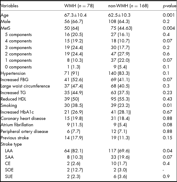
There were 78 cases patients who presented with WMH on FLAIR sequence. The mean age of the patients was 64±10.6 years and 33.3% (82/246) were female. The risk factor profile was 85.8% with hypertension (78.4% previously diagnosed, 7.4% new diagnosis), 44.7% with hyperglycaemia (28.3% previously diagnosed, 16.4% new diagnosis), 48.7% with hyperlipidaemia (7.8% previously diagnosed, 40.9% new diagnosis), 24.9% with high Haemoglobin A1c (HbA1c) and 28% smokers. Among the 246 patients, 50.8% (125 subjects) fulfilled the diagnostic criterion of MetS of ATP-III. Univariate analyses revealed a significant difference of age (p = .001), MetS [p = .004, OR 2.2, 95% CI 1.2–3.8] and smoking [p = .01, OR 2.1, 95% CI 1.1–3.7] in different WMH severity group (Table 1).
Demographic Variables and Baseline Characters of WMH and Non-WMH Group

Except for P values, values were n (%).
FBG: fasting blood glycemia; TG: triglycerides; HDL: high density lipoprotein; HbA1c: Hemoglobin A1c; LAA: Large-artery arthrosclerosis; SAA: Small artery occlusion lacunar; CE: cardio embolism; SOE: stroke of other determined etiology; SUE: stroke of undetermined etiology.
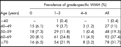
The scores (adding p-WMH and d-WMH together) are as follows: 0 (n = 69, 28%), 1 (n = 57, 23.2%), 2 (n = 63, 25.6%), 3 (n = 33, 13.4%), 4 (n = 16, 6.5%), 5 (n = 5, 2%) and 6 (n = 3, 1.2%). Extend of WMH by age group is listed in Table 2.
Extent of WMH by Age Group (Score Graded using Fazekas Scale)

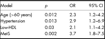
We tested the interrelation of independent factors by spearman rank test. The results showed that age and MetS related to WMH (r = 0.18, p = .005 and r = 0.18, p = .004, respectively). Hypertension is weakly associated with WMH in correlation analysis (r = 0.11, p = .08). Besides MetS is significantly related to all the components of MetS, MetS is also related to female gender (hypertension to MetS r = 0.3, p < 0.001, hyperglycaemia to MetS r = −0.4, p < .001, Low HDL related to MetS r = 0.5, p < .001, high TG to MetS r = 0.5, p < .001, large waistline circumferences to MetS r = 0.5, p < .001, female to MetS r = 0.3, p < .001). Age is related to HbA1c and low HDL-C is related to female gender too (age to HbA1c r = −0.2, p = 0.007, low HDL-C to gender r = 0.3, p < 0.001). Binary logistic regression analyses were used to identify the independent risk factor of WMH. First, we adjusted for age and gender, and age remained significantly associated with WMH [OR 2.3, 95% CI 1.2–4.2] (Table 3). We additional adjusted for all the confounders of vascular risk factors in model 2 and found age, hypertension and low HDL-C are independent risk factors (Table 3). In addition, entering the confounders to the model did not meaningfully change the significance of age. Then, we examined the association between cerebral WMH and MetS. After adjustment for age and gender as covariates, the association between and cerebral WMH was still significant [OR = 3.7, 95% CI 1.8–7.5, p = .002].
Binary Regression of Factors Related to WMH

OR: odds ratio; CI: confidence interval.
Model 1, adjusted for age and gender (Goodness-of-fit statistics: χ2 = 1.9, p = .384).
Model 2 additionally adjusted for hypertension, FBG, HbA1c, smoking, waistline circumference, serum triglyceride, high density lipoprotein cholesterol level (Goodness-of-fit statistics: χ2 = 0, p = 1).
Model 3 additionally adjusting for the MetS (Goodness-of-fit statistics: χ2 = 0.616, p = .735).
A multinomial logistic regression was constructed to evaluate whether there is higher prevalence of MetS in higher WMH grade. After adjusting for age and gender, MetS prevalence is significantly higher in severe WMH group (score 4–6) than none [p = 0.006, OR 5.7, 95%CI 1.6-19.9] and mild WMH [P < 0.001, OR 8.1, 95% CI2.5-15.8]. But there is no significant difference between mild WMH (score 1–3) to none (result not shown).
Discussion
From the result of our study, MetS, as well as hypertension and low HDL-C are associated with WMH in stroke patients. The prevalence of MetS tends to increase with the severity of WMH.
The most important result of our study is that MetS, as a clinical clustering of risk factors is associated with WMD lesion load. To our best knowledge, this is the first study showing that MetS is associated with WMH in stroke patients. It is important to know that MetS and its components specifically related to WMH in stroke patients because it will help us to predict certain subset of patients with WMH having higher chance of stroke than others in future.
WMH has attracted increased attention because it has as high prevalence as 50–100% in old people (de Leeuw et al., Reference de Leeuw, de Groot, Achten, Oudkerk, Ramos, Heijboer and Breteler2001; Launer et al., Reference Launer, Berger, Breteler, Dufouil, Fuhrer, Giampaoli and Hofman2006; Longstreth et al., Reference Longstreth, Manolio, Arnold, Burke, Bryan, Jungreis and Fried1996; Wen & Sachdev, Reference Wen and Sachdev2004). The known risk factors for WMH are old age, female gender, higher blood pressure and diabetes according to previous studies (Basile et al., Reference Basile, Pantoni, Pracucci, Asplund, Chabriat and Erkinjuntti2006; Choi et al., Reference Choi, Cho, Kang, Shin, Park and Lee2009; Dufouil et al., Reference Dufouil, de Kersaint-Gilly, Besançon, Levy, Auffray, Brunnereau and Tzourio2001; Kuller et al., Reference Kuller, Margolis, Gaussoin, Bryan, Kerwin and Limacher2010; Park et al., Reference Park, Yasuda, Toyonaga, Nakabayashi, Nakasato, Nakagomi and Shimizu2007; Varghese et al., Reference Varghese, Chandra, Christopher, Rajeswaran, Prasad, Subasree and Issac2016). Most of these risk factors, together with dyslipidaemia are also the risk factors of stroke. Previous studies had contradictory conclusions as to whether MetS is an independent risk factor of WMH (Dearborn et al., Reference Dearborn, Schneider, Sharrett, Mosley, Bezerra, Knopman and Gottesman2015; Nandigam, Reference Nandigam2008; Wen & Sachdev, Reference Wen and Sachdev2004). Basile et al. (Reference Basile, Pantoni, Pracucci, Asplund, Chabriat and Erkinjuntti2006) found that though MetS is related to WMH in health subjects, as far as stroke patients are concerned, lacunar stroke instead of MetS, is associated with WMH. Similar conclusion can be drawn from the study of Tiehuis et al., (Reference Tiehuis, van der Graaf, Mali, Vincken, Muller and Geerlings2014). Dearborn et al. found that MetS as a whole has no relation to the WMH progression (Dufouil et al., Reference Dufouil, de Kersaint-Gilly, Besançon, Levy, Auffray, Brunnereau and Tzourio2001). Nandigam (Reference Nandigam2008) and Park et al. (Reference Park, Ryoo, Kim, Kim, Chung, Lee and Bang2012) found that in healthy subjects MetS is significantly associated with WMH. Different studies had different recruitment criteria and include different subjects, which is one of the reasons that conclusions contradict to each other. In addition, the method to define WMH severity is also different. That leads to conflicting results and directly comparing the result is difficult, e.g., Tiehuis et al. evaluated WMH severity by volumetric analysis while others used various vision scales. WMH in general healthy population is usually asymptomatic while in stroke patients WMH is related closely to stroke events, which means different significance of WMH in different population. WMH is a neuroimaging finding that is caused by pathologic changes such as demyelination, gliosis, vessel lipohyalinosis and disturbed blood–brain exchanges (Hachinski, Potter, & Merskey, Reference Hachinski, Potter and Merskey1987). Hyperinsulinemia and insulin resistance, which is the mechanism underlying MetS leads to overproduction of reactive oxygen species (ROS) and widespread brain microangiopathy (Park et al., Reference Park, Yasuda, Toyonaga, Nakabayashi, Nakasato, Nakagomi and Shimizu2007). It seems that MetS and WMH have a common pathological mechanism that impairs the microcirculation of brain.
Age and hypertension are known risk factors of WMH in previous studies (Basile et al., Reference Basile, Pantoni, Pracucci, Asplund, Chabriat and Erkinjuntti2006; Choi et al., Reference Choi, Cho, Kang, Shin, Park and Lee2009; Liao et al., Reference Liao, Cooper, Cai, Toole, Bryan, Burke and Heiss1997; Park et al., Reference Park, Yasuda, Toyonaga, Nakabayashi, Nakasato, Nakagomi and Shimizu2007; Varghese et al., Reference Varghese, Chandra, Christopher, Rajeswaran, Prasad, Subasree and Issac2016; Wen & Sachdev, Reference Wen and Sachdev2004). Our findings are consistent with previous conclusions and add to mounting evidence that age and hypertension are risk factors for WMH in all subsets of patients. The association of other elements of MetS like hyperglycaemia, dyslipidaemia with WMH is controversial and large waist circumference has found no relevance to WMH in past studies (Bokura, Yamaguchi, Iijima, Nagai, & Oguro, Reference Bokura, Yamaguchi, Iijima, Nagai and Oguro2008; de Leeuw et al., Reference de Leeuw, de Groot, Achten, Oudkerk, Ramos, Heijboer and Breteler2001; Lucatelli et al., Reference Lucatelli, Montisci, Sanfilippo, Sacconi, Suri, Catalano and Saba2016; Rozanski et al., Reference Rozanski, Richter, Grittner, Endres, Fiebach and Jungehulsing2014). We found that low HDL-C cholesterol is associated with WMH. This conclusion is in line with the research of Willey et al. (Reference Willey, Gardener, Moon, Yoshita, DeCarli, Cheung and Wright2014). Though it is uncertain whether all components of lipid profile relate to WMH, it has been found that WMH is associated with cholesterol regulation through some genes, e.g., apoE (Godin et al., Reference Godin, Tzourio, Maillard, Alpérovitch, Mazoyer and Dufouil2009; Nebes et al., Reference Nebes, Vora, Meltzer, Fukui, Williams, Kamboh and Reynolds2001).
We found that in univariate analysis, smoking ratio is significantly different in WMH and non-WMH groups. However, in regression analyses, the relation of smoking to WMH is not significant any more. One of the possible reasons may be age difference between two groups though the difference had no statistical significance. It could also be possible that patients who have MetS tend to have more unhealthy life habits like smoking, since both of them are higher in WMH group. Therefore, though smoking seems relating to WMH, it is not an independent risk factor of WMH in our study. Basile et al. found that smoking is related to severe WMH but not with mild to moderate grade (Wen & Sachdev, Reference Wen and Sachdev2004). Cho et al. (Reference Cho, Kim, Kim, Ye, Kim, Jung and Seo2016) found that there was no association between smoking and the severity of WMH. As far as the all degree of WMH as a whole, there is no evidence to show there is a relation between WMH and smoking.
Our study has some limitations. First, the visual scale we have taken may underestimate the WMH burden comparing to volumetric analysis (Furie & Smith, Reference Furie and Smith2007). However, the visual scale is more practical in clinical settings and is a widely accepted and used method to study WMH worldwide. In addition, neuroimaging definition and rating scale of WMH is still controversial. The study of Bokura investigated pWMH and dWMH separately, but the neuroimaging definition of these two WMHs is unclear to date (Hachinski et al., Reference Hachinski, Potter and Merskey1987). What is more, PWMH and DWMH usually co-exist in the LA patient clinically and it is difficult for neurologists to clearly diagnose one of the two types by visual rating scale. So, we use the scheme to add pWMH and dWMH together to define WMH or non-WMH. This method is more practical and more consistent among different observers. Second, our study is a cross-sectional study and has relatively small sample size. It should be cautious to say there is a causal relationship between MetS and WMH in stroke patients. A longitudinal study with larger sample size is needed to clarify the attribution of MetS and its components to WMH.
Conclusion
From the result of our study, after adjusting for age, MetS as well as its components hypertension and low HDL-C, is associated with WMH in stroke patients. The prevalence of MetS increases with ascending grade levels of WMH severity.
Financial Support
This study was supported by Wenzhou Science and Technology Bureau (Y20120154).
Conflicts of Interest
None.
Ethical Standards
The authors assert that all procedures contributing to this work comply with the ethical standards of the relevant national and institutional committees on human experimentation and with the Helsinki Declaration of 1975, as revised in 2008.