INTRODUCTION
Strongyloidiasis is an infection caused by the parasitic nematode Strongyloides stercoralis. This parasitic infection is found in tropical and subtropical regions and also in temperate areas where migrants travel from impoverished regions to more affluent countries (Genta et al. Reference Genta, Weesner, Douce, Huitger-O'Connor and Walzer1987; Olsen et al. Reference Olsen, van Lieshout, Marti, Polderman, Polman, Steinmann, Stothard, Thybo, Verweij and Magnussen2009; Gonzalez et al. Reference Gonzalez, Gallo, Valls, Munoz, Puyol, Pinazo, Mas and Gascon2010). It has been estimated that 30–100 million people are infected globally (Bethony et al. Reference Bethony, Brooker, Albonico, Geiger, Loukas, Diemert and Hotez2006). The prevalence of strongyloidiasis in temperate regions is estimated to be below 1%, while prevalences up to 25% and above, have been found in tropical areas (Pawlowski, Reference Pawlowski and Grove1989; Yelifari et al. Reference Yelifari, Bloch, Magnussen, van Lieshout, Dery, Anemana, Agongo and Polderman2005; Steinmann et al. Reference Steinmann, Zhou, Du, Jiang, Wang, Wang, Li, Marti and Utzinger2007; Olsen et al. Reference Olsen, van Lieshout, Marti, Polderman, Polman, Steinmann, Stothard, Thybo, Verweij and Magnussen2009; Gonzalez et al. Reference Gonzalez, Gallo, Valls, Munoz, Puyol, Pinazo, Mas and Gascon2010). The true prevalence of strongyloidiasis in a community is difficult to determine because of the chronic subclinical nature, non-specific clinical features of this parasitic infection and the low sensitivity of current laboratory diagnosis (Genta et al. Reference Genta, Weesner, Douce, Huitger-O'Connor and Walzer1987; Dreyer et al. Reference Dreyer, Fernandes-Silva, Alves, Rocha, Albuquerque and Addiss1996; Uparanukraw et al. Reference Uparanukraw, Phongsri and Morakote1999; Steinmann et al. Reference Steinmann, Zhou, Du, Jiang, Wang, Wang, Li, Marti and Utzinger2007; Stothard et al. Reference Stothard, Pleasant, Oguttu, Adriko, Galimaka, Ruggiana, Kazibwe and Kabatereine2008; Agrawal et al. Reference Agrawal, Agarwal and Ghoshal2009; Knopp et al. Reference Knopp, Mohammed, Rollinson, Stothard, Khamis, Utzinger and Marti2009; Olsen et al. Reference Olsen, van Lieshout, Marti, Polderman, Polman, Steinmann, Stothard, Thybo, Verweij and Magnussen2009). Diagnosis of S. stercoralis relies basically on stool examination, which is less sensitive in chronic cases with low larval output (Gill and Bell, Reference Gill and Bell1979; Pelletier, Reference Pelletier1984). The detection of serum antibodies may facilitate a diagnosis, although serology is highly sensitive but specificity can be low. A false positive result may occur due to either past infection or cross reaction with other helminth infections especially filariasis (Gam et al. Reference Gam, Neva and Krotoski1987; Conway et al. Reference Conway, Bailey, Lindo, Robinson, Bundy and Bianco1993; Lindo et al. Reference Lindo, Conway, Atkins, Bianco, Robinson and Bundy1994; Olsen et al. Reference Olsen, van Lieshout, Marti, Polderman, Polman, Steinmann, Stothard, Thybo, Verweij and Magnussen2009); this is reported to be up to 8–16% of cases (Ganesh and Cruz, Reference Ganesh and Cruz2011).
Strongyloides stercoralis, unlike other nematodes, can remain in its host for decades with successive prolonged periods of active infection after the original exposure due to the internal autoinfection phenomenon. This may eventually lead to hyperinfection syndrome (HS) or disseminated infection where high worm burdens invade major organs, causing sepsis and death (Igra-Siegman et al. Reference Igra-Siegman, Kapila, Sen, Kaminski and Louria1981; Hakim and Genta, Reference Hakim and Genta1986; Siddiqui and Berk, Reference Siddiqui and Berk2001; Carvalho and Da Fonseca Porto, Reference Carvalho and Da Fonseca Porto2004; Vadlamudi et al. Reference Vadlamudi, Chi and Krishnaswamy2006; Marcos et al. Reference Marcos, Terashima, Dupont and Gotuzzo2008). The epidemiological association of concurrent S. stercoralis infection in patients infected with human T lymphotrophic virus type I (HTLV-I) has been documented in Japan, Jamaica and in Australia (Nakada et al. Reference Nakada, Kohakura, Komoda and Hinuma1984; Robinson et al. Reference Robinson, Lindo, Neva, Gam, Vogel, Terry and Cooper1994; Einsiedel and Fernandes, Reference Einsiedel and Fernandes2008). HTLV-1 impairs the host immunity thus reducing the efficacy of treatment for strongyloidiasis (Satoh et al. Reference Satoh, Toma, Sato, Takara, Shiroma, Kiyuna and Hirayama2002, Reference Satoh, Kiyuna, Shiroma, Toma, Kokaze and Sato2003; Terashima et al. Reference Terashima, Alvarez, Tello, Infante, Freedman and Gotuzzo2002; Hirata et al. Reference Hirata, Uchima, Kishimoto, Zaha, Kinjo, Hokama, Sakugawa, Kinjo and Fujita2006).
Poor sanitation and hygiene, resulting in faecal contamination of the soil, and warm moist tropical conditions are optimal for survival of S. stercoralis (Hall et al. Reference Hall, Conway, Anwar and Rahman1994; Borda et al. Reference Borda, Rea, Rosa and Maidana1996; Menezes et al. Reference Menezes, Lima, Freitas, Rocha, Silva and Dolabella2008; Gamboa et al. Reference Gamboa, Kozubsky, Costas, Garraza, Cardozo, Susevich, Magistrello and Navone2009). There are scant data on strongyloidiasis epidemiology from the developing world especially in tropical countries (Knopp et al. Reference Knopp, Mohammed, Rollinson, Stothard, Khamis, Utzinger and Marti2009; Olsen et al. Reference Olsen, van Lieshout, Marti, Polderman, Polman, Steinmann, Stothard, Thybo, Verweij and Magnussen2009; Glinz et al. Reference Glinz, N'Guessan, Utzinger and N'Goran2010); some details from East Asia and Thailand have been provided (Steinmann et al. Reference Steinmann, Zhou, Du, Jiang, Wang, Wang, Li, Marti and Utzinger2007). Two studies in Bangladesh performed over 15 years ago have identified several environmental factors along with some health-related behaviour as risk factors based on stool examination (Hall et al. Reference Hall, Conway, Anwar and Rahman1994; Conway et al. Reference Conway, Hall, Anwar, Rahman and Bundy1995).
The aim of the current study was to determine the seroprevalence of strongyloidiasis and to identify individual risk factors in 2 settings of Bangladesh. In addition, HTLV-1 antibodies were measured to determine the presence of concurrent infection with S. stercoralis in Bangladesh.
MATERIALS AND METHODS
Sample population
Two groups of subjects were selected for this study. Group A were residents living in a slum area (Korail) situated at Mohakhali, an eastern suburb of Dhaka. Group B were healthy blood donors who were residents of Dhaka city centre. In group A, 1 individual in every fifth house in the slum had a blood sample collected together with a completed questionnaire. Thus 1004 samples were randomly taken from the slum's population of approximately 40 000 at the time of this study (Asia Foundation, Bangladesh). Within each chosen household blood was taken from a respondent over 15 years of age. In group B, a total of 299 serum samples donated by the city dwellers on a given day were collected from a blood bank (Quantum Foundation, Bangladesh). No demographic information (questionnaire) was available from group B. Sera collected from both groups were tested for antibodies against Strongyloides antigen.
Ethical clearance was approved by the National Research Ethics Committee, Bangladesh Medical Research Council (BMRC; Dhaka, Bangladesh; Ref: BMRC/NREC/2007-2010/1256; Dated 20-01-2009). As most of the participants were illiterate, consent and completion of a questionnaire were obtained verbally from each respondent in group A at the time of collection. Follow-up treatment of infected individuals was not achieved because participants could not be located at the end of the study.
Strongyloides ELISA
Sera separated from freshly collected blood were stored at −20 °C prior to testing. IgG against Strongyloides antigen was detected by an indirect enzyme-linked immunosorbent assay (ELISA) using soluble antigen from Strongyloides ratti third stage larvae (L3) (Grove, Reference Grove and Grove1989). Microtitre plates (Maxisorb; Nunc, Thermofisher, Victoria, Australia) were coated with S. ratti antigen in carbonate buffer (pH 9·6) and incubated overnight at 4 °C. All the IgG-positive reactive sera were then screened for isotypes IgG1 and IgG4.
Detection of total IgG
The antibody assay was performed as described previously (Grove, Reference Grove and Grove1989; Rodrigues et al. Reference Rodrigues, de Oliveira, Sopelete, Silva, Campos, Taketomi and Costa-Cruz2007). Briefly, antigen-coated plates were blocked with phosphate-buffered saline (PBS) with 0·05% Tween and 0·5% skim milk for 30 min; sera (1:500) were added and tested in duplicate. Plates were incubated for 1 h at 37 °C. After washing, rabbit anti-human IgG conjugated with horseradish peroxidase (HRP; Bio-Rad, NSW, Australia) was added and incubated for 1 h. Washed plates were developed in TMB substrate (Elisa Systems Pty. Ltd, Queensland, Australia) for 5 min and stopped by addition of 1 M phosphoric acid. The sensitivity and specificity of this method have been previously determined to be >85% (Grove, Reference Grove and Grove1989).
Detection of isotypes IgG1 and IgG4
Microtitre plates were incubated for 1 h at 37 °C with serum diluted in blotto (2% skim milk powder in 0·1% PBS Tween) at 1:100 for IgG1 and 1:5 for IgG4. Washed plates were subsequently incubated with biotinylated monoclonal antibodies specific for either human IgG1 (1:200; Invitrogen, Victoria, Australia) or IgG4 (1:20; Invitrogen, Victoria, Australia) for 1 h. Plates were washed and streptavidin conjugated to HRP (Merck Pty. Ltd, Victoria, Australia) was added at 1:1000 dilution followed by 45 min incubation at 37 °C. TMB substrate (Elisa Systems Pty. Ltd, Queensland, Australia) was developed for 10 min and stopped with 1 M phosphoric acid. Absorbance levels greater than 0·17 for IgG1 and greater than 0·18 for IgG4 were regarded as positive. These cut-off values were calculated by adding 2 standard deviations above the mean of negative samples (n = 70).
Risk factors associated with strongyloidiasis
Respondents from group A (slum residents) who provided a blood sample also completed a questionnaire covering demographic, life style, socio-economic and medical history. Data collected included: sex, age, level of education, occupation, average monthly income, number of family members, type of toilet used; aspects of personal hygiene such as wearing shoes, trimming nails regularly and washing hands with soap after defecation; and history of diarrhoea (in respondent and household) in the past 6 months. These factors were then evaluated against seroreactivity of each blood specimen.
Other serological studies
Detection of filarial antibody
All positive serum samples from groups A and B were also tested for the presence of lymphatic filariasis antibody. An indirect ELISA for the detection of Bm14-specific IgG4 antibody (Cellabs, NSW, Australia) was used and results were interpreted according to the manufacturer's instruction.
Detection of HTLV-I and II
Sera were also tested for HTLV-I and II antibodies, using the Murex HTLV I + II (Abbott, NSW, Australia) assay and results were interpreted according to the manufacturer's instructions.
Statistical analysis
Statistical package, SPSS version 17 was used (i) to compare the prevalence of infection between groups A and B, (ii) to determine significant risk factors from the questionnaire, and (iii) to find out the association, if any, of each with positive serology using χ 2 statistics. Levels (as shown by optical density; OD values) of IgG, IgG1 and IgG4 antibodies to S. ratti in group A were compared by Pearson's correlations statistics. Logistic regression was used to compute the risk of infection with age, entered as a continuous variable. Values of P < 0·05 were considered statistically significant.
RESULTS
Detection of IgG (total)
The Strongyloides antibody test was positive in 222 of 1004 (22·1%) sera from group A and 15 of 299 (5·0%) sera from group B subjects. Slum residents are 4-fold more likely to have strongyloidiasis than city dwellers (χ 2 = 44·10, odds ratios (OR) = 5·38, 95% confidence interval (CI) = 3·13–9·20, P < 0·001).
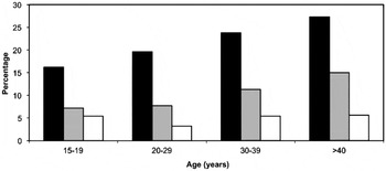
In group A, there was no difference found in prevalence rate between males (108/492) and females (114/512). The seroprevalence rose progressively with age (Fig. 1) and the risk of being seropositive increased each year (OR = 1·02 for each year increase, 95% CI = 1·01–1·03, P = 0·004). Thus, the lowest and highest prevalence of 16·2% and 27·3% were observed in group 15–19 years and in >40 years, respectively.
Comparison of total IgG response (black), IgG1 response (grey) and IgG4 response (white) at different ages in group A.

Comparisons of prevalence according to age and sex between groups A and B could not be made as no data were available from the latter group.
Detection of isotype IgG1 and IgG4
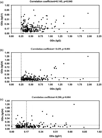
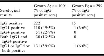
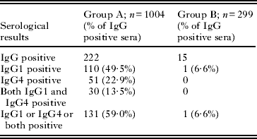
In group A, IgG1, IgG4 and both subclasses were detected in 50%, 23% and 13·5% of the total IgG positive sera, respectively. In group B, only 1 serum specimen had detectable IgG1 and none had IgG4 (Table 1). Similar to total IgG, the prevalence of IgG1 increased with age in group A (logistic regression considering age as a continuous variable: OR = 1·03, 95% CI = 1·01–1·05, P < 0·001). Thus the lowest percentage of reactive IgG1 was found in the 15–19 years age group (7·2%) and the highest in >40 years age group (15·0%) (Fig. 1). IgG4 did not show this trend with age. Furthermore, there was a positive correlation between the level of IgG and IgG isotype (IgG1 and IgG4) reactions in group A (Fig. 2) determined by Pearson's correlations statistics (P < 0·05).
Pearson's correlations between the levels of specific IgG and IgG1 (a), IgG and IgG4 (b) and IgG1 and IgG4 (c) antibodies to Strongyloides antigen in serum samples of respondents in group A. Dotted lines represent the cut-off levels.

IgG1 and IgG4 reactive to Strongyloides antigen in groups A and B
Risk factors associated with Strongyloides-positive sera in group A
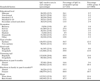
Socio-demographic information was collected from respondents in group A only (Table 2). There were no correlations of either socio-economic or personal hygiene factors to the presence of antibody reactive to Strongyloides antigen. A strong relationship was found between positive IgG and having a family member who had diarrhoea in the past 6 months (P < 0·01); the seropositive and seronegative cases were 84·7% and 75·7%, respectively. However, those respondents with a history of diarrhoea did not have a significant association with (83·3%) or without (81·6%) antibody reactivity. In addition, within the seropositive group (n = 222) people who completed their secondary school were less likely to have strongyloidiasis compared to people who had not completed their secondary school. This difference was again not statistically significant.
Life-style factors associated with total IgG reactive to Strongyloides antigen in group A

** P < 0·01 = Significant association with seroprevalence.
Other serological results
None of the sera seropositive to Strongyloides antigen in either group had detectable HTLV I or II antibodies. Five sera from Strongyloides seropositive respondents in group A had filarial antibody reactions, whereas none of the sera had detectable filarial antibody in group B.
DISCUSSION
To our knowledge, this is the first serological study for strongyloidiasis conducted in Bangladesh. In total, 1004 samples were collected from a slum (group A) by choosing 1 volunteer in every fifth house and in group B 299 sera were collected from a blood bank on a single given day. The study provides serological evidence that strongyloidiasis is present in Dhaka city; a significantly higher (P < 0·001) seroprevalence was found among slum dwellers (group A) compared to socio-economically better-off city dwellers (group B). Indeed, group B was comprised of healthy blood donors living in fixed housing in a central part of Dhaka.
No sex difference in seroprevalence was observed in this study which agrees with some previous studies (Douce et al. Reference Douce, Brown, Khamboonruang, Walzer and Genta1987; Gyorkos et al. Reference Gyorkos, Genta, Viens and MacLean1990; Lindo et al. Reference Lindo, Robinson, Terry, Vogel, Gam, Neva and Bundy1995; Yori et al. Reference Yori, Kosek, Gilman, Cordova, Bern, Chavez, Olortegui, Montalvan, Sanchez, Worthen, Worthen, Leung and Ore2006). However, other studies have reported a higher number of infected males compared to females in coprological examination when individuals were either farmers or refugees from rural areas (Faust and Giraldo, Reference Faust and Giraldo1960; Arakaki et al. Reference Arakaki, Kohakura, Asato, Ikeshiro, Nakamura and Iwanaga1992; Marnell et al. Reference Marnell, Guillet and Holland1992). Perhaps both males and females living in slum conditions of Dhaka have an equal chance of infection around their residence with little influence of the type of work they do.
The IgG seroprevalence in group A increased by 1·8% for each year of age (P = 0·004), which is congruent with studies from a north-eastern Amazon rural community in Peru (Yori et al. Reference Yori, Kosek, Gilman, Cordova, Bern, Chavez, Olortegui, Montalvan, Sanchez, Worthen, Worthen, Leung and Ore2006), Jamaica (Lindo et al. Reference Lindo, Robinson, Terry, Vogel, Gam, Neva and Bundy1995) and south-central Côte d'Ivoire (Becker et al. Reference Becker, Sieto, Silué, Adjossan, Koné, Hatz, Kern, N'Goran and Utzinger2011). Since autoinfection maintains the parasitic infection in the human host, then progressive increases of infection with age is suggestive that strongyloidiasis has a cumulative infection rate in a population. In contrast, age showed no association with serological evidence of infection in a population group in Thailand (Douce et al. Reference Douce, Brown, Khamboonruang, Walzer and Genta1987). In our study the highest prevalence was observed in the oldest age group (>40 years), whereas higher prevalence is documented in the 20–29 years age group in Southeast Asian refugees (Gyorkos et al. Reference Gyorkos, Genta, Viens and MacLean1990). The disparity of seroprevalence amongst age groups suggests that the exposure to this parasitic infection differs in different populations. In studies of fecal carriage of S. stercoralis (copro-prevalence), the younger age group had a higher chance of being infected; prevalence of 17·8%, 44·0% and 8·1% in children less than 12 years of age, have been observed in Bangladesh, Sudan and Thailand, respectively (Sornmani et al. Reference Sornmani, Vivatanasesth, Bunnag, Intarakhao and Harinasuta1973; Marnell et al. Reference Marnell, Guillet and Holland1992; Hall et al. Reference Hall, Conway, Anwar and Rahman1994). In Southeast Asian refugees, children aged 10–19 years were found to have prevalence of 32·5% (Gyorkos et al. Reference Gyorkos, Genta, Viens and MacLean1990). Respondents under 15 years of age were excluded from our study and the serological pattern in children could not be determined. In addition, follow-up coprological examination was not done because respondents in the slums could not be found upon our return so no stool specimens were collected from either of our studied groups. Association of age and infection prevalence in stool examination thus, could not be achieved.
Non-specific antibody (IgG) reaction in ELISA in detecting Strongyloides infection includes the possibility of cross-reaction with other helminths such as filarial infection (Grove, Reference Grove and Grove1989; Conway et al. Reference Conway, Bailey, Lindo, Robinson, Bundy and Bianco1993; Lindo et al. Reference Lindo, Conway, Atkins, Bianco, Robinson and Bundy1994; van Doorn et al. Reference van Doorn, Koelewijn, Hofwegen, Gilis, Wetsteyn, Wismans, Sarfati, Vervoort and van Gool2007; Olsen et al. Reference Olsen, van Lieshout, Marti, Polderman, Polman, Steinmann, Stothard, Thybo, Verweij and Magnussen2009). Previous studies have shown that the presence of IgG1 and IgG4 is more specific than the total IgG response for detection of strongyloidiasis in humans (Conway et al. Reference Conway, Lindo, Robinson, Bundy and Bianco1994; Lindo et al. Reference Lindo, Conway, Atkins, Bianco, Robinson and Bundy1994; Rodrigues et al. Reference Rodrigues, de Oliveira, Sopelete, Silva, Campos, Taketomi and Costa-Cruz2007), whereas detection of IgG2 and IgG3 is less sensitive (Conway et al. Reference Conway, Lindo, Robinson, Bundy and Bianco1994). IgG4 is found to be specific in chronic infection and works as a modulator in the IgE-mediated immune response (Atkins et al. Reference Atkins, Lindo, Lee, Conway, Bailey, Robinson and Bundy1997; Satoh et al. Reference Satoh, Toma, Sato, Kikuchi, Takara, Shiroma, Kiyuna and Hirayama1999; Rodrigues et al. Reference Rodrigues, de Oliveira, Sopelete, Silva, Campos, Taketomi and Costa-Cruz2007). In our study, the level of total IgG and isotype reaction showed a significant positive relationship (P < 0·05) in group A. This agrees with a recent study in Brazil (Rodrigues et al. Reference Rodrigues, de Oliveira, Sopelete, Silva, Campos, Taketomi and Costa-Cruz2007), where it was found that 3·3% and 23·3% of sera from patients with intestinal helminth infections other than S. stercoralis also showed reaction against IgG1 and IgG4 respectively. From the total IgG sera reactive to S. ratti, 50% had IgG1 binding and 23% had IgG4 binding in group A. In group B, only 1 serum from the IgG positive samples had reactive IgG1 whereas no sera had IgG4 reaction. All sera reactive to Strongyloides antigen from group A and group B were tested for antibodies to a recombinant antigen Bm14; this commercial assay has a sensitivity of 91% and 96% towards Wuchereria bancrofti and Brugia infections respectively (Lammie et al. Reference Lammie, Weil, Noordin, Kaliraj, Steel, Goodman, Lakshmikanthan and Ottesen2004). Five sera in group A showed filarial antibody responses; 3 of these had no detectable IgG1 and IgG4 antibodies to Strongyloides antigen which suggests a cross-reactive filarial infection, 2 sera had both IgG1 and IgG4 reactions indicating either cross-reactive filarial infections or dual infections of filarial and S. stercoralis. A case of acute S. stercoralis infection does not necessarily display positive serology; false negative serology is also known to occur in immunosuppressed patients (Repetto et al. Reference Repetto, Duran, Lasala and Gonzalez-Cappa2010). Thus delayed seroconversion leads to false negative results (Repetto et al. Reference Repetto, Duran, Lasala and Gonzalez-Cappa2010; Baaten et al. Reference Baaten, Sonder, van Gool, Kint and van den Hoek2011); those participants in our study who were recently infected or are in an immunosuppressed state with S. stercoralis may be missed. A recent serodiagnostic test involving measurement of IgG avidity can identify those who have recently seroconverted (Gonzaga et al. Reference Gonzaga, Ribeiro, Feliciano, Manhani, Silva, Ueta and Costa-Cruz2011); this, however, still does not detect acute cases of strongyloidiasis where serum antibodies are not present.
Socio-economic and personal hygiene factors could not be associated with the presence of antibody to Strongyloides antigen in our study. An earlier survey based on a household cluster in Bangladesh showed that using community latrines, living in a house with either earth floor or metal roof, having 5 or more family members, promiscuous defecation by children, earning monthly incomes of less than BDT 1040 (US $13), and poor literacy of the respondents were associated with S. stercoralis infection in stool (Hall et al. Reference Hall, Conway, Anwar and Rahman1994). Thus fecal analysis seemed to be more associated with environmental risk factors. However, stool examination underestimates the infection prevalence especially in chronic cases. Detection of antibody to Strongyloides antigen identifies chronic subclinical infection; thus the relationship between health aspects and environmental condition in individuals may not necessarily correlate with serology. We found that the higher the education level the lower the chance of being seropositive but the difference was not statistically significant.
Interestingly, a significant association was found with seropositive respondents when their family members had had a history of diarrhoea over the past 6 months; a history of diarrhoea in respondents themselves had no association with detectable antibodies. Furthermore, surveys in Peru and an endemic area in Jamaica found that wearing of shoes occasionally or not at all and sharing the bedroom with reference cases were associated with strongyloidiasis determined both by coprological examination and serology (Lindo et al. Reference Lindo, Robinson, Terry, Vogel, Gam, Neva and Bundy1995; Yori et al. Reference Yori, Kosek, Gilman, Cordova, Bern, Chavez, Olortegui, Montalvan, Sanchez, Worthen, Worthen, Leung and Ore2006). Thus, the association of risk factors with infection depends both on the level of disease endemicity and environmental factors that can enhance transmission between individuals.
Co-infection of HTLV-1 and S. stercoralis has been documented in Japan (Zaha et al. Reference Zaha, Hirata, Uchima, Kinjo and Saito2004), Jamaica (Robinson et al. Reference Robinson, Lindo, Neva, Gam, Vogel, Terry and Cooper1994), Brazil (Chieffi et al. Reference Chieffi, Chiattone, Feltrim, Alves and Paschoalotti2000; Porto et al. Reference Porto, Alcantara, Leal, Castro and Carvalho2005) and Australia (Einsiedel and Fernandes, Reference Einsiedel and Fernandes2008). Impaired immunity in cases with concurrent infections of HTLV-1 and strongyloidiasis can lead to HS (Newton et al. Reference Newton, Limpuangthip, Greenberg, Gam and Neva1992; Gotuzzo et al. Reference Gotuzzo, Terashima, Alvarez, Tello, Infante, Watts and Freedman1999; Adedayo et al. Reference Adedayo, Grell and Bellot2002; Hirata et al. Reference Hirata, Uchima, Kishimoto, Zaha, Kinjo, Hokama, Sakugawa, Kinjo and Fujita2006). Furthermore, difficulty in treating strongyloidiasis in those with an impaired immune system will eventually lead to treatment resistance (Sato et al. Reference Sato, Shiroma, Kiyuna, Toma and Kobayashi1994; Satoh et al. Reference Satoh, Toma, Sato, Takara, Shiroma, Kiyuna and Hirayama2002; Terashima et al. Reference Terashima, Alvarez, Tello, Infante, Freedman and Gotuzzo2002). Co-infection was not detected in our study. Thus development into either HS or disseminated disease, in this population, is unlikely to be due to concurrent infection with HTLV-I and its immunosuppressive action.
From our serological findings using total IgG and IgG isotypes we cannot say whether infection in the slum residents took place in or prior to arrival in Dhaka. Movement of people into squalid conditions is likely to predispose to infection. The epidemiological data used in this study was limited to those currently residing in the city and several aspects of our study require broader investigations into rural villages to provide a satisfactory explanation of our findings.
ACKNOWLEDGEMENTS
We gratefully acknowledge the assistance of Dr Brian O'Toole for data analysis and the skilled technical assistance of John Clancy, ICPMR, Westmead Hospital, NSW, Australia. This research was supported by an Australian Leadership Scholarship (ALA) Award funded by the Australian Agency for International Development (Aus AID) and part by the Centre for Infectious Diseases and Microbiology-Public Health. Part of this work was presented at the XII International Congress of Parasitology (ICOPA 2010), Melbourne, Australia.