INTRODUCTION
The genus Antho Gray, Reference Gray1867 (Demospongiae, Poecilosclerida, Microcionina, Microcionidae, Ophlitaspongiinae) has been reported from virtually all regions of the world oceans, excepting Antarctica (Van Soest et al., Reference Van Soest, Boury-Esnault, Hooper, Rützler, De Voogd, Alvarez de Glasby, Hajdu, Pisera, Manconi, Schoenberg, Janussen, Tabachnick, Klautau, Picton, Kelly, Vacelet, Dohrmann, Díaz and Cárdenas2014). In Hooper's revision of Antho (1996, 2002) a large number of genera previously proposed have been sunk into synonymy of Antho s.l. In most cases these synonymies have met with little resistance in the taxonomic community, as the type species of the genera concerned had obvious morphological similarities. To date, the World Porifera Database (Van Soest et al., 2014) lists 54 accepted species of Antho. To facilitate recognition of the species among this large number, Hooper (Reference Hooper1996, Reference Hooper, Hooper and Van Soest2002) subdivided the genus into three subgenera, to which a fourth was added recently (Van Soest et al., Reference Van Soest, Beglinger and De Voogd2013). These subgenera are distinguished by presence or absence of spicular and skeletal characters: echinating acanthostyles, axially condensed skeletons, microsclere complement and peculiar dumbbell-shaped strongyles. The monophyly of these subgenera, Antho (Antho) Gray, Reference Gray1867, Antho (Acarnia) Gray, Reference Gray1867, Antho (Isopenectya) Hallmann, Reference Hallmann1920 and Antho (Plocamia) Schmidt, Reference Schmidt1870, remains untested in the absence of a comprehensive molecular evaluation, but the subgenera have obvious practical value for taxonomy of the many species of Antho.
De Laubenfels (Reference De Laubenfels1930, 1932) erected the genus Jia for a Californian species, Jia jia, with a rather confusedly reticulate skeleton of styles, ectosomal subtylostyles, and a microsclere complement of palmate isochelae and toxas to which were added tiny peculiar J-shaped spicules. A second species possessing these microscleres was described from Norwegian waters by Van Soest & Stone (Reference Van Soest and Stone1986). The structure of that species was so obviously similar to members of the genus Antho, in particular Antho coriacea (Bowerbank, 1866) that these authors named it Antho brattegardi. The J-shaped spicule was proposed to be named ‘croca’ (Latin, meaning shepherd's staff or crook, referring to the shape of an inverted ‘J’). Along with this, they reassigned De Laubenfels’ species also to Antho, proposing Jia as a junior synonym. In their discussion, Van Soest & Stone judged the skeletal structure to be of higher value than the possession of these unique microscleres. This synonymy proposal was followed by Hooper (Reference Hooper1996, Reference Hooper, Hooper and Van Soest2002). Subsequently, Desqueyroux-Faúndez & Van Soest (Reference Desqueyroux-Faúndez and Van Soest1997) reported the occurrence of crocae in Galapagos material of specimens identified as Antho lithophoenix (De Laubenfels, Reference De Laubenfels1927, originally as Plocamia), and along with it established the presence of crocae in the Californian type specimen of this species. These authors maintained the status of Jia as junior synonym of Antho, despite their independent occurrence in a third species. Sim & Kim (Reference Sim and Kim1994) reported the North Atlantic Antho brattegardi from the Korean Sea (misprinted as Antho brathesardi).
Below, we report the occurrence of crocae in two additional species, one collected in the Northern Red Sea, the other in Indonesian waters. Along with these descriptions we studied and reviewed the previously reported species and specimens possessing the crocae. This resulted in the observation of several unique features in specimens assigned to previously known species, e.g. Galapagos Antho lithophoenix sensu Desqueyroux-Faúndez & Van Soest, Reference Desqueyroux-Faúndez and Van Soest1997 and Korean Antho brattegardi sensu Sim & Kim, Reference Sim and Kim1994. We conclude that these specimens belong each to undescribed species which are named and redescribed below. Our observations lead us to abandon the notion that crocae can occur in unrelated Antho species in favour of the more likely synapomorphic nature of these unique spicules. Consequently, we propose here the reinstatement of Jia as a valid taxon with subgeneric status within the genus Antho.
MATERIALS AND METHODS
The material studied here is incorporated in the collections of the former Zoological Museum of the University of Amsterdam (ZMA), now part of the Naturalis Biodiversity Center at Leiden, the Netherlands, of the National Museum of Natural History (formerly, United States National Museum, USNM), Smithsonian Institution, Invertebrate Zoology Department at Washington, DC, USA, and of the Hannam University Natural History Museum (HUNHM), Korea. Sponge specimens originated from various localities and collecting efforts, details of which are given with each separate species description below. Specimens were studied from thick sections, light microscopy preparations of dissociated spicules and SEM observations (for technical details of preparation, cf. Van Soest et al., Reference Van Soest, Beglinger and De Voogd2013; Rützler et al., Reference Rützler, Piantoni, Van Soest and Díaz2014). Measurements of the spicules are given as minimum–mean–maximum of 25 spicules of each category for each species, unless otherwise indicated.
RESULTS
SYSTEMATICS
Phylum Porifera
Class Demospongiae
Order Poecilosclerida
Suborder Microcionina
Family Microcionidae
Subfamily Ophlitaspongiinae
Genus Antho Gray, Reference Gray1867
Subgenus Jia De Laubenfels, Reference De Laubenfels1930
DEFINITION
Antho species having smooth J-shaped microscleres (crocae) among the spicule complement.
TYPE SPECIES
Jia jia De Laubenfels, Reference De Laubenfels1930 (by monotypy).
REMARKS
It is here proposed to revive Jia because the presence of the unique crocae in at least six species (see below) indicates that the croca is a likely synapomorphy of a monophyletic group. Antho is defined as having a choanosomal reticulation of (acantho-)styles and/or (acantho-)strongyles upon which (sub-)ectosomal single styles are erected piercing the ectosomal skeleton of subtylostyles. The type species of subgenus Jia is an atypical Antho by having its choanosomal skeleton of smooth or only occasionally spined styles in a virtually confused arrangement. Other members of the subgenus Jia demonstrate a renieroid skeleton, more representative of the genus Antho.
Antho (Jia) jia (De Laubenfels, Reference De Laubenfels1930) comb. nov.
Antho (Jia) jia De Laubenfels, Reference De Laubenfels1930, dried holotype USNM 21510 from Monterey Bay, California (scale bar=1 cm).

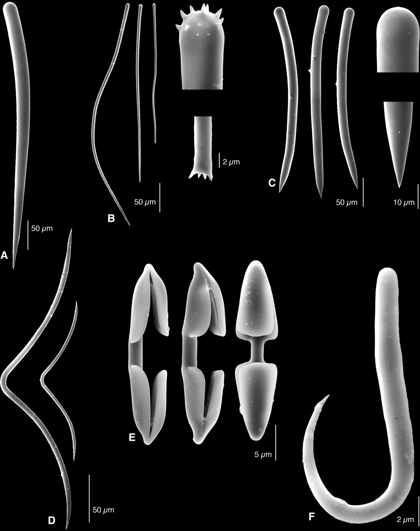
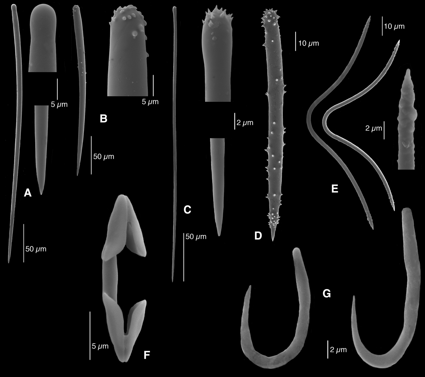
Antho (Jia) jia De Laubenfels, Reference De Laubenfels1930, holotype USNM 21510, SEM images of the spicules: (A) subectosomal style; (B) subtylostyles and details of quasitylote variation; (C) choanosomal styles and details of head and point; (D) toxas; (E) palmate isochelae; (F) croca.
Jia jia De Laubenfels, Reference De Laubenfels1930: 28; De Laubenfels, Reference De Laubenfels1932: 97, figure 58.
Antho (Antho) jia; Hooper Reference Hooper1996: 53, figure 17E & F; Lee et al., Reference Lee, Elvin and Reiswig2007: 70, figure A.
MATERIAL EXAMINED
Holotype USNM 21510, United States, California, Monterey Bay, 700 m, coll. E.F. Ricketts, 9 May 1929.
DESCRIPTION
The holotype (Figure 1, now dry) is a 18 cm2 crust, 1–8 mm thick, without signs of openings on the smooth or (partly) ridged surface; it encrusts a dictyonine hexactinellid sponge, which was dead when collected. Consistency is firm and brittle. The colour now is tan (khaki); in life it was reported to have been with an orange tinge (de Laubenfels, 1932).
Skeleton. There is no apparent ectosomal specialization, except for the abundance of a large category of styles and thin subtylostyles. The choanosome is a multispicular reticulation of thick styles, with some tracts leading toward the surface, accompanied by thin subtylostyles and scattered microscleres are common (toxas, isochelae and crocae).
Spicules. Styles, subtylostyles, short (acantho-)styles, toxas, isochelae and crocae.
Large subectosomal smooth styles (Figure 2A), gently curved, thickest just below the top (round end) or along the top 25% of their lengths, 440–511.2–590 × 24–25.9–28 µm.
Slim subtylostyles (Figure 2B), slightly bent or wavy, with microspined head, a small percentage also with microspined pointed ends (‘quasitylotes’), 240–343.2–445 × 5–6.7–8 µm.
Robust choanosomal styles (Figure 2C), curved and smooth, only a few showing 1–5 small spines along the shaft, the thickest point of the spicules is at the top, followed by a faint constriction below, 300–362.5–520 × 17–20.2–23 µm.
Toxas (Figure 2D), deeply curved, ends smooth or barely rugose, 110–213.5–313 × 2–3.8–5 µm.
Palmate isochelae (Figure 2E), strongly developed, with pointed ends, 25–26.6–30 µm.
Crocae (J-shaped microscleres) (Figure 2F), relatively robust, 16–17.9–19 µm; near the rounded end, they are 1–2 µm thick.
ECOLOGY
Bathyal, from 700 m depth.
DISTRIBUTION
Known only from the type locality, off Monterey, California.
REMARKS
The skeleton lacks the usual basal renieroid reticulation of Antho species, instead of which it has multispicular meshes, some perpendicular tracts, or is confused. Also, the choanosomal spicules are not the usual acanthostyles or acanthostrongyles, but are mostly smooth, robust styles, bearing only a few if any spines. Apart from these differences, the general spicule complement is similar to that of the species described below.
Antho (Jia) brattegardi Van Soest & Stone, Reference Van Soest and Stone1986 comb. nov.
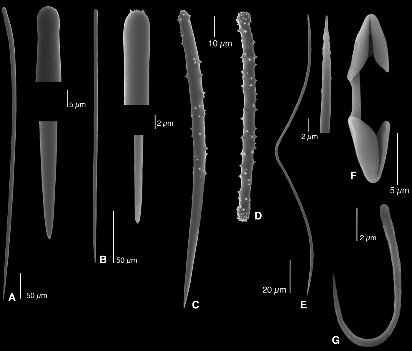
Antho (Jia) brattegardi Van Soest & Stone, Reference Van Soest and Stone1986, SEM images of the spicules of ZMA Por. 10398 from Mingulay, Scotland: (A) subectosomal style and details of head and point; (B) ectosomal subtylostyle and details of head and point; (C) acanthostyle; (D) acanthostrongyle; (E) toxa and detail of end of apex; (F) palmate isochela; (G) croca.
Antho brattegardi Van Soest & Stone, Reference Van Soest and Stone1986: 42, figures 1–3
Antho (Antho) brattegardi; Hooper, Reference Hooper1996: 420; Picton & Goodwin, Reference Picton and Goodwin2007: 1446; Roberts et al., Reference Roberts, Davies, Henry, Dodds, Duineveld, Lavaleye, Maier, Van Soest, Bergman, Huehnerbach, Huvenne, Sinclair, Watmough, Long, Green and Van Haren2009, suppl. 1: 3; Van Soest et al., Reference Van Soest, Boury-Esnault, Vacelet, Dohrmann, Erpenbeck, De Voogd, Santodomingo, Vanhoorne, Kelly and Hooper2012b (on line at http://species-identification.org/species.php?species_group=sponges&menuentry=soorten&id=116&tab=beschrijving); Van Soest & De Voogd, Reference Van Soest and De Voogd2013: table 2.
Antho (Acarnia) brattegardi; Van Soest et al., Reference Van Soest, Boury-Esnault, Hooper, Rützler, De Voogd, Alvarez de Glasby, Hajdu, Pisera, Manconi, Schoenberg, Janussen, Tabachnick, Klautau, Picton, Kelly, Vacelet, Dohrmann, Díaz and Cárdenas2014 (WPD on line).
Not: Antho brathesardi (sic); Sim & Kim, Reference Sim and Kim1994: 22, pl. 9 (missprinted name, different species, see below).
MATERIAL EXAMINED
Holotype ZMA Por. 05190 (with schizoholotype BMNH 1982.9.6.1), Norway, Bergen area, N of Björöy, 60.3333°N 5.175°E, 35–45 m, dredged, coll. Van Soest et al., 13 August 1982; ZMA Por. 20212, United Kingdom, Scotland, Outer Hebrides, Mingulay, 56.8032°N 7.4471°W, 143 m, coll. R.W.M. van Soest, BIOSYS 2006 stat. BX 126/2, 20 July 2006; ZMA Por. 20216a, United Kingdom, Scotland, Outer Hebrides, Mingulay, 56.8033°N 7.4471°W, 82 m, coll. R.W.M. van Soest, BIOSYS 2006 stat. BX 127/4, 20 July 2006; ZMA Por. 20232a, United Kingdom, Scotland, Outer Hebrides, Mingulay, 56.8034°N 7.4468°W, 144 m, coll. R.W.M. van Soest, BIOSYS 2006 stat. BX 172/5, 21 July 2006; ZMA Por. 20398, United Kingdom, Scotland, Outer Hebrides, Mingulay, 56.8243°N 7.3686°W, 128–137 m, coll. R.W.M. van Soest, BIOSYS 2006 stat. DR 182/rest, 22 July 2006.
DESCRIPTION
(Partly taken from Van Soest & Stone, Reference Van Soest and Stone1986). Thinly encrusting on barnacles, corals or stones, lateral size several mm2, thickness 1 mm, oscules not apparent, surface hispid, consistency fragile, crumbly. Colour orange or beige alive, cream in alcohol.
Skeleton. Subectosomal styles protruding far beyond the surface, ectosomal subtylostyles scattered tangentially. Choanosomal skeleton a basal reticulation of acanthostrongyles, with on average 2 spicules at each side. The meshes of the basal skeleton are reinforced by acanthostyles and the proximal ends of the protruding styles, making them anisotropic. Acanthostyles are also echinating the nodes. There is little binding spongin, so the meshwork is not tight. Microscleres are scattered throughout the skeleton.
Spicules. Styles, subtylostyles, acanthostrongyles, acanthostyles, palmate isochelae, toxas and crocae.
Subectosomal styles (Figure 3A), smooth, relatively long, with constricted heads, usually curved, 301–512.7–765 × 7–9.5–12 µm (holotype 442–765 × 8 µm).
Subtylostyles (Figure 3B), usually with faintly spined head, occasionally smooth, straight, large length variation, 204–304.9–435 × 2.5–3.9–5 µm (holotype 220–410 × 3–5 µm).
Acanthostyles (Figure 3C), spined strongly on the head and sparingly along the shaft, 150–199.4–270 × 7–8.6–12 µm (holotype 160–195 × 10 µm).
Acanthostrongyles (Figure 3D), straight or slightly curved, strongly spined on the swollen apices, lighter on the shaft, 90–116.7–135 × 2–6.2–8 µm (holotype 90–130 × 2–8 µm).
Toxas (Figure 3E), deeply curved, apices smooth or faintly rugose in the largest spicules, in a large size range, possibly in two overlapping size categories, 21–101.8–160 µm (holotype 140–160 and 30–40 µm).
Palmate isochelae (Figure 3F), relatively long, 16–18.9–24 µm (holotype 17–24 µm).
Crocae (Figure 3G), relatively large, 9–11.4–14 µm (holotype 10–14 µm).
ECOLOGY
On hard substratum at depths between 30 and 144 m.
DISTRIBUTION
Norway, Scotland, Northern Ireland.
REMARKS
The species was also recorded from Northern Ireland by Picton & Goodwin (Reference Picton and Goodwin2007: 144). They report generally similar habit and spicule sizes, but their crocae were apparently only 7.5–10 µm.
Sim & Kim (Reference Sim and Kim1994: 22, pl. 9 figures 1–4) report the occurrence of this species from South Korea. Their material is a large branching sponge unlike the small crusts described here, and the crocae are measured as 11–20 µm. We describe here the Korean sponge Antho (Jia) ramosa sp. nov., cf. below.
Antho (Jia) lithophoenix (De Laubenfels, Reference De Laubenfels1927) comb. nov.
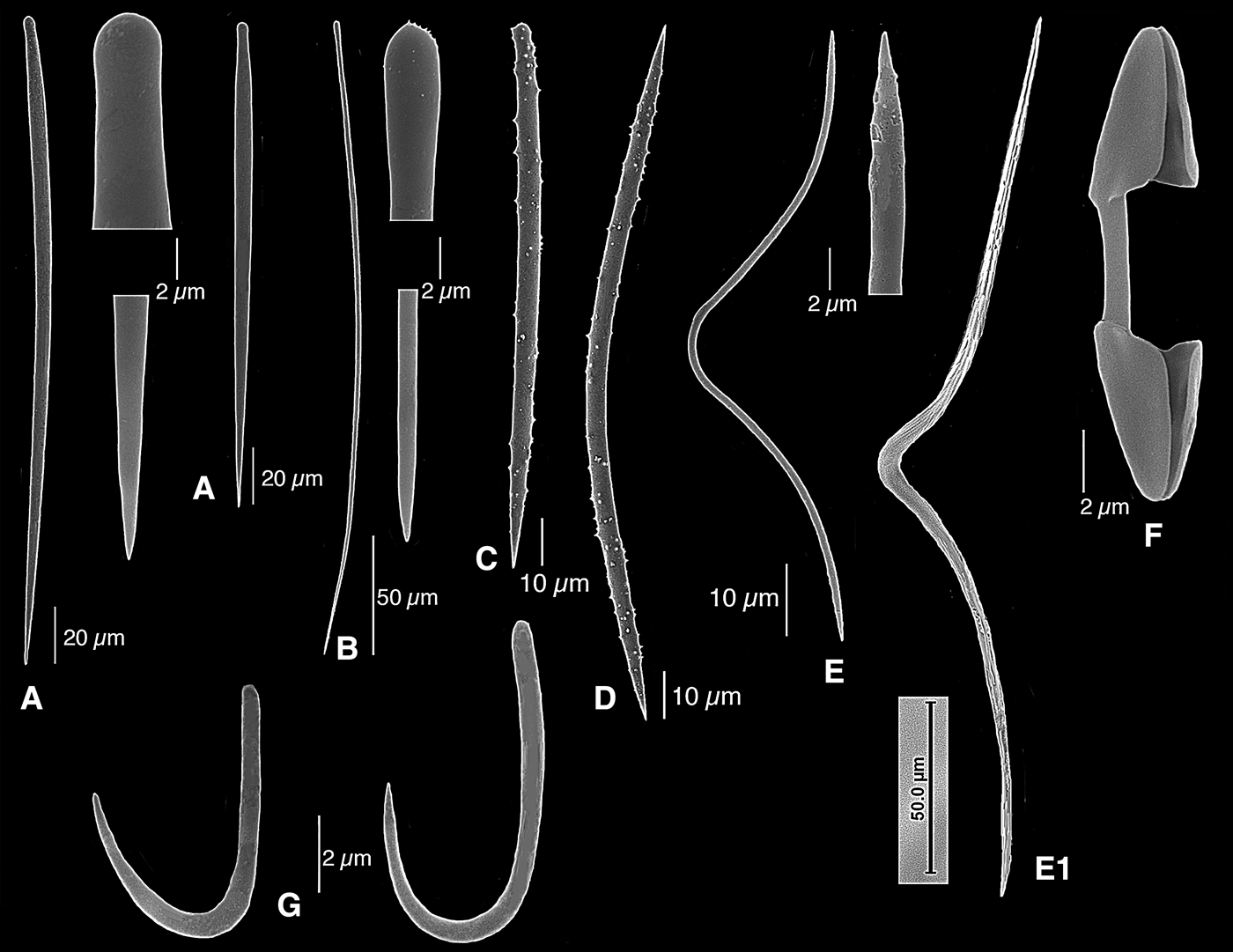
Antho (Jia) lithophoenix (De Laubenfels, Reference De Laubenfels1930), holotype USNM 21460 from Monterey Peninsula, California, SEM images of the spicules: (A) subectosomal style; (B) subtylostyle and details of head and point; (C) quasitylote variation of subtylostyle and details of head and point; (D) acanthostyles; (E) toxas; (F) palmate isochelae; (G) crocae.
Plocamia lithophoenix De Laubenfels, Reference De Laubenfels1927: 263, figures 7 & 8.
Isociona lithophoenix De Laubenfels, Reference De Laubenfels1930: 28; De Laubenfels, Reference De Laubenfels1932: 99, figure 59; Dickinson, Reference Dickinson1945: 23, pl. 35 figures 69–70, pl. 36 figures 71–72, as lithophenix (sic); Lee et al., Reference Lee, Elvin and Reiswig2007: 159.
?Antho lithophoenix sensu Sim & Bakus, Reference Sim and Bakus1986: 11 (spicule size data are different).
Non: Antho (Plocamia) lithophoenix; Desqueyroux-Faúndez & Van Soest, Reference Desqueyroux-Faúndez and Van Soest1997: 438, figures 155–164 (different species, Antho (Jia) galapagosensis sp. nov., see below).
MATERIAL EXAMINED
Holotype USNM 21460, United States, California, Pacific Grove, intertidal, coll. de Laubenfels, July 1925.
DESCRIPTION
Encrusting, lumpy, about 80 cm2 in coverage, 10 mm thick (only a 1.5 cm2 fragment remains). Consistency is firm but brittle, no openings visible on the remaining fragment. Colour in alcohol is pale ochre, was brilliant vermillion in life (per original author).
Skeleton. The ectosome is structured similar to the choanosome, but there is an abundance of larger, sparsely spined styles accompanied by slim subtylostyles. The choanosome is a multispicular meshwork of acanthostyles, with two, rarely three, spicules on each side, and in places there are multispicular tracts leading toward the surface. Subtylostyles too occur alongside the meshes and there are clusters of isochelae throughout the tissue, along with toxas and fairly rare crocae.
Spicules. Styles, subtylostyles, acanthostyles, toxas, palmate isochelae, crocae.
Subectosomal styles slightly curved (Figure 4A), with microspined tops and a few single spines along the shaft, 165–209.0 –260 × 11–13.6–16 µm.
Subtylostyles (Figure 4B), slim and straight, with microspined heads and sharp points, 210–277.7–330 × 5–5.5–7 µm; a smaller, rarer variety – resembling quasitylotes reported from some Clathria species (cf. Hooper, Reference Hooper1996) – has similar microspined tyles but also a few microspines ornamenting the points, comparable to similar spicules seen in the genotype, but more common, approaching 40% of all subtylostyles, 145–167.6–200 × 4–4.9–6 µm.
Robust, heavily spined acanthostyles (Figure 4D), generally straight, with spines in dense clusters near the rounded top, giving the appearance of acanthotylostyles; a few, rare modifications approach the shape of acanthostrongyles, 110–152.5 –334 × 15–16.3–19 µm.
Toxas (Figure 4E), deeply curved, of great size range but not separable into size classes, 30–113.9–200 µm length; they vary 1–2 µm in thickness.
Palmate isochelae (Figure 4F), 21–23.4–25 µm.
Crocae (Figure 4G) are rare but have the typical J-shape; they measure 7–10 µm, with 1 µm thickness near the rounded end.
ECOLOGY
Intertidal, encrusting rocks.
DISTRIBUTION
Only known from the type locality, the intertidal zone of Pacific Grove, Monterey Peninsula, California.
REMARKS
De Laubenfels’ figures (1927: figure 8, and 1932: figure 59) show some acanthostrongylote, even -tylote, modifications of the acanthostyles. The former are extremely rare, the latter we never encountered in the original preparations of this author, nor in a subsequent one made by us. A few of the thicker acanthostyles, however, have heavy spination near the point and may give the impression of strongylote shape, but never as pronounced as in the new species, Antho (Jia) galapagosensis sp. nov. described below, which was previously mistaken for it.
Antho (Jia) galapagosensis sp. nov.
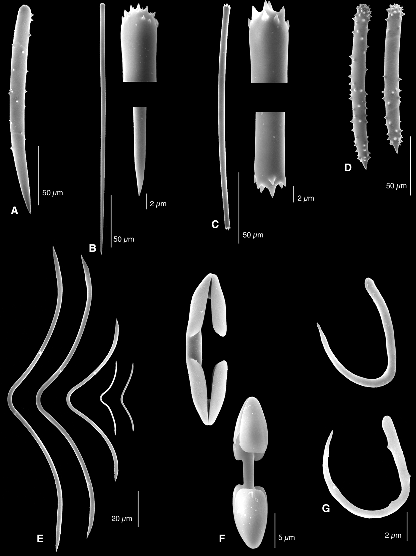
Antho (Jia) galapagosensis sp. nov., holotype USNM 37920 from Gardner Island, Galapagos, SEM images of the spicules: (A) subectosomal style and detail of head; (B) subtylostyle and details of head and point; (C) acanthostyles; (D) acantho(tylo)strongyles; (E) toxas with irregularly swollen apices; (F) palmate isochelae; (G) crocae (with relatively shallow curve).
Antho (Plocamia) lithophoenix; Desqueyroux-Faúndez & Van Soest, Reference Desqueyroux-Faúndez and Van Soest1997: 438, figures 155–164 (not: Plocamia lithophoenix De Laubenfels, Reference De Laubenfels1927, see above).
TYPE MATERIAL
Holotype USNM 37920, Ecuador, Galapagos Islands, Gardner Island (=Isla Española), 22 m, coll. W.D. Hope, stat. 23, February 1978 (ZMA Por. 11239, fragment of holotype).
Paratype ZMA Por. 11240, Ecuador, Galapagos Islands, James Island (= Isla Santiago), 0.2°S 90.8667°W, 34 m, rock dredge, coll. SEPBOP ‘Anton Bruun’ stat. 18B-794E, 24 September 1966.
DESCRIPTION
(Partly from Desqueyroux-Faúndez & Van Soest, Reference Desqueyroux-Faúndez and Van Soest1997.) Thinly or massively encrusting, fragmented, size (l × b × h) up to 2.8 × 1.6 × 0.7 cm. Surface punctate or minutely conulose, no apparent oscules. Consistency firm, crumbly. Colour in alcohol pale reddish to yellow-beige.
Skeleton. Single thick styles surrounded by bouquets of subtylostyles form the ectosomal skeleton. Choanosomal skeleton an isotropic tight-meshed reticulation (meshes about 100 µm, sides two spicules thick) of acanthostrongyles, echinated at the nodes by acanthostyles.
Spicules. Styles, subtylostyles, acanthostyles, acanthostrongyles, toxas, palmate isochelae, crocae.
Subectosomal styles (Figure 5A), relatively short and fat, heads with prominent spines, shaft smooth, usually curved, sharply pointed, 195–292.4–345 × 13–15.1–18 µm.
Subtylostyles (Figure 5B), with microspined or smooth heads, straight, variable in length, 171–263.8–358 × 1.5–2.4–3 µm.
Acanthostyles (Figure 5C), usually slightly curved, spined most heavily on the head, but further spines distributed evenly over the shaft, 138–160.2–180 × 7–9.8–12 µm.
Acanthostrongyles (Figure 5D), spined all over, but more densely spined on both endings, straight, 116–124.2–149 × 8–10.8–12 µm.
Toxas (Figure 5E), deeply curved, with rugose endings and swellings a short distance from the apices, variable in length, 22–107.2–246 µm.
Palmate isochelae (Figure 5F), relatively narrow, 19–20.1–22 µm.
Crocae (Figure 5G), relatively thin and with shallow curve, 10–10.8–12 µm.
ECOLOGY
Occurring on hard substratum at intermediate depths, 22–34 m.
DISTRIBUTION
Known only from the Galapagos Islands, Gardner & James Islands.
ETYMOLOGY
The name reflects the type locality.
REMARKS
Desqueyroux-Faúndez & Van Soest (Reference Desqueyroux-Faúndez and Van Soest1997) assigned this material to Antho lithophoenix, but that species does not have proper acanthostrongyles as a separate spicule category next to the acanthostyles. Also the styles of A. lithophoenix have some spines along their shafts, and a proportion of the subtylostyles are quasitylotes, which has not been observed in the Galapagos material. These differences are here interpreted as evidence of specific distinctness.
Antho (Jia) lithisticola sp. nov.
Antho (Jia) lithisticola sp. nov., holotype ZMA Por. 07716b from Sumba, Indonesia: (A) habit (fragments scraped off from lithistid shown in B, scale bar=1 cm); (B) lithistid Scleritoderma nodosum on which the holotype was growing (scale bar=1 cm); (C) light microscopic view of surface skeleton (scale bar=200 µm); (D) detail of choanosomal skeleton (scale bar=100 µm).
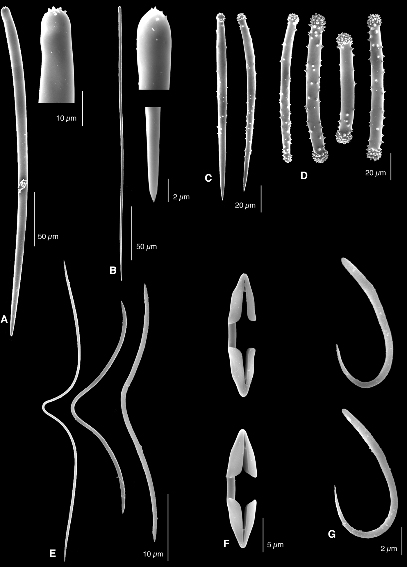
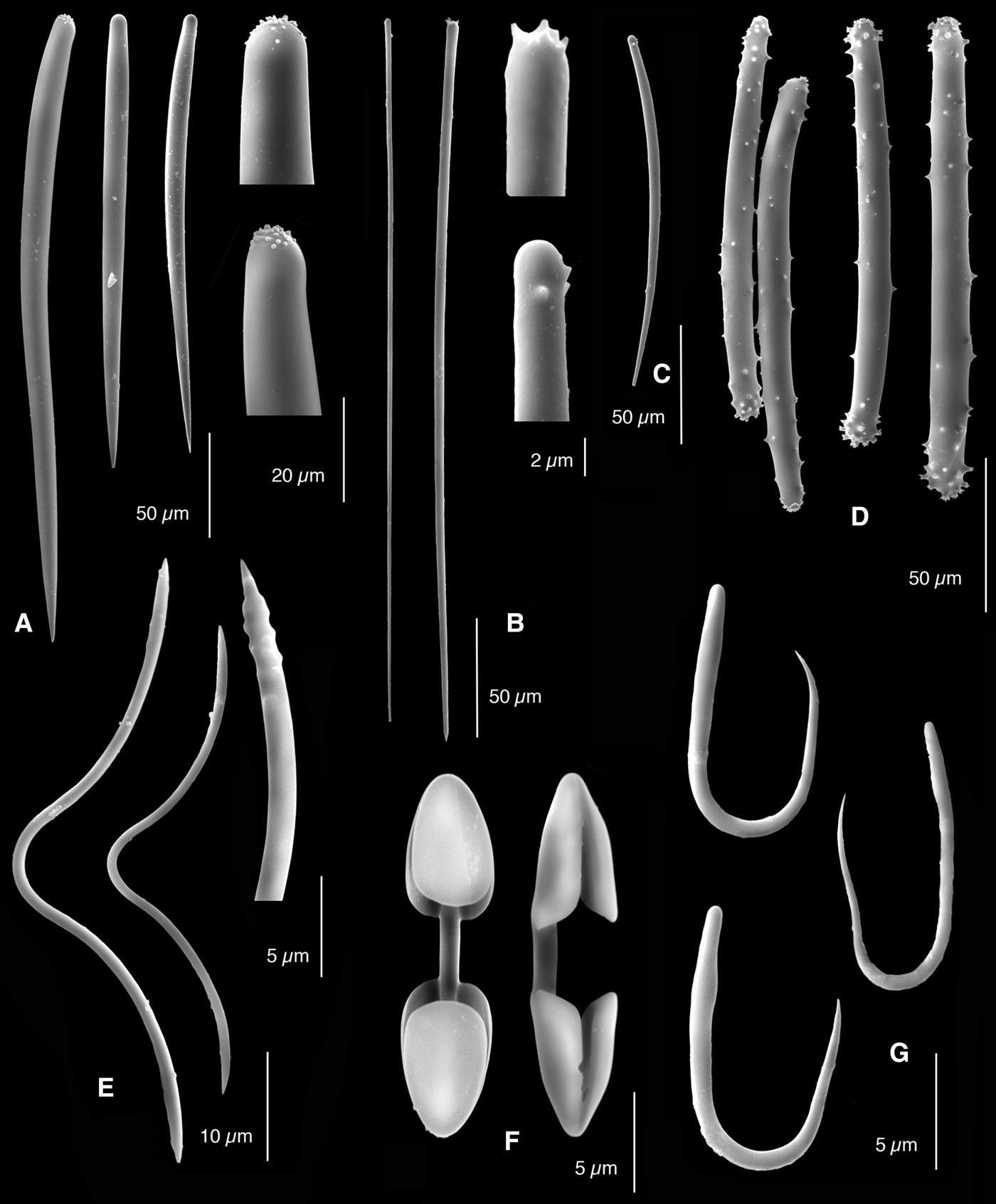
Antho (Jia) lithisticola sp. nov., holotype ZMA Por. 07716b, SEM images of spicules: (A) smooth subectosomal style and details of head and point; (B) shorter subectosomal/choanosomal style with some spination on head and shaft and detail of head; (C) subtylostyle and detail of head and point; (D) acanthostyle; (E) toxas and detail of ending of toxa; (F) palmate isochela; (G) crocae.
TYPE MATERIAL
Holotype ZMA Por. 07716b, Indonesia, Nusa Tenggara, Sumba, dredged, 9.0333° S 120.725°E, 50 m, sandy bottom, encrusting a large lithistid, coll. R.W.M. van Soest, Indonesian Dutch Snellius II Expedition, RV ‘Tyro’, stat. 061/V/31, 15 September 1984.
DESCRIPTION
Thin encrustation (Figure 6A) on a large specimen of Scleritoderma nodosa Sollas, 1888 (Figure 6B). Colour noted as dark red, but since several other encrusting sponges were present on the same lithistid sponge this is uncertain. Several patches were scraped off, together having a lateral expansion of several cm2, thickness less than 1 mm. Oscules (Figure 6C) small and indistinct, one was found to have a diameter of 200 µm. Surface microhispid. Consistency soft.
Skeleton. Ectosomal region shows long smooth styles, rooted in the choanosomal skeleton, protrude from the surface, surrounded by subtylostyles partly arranged tangentially, partly confused. The choanosomal skeleton (Figure 6D) shows a tight-meshed isotropical reticulation of acanthostyles, usually two spicules at the side; mesh sizes 75–150 µm. In the peripheral parts this basal reticulation is penetrated by shorter smooth styles that may have a spined head and occasional spines along the shaft. Microscleres are abundant in all parts of the skeleton.
Spicules. Styles, subtylostyles, acanthostyles, toxas, palmate isochelae and crocae.
Subectosomal styles, predominantly smooth (Figure 7A), but 20% of them bear spines on the head and more rarely a few spines along the shaft (Figure 7B), usually subterminally constricted (having a ‘neck’), 189–331.1–438 × 7–8.7–10 µm.
Subtylostyles (Figure 7C) with microspined heads, occasionally smooth, straight, in a large size range, 195–269.9–330 × 1.5–2.5–4 µm.
Acanthostyles (Figure 7D), spined all-over, but with dense spination on head and point, 105–119.7–153 × 5–9.3–12 µm.
Toxas (Figure 7E), deeply curved, ends rugose or microspined, in a large size range, 45–88.6–132 µm.
Palmate isochelae (Figure 7F), relatively narrow, 14–17.3–20 µm.
Crocae (Figure 7G), variable in size and outline, relatively thick, 7–9.2–11 × 0.5–1.5 µm.
ECOLOGY
Deeper water, approximately at 50 m depth, epibiontic on sandy bottom below the reefs.
DISTRIBUTION
Known only from the type locality, NE coast of Sumba, Indonesia.
ETYMOLOGY
The name refers to the large lithistid on which the new species was found to grow.
REMARKS
Antho (J.) lithophoenix shows similarities with A. (J.) lithisticola sp. nov. as it shares the spines along the shaft in the styles, and it also lacks true acanthostrongyles. The acanthostyles in that species are much more robust (approximately twice as thick), and there are quasitylotes lacking from the present new species. The new species resembles A. (J.) galapagosensis sp. nov. in its tight-meshed isotropical choanosomal reticulation, but differs sharply from that species in the lack of acanthostrongyles. Antho (J.) brattegardi likewise has acanthostrongyles, and also differs in having much longer subectosomal styles. Antho (Jia) jia has smooth styles in the choanosomal reticulation. Differences with the new species from the Gulf of Aqaba, Antho (Jia) wunschorum sp. nov. are discussed below.
Antho (Jia) wunschorum sp. nov.
Antho (Jia) wunschorum sp. nov., holotype ZMA Por. 13651 from Gulf of Aqaba, Red Sea: (A) habit in situ; (B) preserved holotype (scale bar=1 cm); (C) light microscopy cross section of skeleton (scale bar=200 µm); (D) light microscopy view of ectosomal subtylostyles and microscleres including toxas, palmate isochelae and crocae (scale bar=50 µm).
Antho (Jia) wunschorum sp. nov., holotype ZMA Por. 13651, SEM images of spicules: (A) subectosomal style with details of head and point, and shorter subectosomal/choanosomal style; (B) subtylostyle and details of head and point; (C) acanthostyle; (D) rare acanthoxea; (E) normal toxa, and detail of ending; (e1) rare giant ‘oxeote’ tox (light microscopy image); (F) palmate isochela; (G) crocae.
TYPE MATERIAL
Holotype ZMA Por. 13651, Israel, Red Sea, Gulf of Aqaba, Canyon, cave, 10 m, approximately 29.5°N 34.9°E, coll. Mark and Iris Wunsch, # AQ34, 2 July 1998.
DESCRIPTION
Thin encrustation on limestone substratum (Figure 8A), less than 2 mm thick. Colour red alive, and pale yellow in preservation. Preserved fragment (Figure 8B) measures 3 × 2 × 0.3 cm, but size in situ about three times that of the fragment. Surface punctate with venal pattern: scattered oscules of several mm in diameter are situated at the confluence of slightly swollen canals, set off against the red surface by greyish white lining. Consistency soft.
Skeleton (Figure 8C). The ectosomal skeleton is barely developed, but recognizable by tangentially arranged subtylostyles. Subectosomal and choanosomal skeleton have distinctly anisotropic aspect, with single subectosomal smooth styles continued as spicule lines, partially made up of shorter smooth styles and acanthostyles, into the basal skeleton. These primary lines are connected by single or occasionally two acanthostyles (as well as by rare acanthoxeas). Mesh sizes approximately 200 × 150 µm. Microscleres are scattered throughout the skeleton (Figure 8D).
Spicules. Styles, subtylostyles, acanthostyles, acanthoxeas, palmate isochelae, toxas (including rare ‘oxeote’ toxas), and crocae.
Subectosomal styles (Figure 9A), smooth, relatively thin, constricted subterminally, in a large size range, 147–185.3–225 × 5–5.9–7 µm.
Subtylostyles (Figure 9B), with smooth or faintly microspined heads, with thin, straight or occasionally curved shafts, 192–246.0–302 × 1.5–2.1–2.5 µm.
Acanthostyles (Figure 9C), fusiform, relatively thin, spination reduced, with limited length range, 114–122.1–129 × 4–5.1–7 µm.
Acanthoxeas (Figure 9D), curved, possibly derived from acanthostyles, rare, only three were found: 146–158 × 4–4.5 µm.
Toxas, in two categories: deeply curved (Figure 9E) with smooth or slightly roughened apices, in a wide size range, 42–103.7–150 µm, and rare giant ‘oxeote toxas’ (Figure 9E1) with small shallow curve, thick shaft and straight ends, only two were found: 290–302 × 5–6 µm.
Palmate isochelae (Figure 9F), rather narrow and relatively small, 12–15.1–18 µm.
Crocae (Figure 9G), relatively small and thin, 7–8.7–11 µm.
ECOLOGY
In marine cave at 10 m depth.
DISTRIBUTION
Known only from the type locality in the northern Gulf of Aqaba.
ETYMOLOGY
Named after Mark and Iris Wunsch, formerly of the University of Bremen, Germany, currently of Greencoast Media, Canada, for providing a rich collection of sponge fragments from the Gulf of Aqaba, Red Sea.
REMARKS
The characteristic features of this new species from the Gulf of Aqaba are the low spicular density and relatively small and thin spiculation, making it strikingly different from all above described species. The new species shares the thinly encrusting habit and lack of acanthostrongyles with A. (J.) lithisticola sp. nov., but is otherwise rather strongly different from that species. Its skeleton is much less tightly meshed and is in fact not renieroid, but anisotropic. In this aspect it resembles branching Antho species, such as the recently redescribed North Atlantic Antho (Antho) paradoxa (Babiç, Reference Babiç1922), see Van Soest et al. (Reference Van Soest, Beglinger and De Voogd2013), and the new species from Korea, Antho (Jia) ramosa sp. nov. described below. Like those species A. (J.) wunschorum sp. nov. has the skeleton loosely constructed with subectosomal styles penetrating into the choanosomal skeleton. This causes the skeleton to look anisotropic, with primary ascending spicule tracts different from the connecting tracts, unlike the renieroid skeletons found in most Antho species, including the type species Antho (Antho) involvens (Schmidt, Reference Schmidt1862). Further differences with A. (J.) lithisticola sp. nov. are the fusiform shape and the lesser thickness of the acanthostyles. The subectosomal styles show the absence of any spination, in contrast to those of A. (J.) lithisticola sp. nov., where about 20% is spined. Moreover, the styles of A. (J.) wunschorum sp. nov. are distinctly shorter than in the Indonesian species. We detected in the present species several very large and robust ‘oxeote’ toxas with a shallow curve and smooth apices, a type of toxa known from Antho (Antho) paradoxa and Celtic Seas’ Antho (Antho) granditoxa Picton & Goodwin, Reference Picton and Goodwin2007. Curiously, also several acanthoxea-like modifications of the acanthostyles were found among the spicules in this species. Such acanthoxeas are known from Mediterranean Antho (Antho) oxeifera (Ferrer Hernández, Reference Ferrer Hernández1921) (see redescription by Uriz & Maldonado, Reference Uriz and Maldonado1993).
Antho (Jia) ramosa sp. nov.
Figures 10 & 11
Antho (Jia) ramosa sp. nov. holotype HUNHM Por. 115 from South Korea: (A) habit (scale bar=1 cm), (B) SEM cross–section of peripheral skeleton (scale bar=500 µm).
Antho (Jia) ramosa sp. nov., SEM images of the spicules taken from the holotype (H) and paratype (P), (A) various subectosomal styles and details of heads (all from P), (B) ectosomal subtylostyles and details of heads (all from P), (C) acanthostyle (P) (please note scale differs from d), (D) various acanthostrongyles (all from P), (E) toxas, left (P), right (H), and detail of rugose ending H), (F) palmate isochelae from front (H) and side (P), (G) various crocae, upper (P), lower (H).
Antho brathesardi (sic); Sim & Kim, Reference Sim and Kim1994: 22, pl. 9, figures 1–4.
TYPE MATERIAL
Holotype HUNHM Por. 115, Korea, Jejudo island, Segwip’o, fish net, 7 September 1985.
Paratype NIBRI V0000304650, Korea, Jeollanam-do, Shinan-gun, Heuksan-myeon, Gageodo Island, 15 m, SCUBA, 20 July 2007; a fragment of the Korean paratype, including a microscopic preparation and SEM stubs are kept in the Smithsonian Institution under reg. nr. USNM 1215280.
DESCRIPTION
(Partially from Sim & Kim, Reference Sim and Kim1994.) Erect arborescent sponge (Figure 10A). Overall size of holotype 21 × 5 cm, individual branches 4 mm in diameter. Surface velvety. Consistency elastic. Colour in alcohol yellowish brown.
Skeleton (Figure 10B). Ectosomal skeleton consists of the protruding ends of subectosomal styles, surrounded by partially erect, partially tangentially arranged subtylostyles. Choanosomal skeleton a rather confused renieroid reticulation of acanthostrongyles echinated at the nodes by acanthostyles.
Spicules (Figure 11). Styles, subtylostyles, acanthostyles, acanthostrongyles, palmate isochelae, toxas and crocae.
Subectosomal styles (Figure 11A), faintly constricted below the head, shaft smooth, but usually with a few short spines on the head. Size in holotype: 350–600 × 7–16 µm, in paratype: 220–620 × 10–25 µm.
Subtylostyles (Figure 11B), with microspined heads. Size in holotype: 250–500 × 3–5 µm, in paratype 140–530 × 3–5 µm.
Acanthostyles (Figure 11C), thin, curved, very lightly spined; size in the holotype: 130–190 × 7–10, only a few were found in the paratype: 190–230 × 8–11 µm.
Acanthostrongyles (Figure 11D), shaft lightly spined, heads usually heavily spined, curved. Size in holotype: 140–150 × 7–12 µm, in paratype: 140–150 × 15–20 µm.
Palmate isochelae (Figure 11E). Size in holotype: 15–20 µm, in paratype: 15–20 µm.
Toxas (Figure 11F), deeply curved, with rugose ends, in a large size range of which only the smaller are depicted in Figure 11. Sizes of small toxas in holotype: 35–85 µm, in paratype: 40–80 µm, sizes of large toxas, in holotype: 140–220 µm, in paratype: 123–165 µm.
Crocae (Figure 11G), with relatively long thin ends. Sizes in holotype: 11–20 µm, in paratype: 11–13 µm.
ECOLOGY
Found in diving depth down to 15 m.
DISTRIBUTION
South of the Korean mainland, in the Yellow Sea. Known to occur at the S coast of Jeju Island (Jeju-do) and at Gageodo Island.
ETYMOLOGY
The name reflects the branching shape.
REMARKS
Some of the spicule sizes of the two specimens reported here show a considerable range, which probably reflects some geographic variability, as was also found between specimens of A. (J.) brattegardi from Norway and Northern Ireland (cf. above). The species is unique among Antho (Jia) species by being branching erect, whereas all other species are encrusting. Although the spiculation of the new species is indeed overall closest to Antho (Jia) brattegardi the Korean species differs by the strongly deviating habit, considerably shorter subectosomal styles (up to 765 µm in brattegardi) with spined heads (predominantly smooth in A. (J.) brattegardi), and considerably longer subtylostyles (only up to about 400 µm in A. (J.) brattegardi).
DISCUSSION
The crocae of all studied specimens are very similar in shape and size, with very little morphological or meristic variation. In attempts to connect these enigmatic forms to the poecilosclerid spicule complement, the crocae were likened to the microscleres of Forcepia (Leptolabis) luciensis (Topsent, Reference Topsent1889 as Dendoryx) by De Laubenfels (Reference De Laubenfels1932: 99). These are indeed peculiar microscleres but are in fact curled forceps possessing spines (not sigmas as De Laubenfels maintained). They do not resemble crocae, which is clear when one compares the crocae in the present study with the luciensis microscleres depicted in Van Soest, Reference Van Soest, Hooper and Van Soest2002: 537, Figure 5C–E. The association with sigmas is persistent: Hooper (Reference Hooper1996: 53) and Desqueyroux-Faúndez & Van Soest (Reference Desqueyroux-Faúndez and Van Soest1997: 440) use the term sigmoid, whereas Van Soest & Stone (Reference Van Soest and Stone1986: 43) suggested resemblance to small diancistras of Hamacantha (Vomerula) hyaloderma (De Laubenfels, Reference De Laubenfels1932 as Zygherpe). Neither suggestion comes even close to the croca shape. We conclude that its origin is currently unclear and it must be considered a unique spicule type. This in turn is a strong indication that all species possessing these spicules share a common ancestor and form a monophyletic group of Antho.
The additional characters of the species of Antho (Jia) cut right across the characters to distinguish the two main groups of Antho, viz. Antho (Antho) (only acanthostyles, no acanthostrongyles) and Antho (Acarnia) (acanthostrongyles, with or without acanthostyles). Such general spicule characters are likely not indicative of monophyletic groups, as Antho (Jia) jia, Antho (Jia) lithophoenix, Antho (Jia) lithisticola sp. nov. and Antho (Jia) wunschorum sp. nov. are Antho (Antho)-like and Antho (Jia) brattegardi, Antho (Jia) galapagosensis sp. nov., and Antho (Jia) ramosa sp. nov. are clearly Antho (Acarnia)-like. The situation in Antho resembles that in Mycale by showing a number of subgenera with clear apomorphies and a large complement of species assigned to poorly defined ‘general’ subgenera overlapping in their characters with the well-defined groups.
The distribution of the subgenus Jia is now circumglobal in all three oceans (Figure 12), but so far species from the warmer Atlantic regions are lacking. The depth range of 10–700 m and horizontal as well as vertical temperature ranges (cold temperate to tropical) are considerable. These observations appear to differ somewhat from more general patterns exhibited by widespread sponge (sub-)genera (cf. Van Soest et al., 2012). Possibly, crocae were overlooked in known species of Antho, because they are small and often rare, demonstrated by the discovery of crocae in Antho (J.) lithophoenix. Further undescribed species new to science are also expected, as can be concluded by the present new species found in the Red Sea and Indonesia. Both phenomena indicate that more members of Antho (Jia) await to be discovered.
Distribution of Antho (Jia) species over the marine ecoregions of the world (Spalding et al., Reference Spalding, Fox, Allen, Davidson, Ferdaña, Finlayson, Halpern, Jorge, Lombana, Lourie, Martin, McManus, Molnar, Recchia and Robertson2007).
KEY TO THE SPECIES OF ANTHO (JIA)
The key is illustrated and supported by Figures 1–11 and Table 1.
1. – Acanthostrongyles commonly present in the basal reticulation, next to a separate category of acanthostyles .................. 2
– Predominantly acanthostyles in the basal reticulation; acanthostrongyles absent or rare ..................... 4
2. – Subectosomal styles <350 µm in length .................... A. (J.) galapagosensis sp. nov.
– Subectosomal styles exceeding 350 µm, may be as long as 600–700 µm .................... 3
3. – Growth form thinly encrusting ............. A. (J.) brattegardi
– Growth form branching erect .......... A. (J.) ramosa sp. nov.
4. – Choanosomal styles smooth or with only a few spines on the shaft; subectosomal and choanosomal styles barely differentiated .......... A. (Jia) jia
– Choanosomal styles spined all over, clearly differentiated from subectosomal smooth styles ......... 5
5. – Choanosomal skeleton tight-meshed, isotropic; acanthostyles robust, up to 153 × 12 µm ...... 6
– Choanosomal skeleton lax, anisotropic; acanthostyles fusiform, thin, only up to 130 × 7 µm ............. A. (J.) wunschorum sp. nov.
6. – Ectosomal subtylostyles show a large proportion of quasitylotes; acanthostyles up to 19 µm in thickness .......... A. (J.) lithophoenix
– Subtylostyles are all normal; acanthstyles only up to 12 µm in thickness ................ A. (J.) lithisticola sp. nov.
Spicule size data (μm) on all Antho (Jia) species discussed in the text
sm, smooth, sp. spined.
ACKNOWLEDGEMENTS
Elly Beglinger (Department of Collections, Naturalis Biodiversity Centre) assisted with the SEM preparation in Leiden. We thank Yolanda Villacampa and Freya Goetz (Department of Invertebrate Zoology, Smithsonian Institution) for producing SEM images and Molly Kelly Ryan (Smithsonian volunteer) for assistance in preparing the final figures. Mark and Iris Wunsch collected material from the Red Sea and provided an in situ photograph.