Over recent decades, marked improvement in the survival of patients with CHD occurred due to advancements in surgical technique and post-operative management. However, some groups of CHD children are more difficult to manage due to severe co-morbidities or complex cardiac lesions that often lead to palliative surgery. Vascular compression of the airway is one such co-morbidity, seen in 1–2% of children undergoing CHD surgery.Reference McLaren, Elliott and Roebuck1 The airway obstruction and the pathophysiologic effect of underlying CHD may be mutually exacerbating, explaining the higher morbidity and mortality compared to those without CHD.
Cardiovascular causes of tracheobronchial airway compression may occur before or after CHD surgery.Reference McLaren, Elliott and Roebuck1,Reference Davis, Tucker and Russo2 The clinical presentation of vascular airway compression can be variable and non-specific, making it a great challenge to diagnose.Reference Bonnard, Auber, Fourcade, Marchac, Emond and Révillon3 In developing or middle-income countries, a delay in CHD surgery due to long waiting times may further complicate the management. Many of these patients may require respiratory support due to respiratory distress, potentially increasing the risk of morbidity during the perioperative period. Therefore, the presence of vascular compression of the airway in children with CHD should be diagnosed and intervene early to improve the overall outcome.Reference Suh, Kim and Kwon4
CT has emerged as the preferred imaging investigation for diagnosing and characterising vascular causes of airway compression.Reference Etesami, Ashwath, Kanne, Gilkeson and Rajiah5 It is performed in symptomatic patients with a suspected vascular compression of the airway or to confirm a vascular ring or pulmonary artery sling and assist in the pre-operative planning. The rapid acquisition of CT scan without the critical need for general anaesthesia and readily available imaging technique in many tertiary hospitals are the advantages that CT scan has over MRI.
Currently, most studies mainly focused on the VR or PA sling, and there are limited data on tracheobronchial airway compression related to underlying CHD and cardiac interventions that were performed. Therefore, the purpose of this study is to evaluate the cardiovascular causes of tracheobronchial compression in children with CHD using a CT scan and to determine the clinical manifestations and associated morbidity and mortality among children with CHD.
Materials and methods
We retrospectively reviewed clinical records, including the CT scan images at National Heart Institute from January 2007 to December 2017. National Heart Institute is one of the leading referral centres for patients with CHD in Malaysia. Patients with a diagnosis of the vascular ring, pulmonary artery sling, or any CT scan evidence of cardiovascular causes of tracheobronchial airway compression were included in this study. The CT scan images were read and reported by an experienced consultant paediatric cardiologist specialises in imaging (HA).
The causes of tracheobronchial compression were divided into three groups:
-
Group I: Vascular ring/pulmonary artery sling
-
Group II: Abnormally enlarged or malposition of cardiovascular structure due to underlying CHD (post-CHD surgery patient who already noted to have vascular tracheobronchial compression before surgery was also included in this group)
-
Group III: Post-CHD surgery (vascular tracheobronchial compression that only developed after surgery/intervention).
The exclusion criteria were patients aged more than 18-year-old or with non-cardiovascular causes of the trachea or oesophageal compression from the CT scan images. Demographic and clinical data which include age at presentation, age at diagnosis, gender, weight, and height, presenting symptoms, diagnostic modalities, for airway compressed by the cardiovascular structures (trachea, right or left main bronchus), further classification according to the cardiovascular structure that cause compression in group II (compression from enlarged or malposition of the pulmonary artery, ascending or descending aorta, patent ductus arteriosus and left atrium), presence of airway abnormalities like congenital tracheal stenosis or tracheobronchomalacia and associated congenital heart anomalies and other co-morbidities, were collected. Details of interventions were recorded that included surgical or non-surgical treatment, age of surgical intervention, duration of hospital stay, mortality, and cause of death.
Statistical analysis
All statistical analyses were performed using Statistical Package for the Social Science (SPSS) Statistics 22.0 (SPSS Inc. Chicago, IL, USA). Descriptive statistics were used to analyse the clinical characteristic of the study cohorts. Continuous data with a Gaussian distribution were presented as mean (standard deviation), while data with non-Gaussian distribution were presented as median (interquartile range). Categorical data were presented as frequencies, percentages, and cumulative frequencies as appropriate.
Results
Demographic and clinical characteristics
Eighty-one cases were found to have CT scan evidence of vascular tracheobronchial compression between January 2007 and December 2017. During this period, the total number of children who underwent CT scans was 810. Hence, the prevalence of tracheobronchial compression was 10% among patients who underwent a CT scan. The main indications for CT scan evaluation were vascular ring/pulmonary artery sling or suspected vascular causes of airway compression. In seven (8.6%) of the patients, evidence of airway compression was incidentally found during a CT scan for the underlying cardiac lesion.
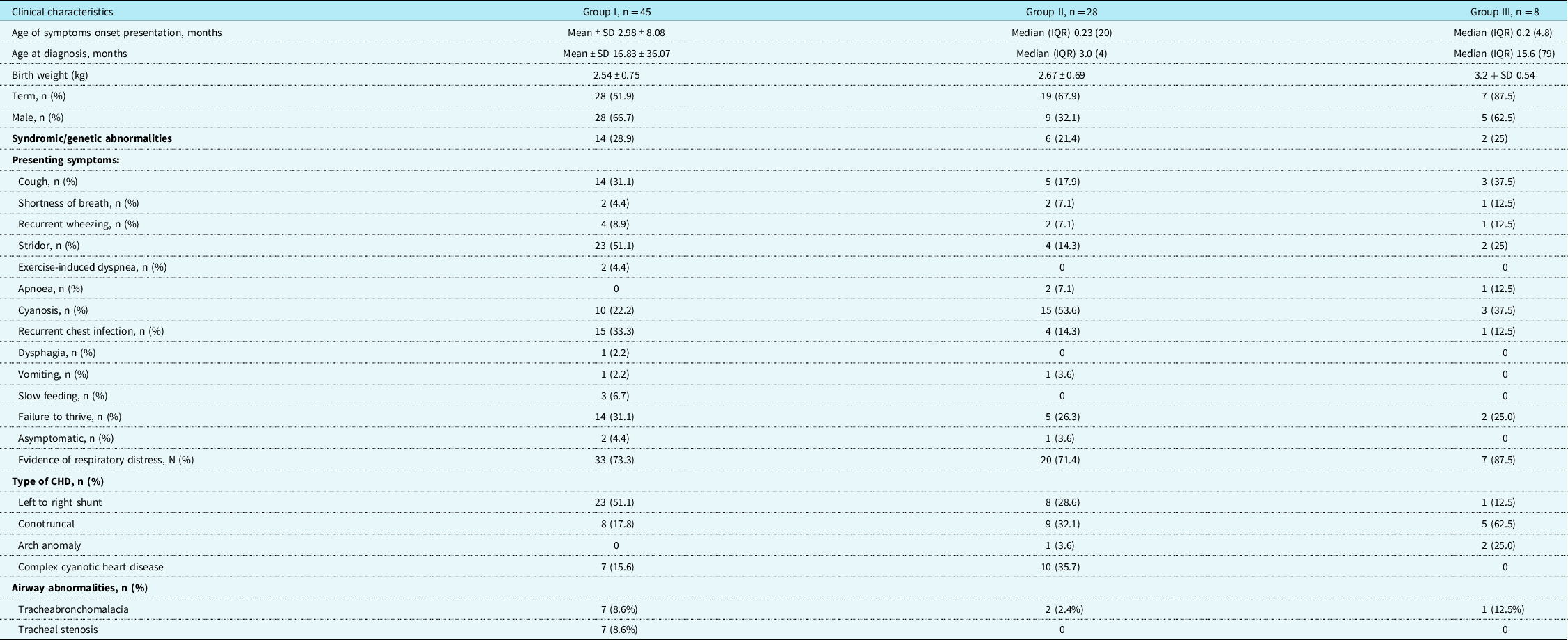
Table 1 demonstrates the demographic and clinical characteristics of the study population based on the three groups’ classification, as explained above.
Demographic and clinical characteristics of patients with vascular tracheobronchial compression of the airways

The presentation age was noted to be in early infancy in all the groups but much lower in groups II and III. Age of diagnosis in patients in groups I and III was delayed with a mean of 16.8 months and median of 15.6 months, respectively, compared to group II with a median age of diagnosis of 3 months.
About 84.5% of patients in group I had underlying CHD, most commonly left to right shunt lesions (51.1%), and followed by conotruncal lesions (17.8%) and complex cyanotic heart disease (15.6%). Complex cyanotic heart disease (35.7%) and conotruncal lesion (32.1%) were more common in group II. Whereas in group III, the conotruncal lesion (62.5%) and arch anomaly (25%) were more prominent. In group I, 14 (28.9%) cases were syndromic or had genetic abnormalities. The syndromes reported were four cases of Di George syndrome, two cases of Down syndrome, four cases of VACTERL association, and four cases of dysmorphic features with no specific genetic abnormalities. Di George syndrome was found in three cases in group II and one case in group III.
The most common clinical presentation in group I was stridor (51.1%), followed by recurrent chest infections (33.3%) and cough (31.1%). Only one patient had dysphagia. In group II, only four (14.3%) cases were noted to have stridor, but more presented with cyanosis in 15 (53.6%) cases due to underlying CHD. In group III, the prominent symptoms were cough (37.5%), shortness of breath (12.5%), wheezing (12.5%), and apnoea (12.5%). Evidence of respiratory distress and failure to thrive were almost equally reported in all three groups.
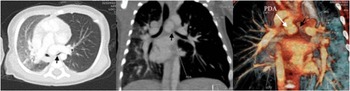
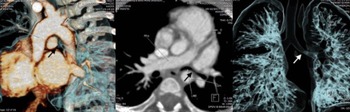
In children with CHD, vascular tracheobronchial compression was suspected in patients with persistent lung atelectasis or who had failed extubation. Three of the eight patients in group III were noted to have a persistent collapse of the left lung, and two cases were found to have hypoplasia of the left lung related to significant compression of the left main bronchus (data not shown). Figure 1 belonged to a patient with underlying double-inlet left ventricle and pulmonary atresia who needed further investigation because of failed extubation twice. Her CT scan showed partial compression of the left main bronchus by the large patent ductus arteriosus with hyperinflation of the left lung.
CT scan belonged to a 2-month-old infant with underlying double-inlet left ventricle and pulmonary atresia who failed extubation twice. Her CT scan showed partial compression of the left main bronchus (arrow) by the large PDA with hyperinflation of left lung and area of consolidation on the right lung.

Echocardiography detected only 17 of 45 (37.8%) vascular ring and pulmonary artery sling cases for the diagnostic modalities. Two patients had barium swallow performed; one of the patients, a case of right aortic arch/aberrant left subclavian artery with ligamentum arteriosum, was not able to tolerate a solid diet since the patient started weaning. The barium swallow revealed indentation at the posterior oesophagus at the level of T4/T5. His symptoms resolved after the division of the ligamentum arteriosum, and reimplantation of left subclavian artery to the left common carotid artery was performed. Bronchoscopy was performed in 25 (30.9%) of the study population. Tracheobronchomalacia was found in 10 (12.3%) of the cases, and 7 (8.6 %) patients had tracheal stenosis that was seen mainly in group I.
Cardiovascular causes of tracheobronchial airway compression
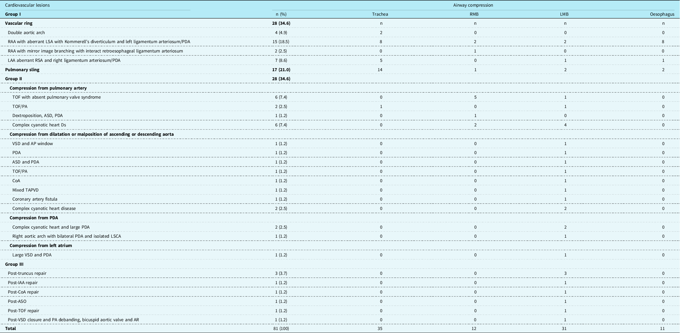
The cardiovascular causes of tracheobronchial airway compression are depicted in Table 2. Tracheobronchial compression was mostly seen in group I with 45 (55.5%) cases, followed by group II, 28 (34.6%) cases, and group III, 8 (9.9%) cases. Right aortic arch with aberrant left subclavian artery that arises from Kommerell’s diverticulum and left ligamentum arteriosum/patent ductus arteriosus was the most common type of vascular ring, seen in 15/28 (53.6%) of the patients. Eight of 15 patients from this group had radiological evidence of oesophageal compression, but only 1 patient had dysphagia symptoms. Both vascular ring and pulmonary artery sling caused compression mainly to the trachea.
Cardiovascular causes of tracheobronchial airways compression

AP = aorto pulmonary; AR = aortic regurgitation; ASD = atrial septal defect; ASO = arterial switch operation; CoA = coarctation of aorta; IAA = Interrupted aortic arch; LAA = left aortic arch; LMB = Left main bronchus; LSCA = left subclavian artery; PA = pulmonary artery; PDA = patent ductus arteriosus; RAA = Right aortic arch; RMB = Right main bronchus; TAPVD = total anomalous pulmonary venous drainage; VSD = ventricular septal defect
External tracheobronchial compression caused by dilated or malposition of cardiovascular structure in group II may occur due to the dilated pulmonary artery, dilated or malposition of the aorta, dilated atrium, or from the ductus arteriosus. The dilated pulmonary artery was seen in 15 (18.5%) of patients, tetralogy of Fallot with absent pulmonary valve syndrome found in 6 cases, and complex cyanotic heart disease in 6 patients, that is, heterotaxy syndrome, dextrocardia and congenitally corrected transposition of the great arteries, ventricular septal defect, and pulmonary stenosis.
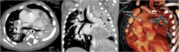
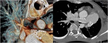
Compression of the airway caused by dilated or malposition of ascending or descending of the aorta, patent arterial duct, and left atrium was found in nine (11.1%), three (3.7%), and one (1.2%) of the patients, respectively. The left main bronchus was the most commonly affected airway structure in group II. Figure 2 demonstrated a case of ventricular septal defect and aortopulmonary window, causing enlargement of the pulmonary artery. The CT scan demonstrated compression of the left main bronchus by the descending aorta and left pulmonary artery causing a collapsed posterior segment of the left lung.
CT scan of 5-month-old girl demonstrates an aortopulmonary window causing massive enlargement of pulmonary artery and aorta. Compression of left main bronchus (arrow) occured by the descending aorta and left pulmonary artery causing collapse of posterior segment of the left lung.

There were eight cases in group III, three cases following truncus repair, one case each following interrupted aortic arch repair, coarctation of aorta repair, tetralogy of Fallot repair, ventricular septal defect closure, pulmonary artery debanding, and arterial switch operation. The left main bronchus was once again the most affected airway structure in this group of patients.
Figure 3 belongs to a 5-month-old girl who had interrupted arch repair during the neonatal period. CT scan showed significantly compressed the distal left main bronchus between the anteriorly displaced descending aorta and right pulmonary artery. Another patient following post-truncus repair developed left bronchus compression from the stented proximal right pulmonary artery. This patient developed significant bilateral proximal pulmonary artery branches stenosis that required stenting about 8 years after the truncus repair (Fig 4).
CT scan belonged to a 5-months-old girl who had interrupted arch repair during the neonatal period. CT scan shows severely compressed distal left main bronchus (arrow) between anteriorly displaced descending aorta and right pulmonary artery.

CT scan belonged to 12-year-old boy who developed significant bilateral proximal PA branches stenosis following truncus repair that required stenting. His CT shows compression of the left bronchus (arrow) from the stented proximal right pulmonary artery. The left lung was hypoplastic with compensatory hyperinflation of the right lung.

Surgical intervention and outcome of surgery
About 52/81 (64 %) of the study population had undergone surgery. During this study period, the total number of CHD surgeries was 12,320 cases. Therefore, the prevalence of children who had CT scan evidence of tracheobronchial compression among patients that underwent CHD surgery was 0.42%.
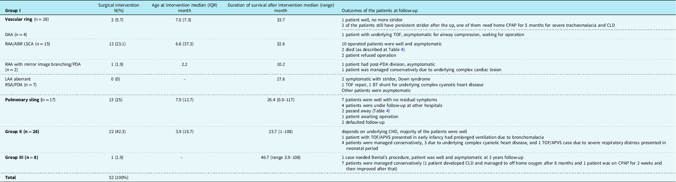
Twenty-nine of 45 cases (64.4%) of patients in group I underwent surgery, mainly involving patients with right aortic arch with aberrant left subclavian artery, pulmonary artery sling, and double aortic arch. The median age at intervention in patients with double aortic arch and right aortic arch with aberrant left subclavian artery was 7 and 6.6 months, respectively. Most patients with left aortic arch with aberrant right subclavian artery/patent ductus arteriosus were managed conservatively as the majority of the patients were asymptomatic. Thirteen of the 17 (76%) of the patients in the pulmonary artery sling cohorts underwent intervention at a median age of 7.9 months. Overall, the outcome of patients in group I in this study was good, but the presence of early airway obstructive symptoms due to severe tracheobronchial obstruction, airway abnormalities, and concomitant complex CHD was associated with prolonged respiratory support requirement and mortality.
Almost 80% (22/28) of patients in group II underwent surgical intervention for the underlying cardiac lesion at a median age of 3.9 months. The surgical outcome of patients in this group depended on underlying cardiac lesions, and most of them made an uneventful recovery with no residual symptoms. One of the patients with tetralogy of Fallot with absent pulmonary valve syndrome developed prolonged ventilation due to severe symptomatic airway obstruction despite undergoing surgical treatment. Four patients were managed conservatively, comprised of three patients with underlying complex cyanotic heart disease and one tetralogy of Fallot with absent pulmonary valve syndrome with severe obstructive airway symptoms developed early in the neonatal period.
The majority of patients in group III were managed conservatively. Three of them required prolonged ventilation. One of these patients posts interrupted aortic arch repair required home oxygen for 8 months due to the development of chronic lung disease and severe bronchomalacia. Only one patient post-ventricular septal defect closure and pulmonary artery debanding needed further surgical intervention due to the presence of bicuspid aortic valve with severe aortic regurgitation and dilated ascending aorta, causing compression to left main bronchus. This patient who had recurrent wheezing underwent Bental’s procedure at the age of 12 years. The operation was uneventful, and the patient remained well and asymptomatic during the follow-up 2 years later.
The summary of the surgical interventions and related outcomes is summarised in Table 3.
Surgical intervention and outcomes among children with vascular tracheobronchial compression

APVS = absent pulmonary valve syndrome; BT = Blalock-Taussig; CLD = Chronic lung disease; CPAP = Continuous positive airway pressure; DAA = Double aortic arch; LAA = Left aortic arch; PDA = patent ductus arteriosus; RAA/ABR LSCA = Right aortic arch with aberrant left subclavian artery; RSCA = right subclavian artery; TOF = Tetralogy of Fallot
Mortality
The cases of mortality and the causes of early deaths are summarised in Table 4. Both pulmonary artery sling cases (patient 1 and 2) presented early with airway obstruction and required complex cardiac surgery. Whereas, in patients with right aortic arch with aberrant left subclavian artery and patent ductus arteriosus, one of the patients had underlying complex CHD (congenitally corrected transposition of the great arteries, ventricular septal defect, and mild pulmonary stenosis) and died at 2 weeks after the patent ductus arteriosus division due to pulmonary hypertension crisis (patient 4). Patient 3 initially underwent patent ductus arteriosus division but later needed additional excision of Kommeral’s diverticulum and reimplantation of left subclavian artery to left common carotid artery, about 9 days after the first operation. Unfortunately, this patient died of multiorgan failure following the second operation. Patient 5 had an underlying complex cardiac lesion (congenitally corrected transposition of the great arteries, ventricular septal defect, and coarctation of aorta) with severe compression of the left main bronchus by a dilated main pulmonary artery, causing collapsed left lung. This patient, unfortunately, died of sepsis while waiting for the operation to be performed.
Mortality cases

AP = aorto pulmonary; ASD = atrial septal defect; CCTGA = Congenitally corrected transposition of the great arteries; COA = coarctation of aorta; IAA = Interrupted aortic arch; LMB = Left main bronchus; LPA = left pulmonary artery; LSCA = left subclavian artery; PA = pulmonary artery; PDA = patent ductus arteriosus; PMVSD = perimembranous ventricular septal defect; PS = pulmonary stenosis; RAA = Right aortic arch; VSD = ventricular septal defect
Discussion
In our study, about 0.42% of children who underwent CHD surgery had problems with vascular tracheobronchial compression, compared with another study that reported 2.1%.Reference An, Choi and Kwon6 This value may have been underestimated as the prevalence of vascular tracheobronchial compression among children who underwent CT scan was about 10%. That can be explained by the notion that some patients were managed conservatively due to only having very mild symptoms. However, in a country with limited resources, it is not uncommon for these affected patients to be managed conservatively due to expected poor surgical outcomes stemming from undergoing a very high-risk surgery and, at the same time, having complex cardiac lesions.
In our study, vascular ring/pulmonary artery sling was the leading cause of vascular compression of the airway, seen in 45 (55.5%) cases. Right aortic arch with aberrant left subclavian artery and patent ductus arteriosus or ligamentum arteriosum was the most common form of vascular ring that caused external airway compression, seen in 15/28 (51.7%) of patients, which is in contrast with other studies that showed that double aortic arch was the commonest surgically corrected vascular ring,Reference Woods, Sharp and Holcomb7–Reference Park9 with only 4/28 (14.2%) cases of double aortic arch seen in our vascular ring cohort. Left to right shunts, conotruncal cardiac lesions, and complex cyanotic heart disease were the main cardiac lesions that caused abnormal enlargement or malposition of the great vessels or cardiac chambers. These cardiac lesions may compromise the infant’s airway and respiratory function by direct external compression of the airway or increased pulmonary artery pressure and interstitial oedema.Reference Davis, Tucker and Russo2
Consistent with other studies, the left main bronchus was the most commonly affected airway structure in groups II and III. The close proximity of the left main bronchus with the pulmonary artery, ascending aorta and left atrium anteriorly, and descending aorta posteriorly may explain its vulnerability to being compressed if there is an enlargement or malposition of these structures.Reference An, Choi and Kwon6,Reference Kussman, Geva and McGowan10
In our series, almost 10% of the vascular tracheobronchial compressions occur following CHD surgery. We found that the occurrence was mainly following truncus repair, post-aortic arch repair, post-arterial switch operation, and tetralogy of Fallot repair, as previously described in other studies.Reference An, Choi and Kwon6,Reference Kussman, Geva and McGowan10 Cardiac surgical procedures involving conduits may result in direct compression of the airway. In comparison, surgical repair of an interrupted arch or severe coarctation of the aorta may distort the anatomic relationship surrounding the tracheobronchial tree if the end-to-end anastomosis is performed close to the left bronchus. A study reported that airway compression was newly detected following aortic arch repair in 33.3% of the patients who did not show any airway compression before surgery.Reference Jhang, Park, Seo, Goo and Gwak11
About 22 (27.2%) of cases in our study population had underlying syndromes or genetic abnormalities. About one-third of these patients had underlying Di George syndrome. This is not uncommon as Di George syndrome is commonly seen in a patient with a right aortic arch, conotruncal lesion, or arch anomaly.Reference Peyvandi, Lupo and Garbarini12 The presence of multiple organ involvements in patients with genetic abnormalities may further increase the risk of morbidity.
The delay in diagnosing vascular tracheobronchial compression as seen in group I patients is not an unusual problem.Reference Kussman, Geva and McGowan10,Reference Suh, Kim and Kwon13 Specific symptoms related to an airway obstruction were not common, such as stridor only occurred in half of the group I patients and less than a quarter of patients in groups II and III, respectively. In this study, only one patient (1.2%) had symptoms of dysphagia. The rarity of symptomatic vascular ring/pulmonary artery sling among children with CHD may explain that in many cases, when assessing the presenting symptoms, vascular compression is not always considered. Furthermore, other symptoms like cough, recurrent chest infection, or respiratory distress could be inaccurately attributed to the underlying CHD. Therefore, a high index of suspicion is essential to look for the possibility of vascular tracheobronchial compression if the underlying cardiac lesion is unable to explain the presenting symptoms for decisive diagnostic steps to be initiated promptly.Reference Kussman, Geva and McGowan10,Reference Yubbu, Devaraj, Sahadan and Latiff14
In children with CHD, the presence of vascular compression of the airway should be suspected if there are dominant or persistent respiratory symptoms. The other important clue is if there is difficulty in weaning off the respiratory support despite successful correction of underlying cardiac defects. Tracheobronchial compression should also be considered in persistent lung collapse cases caused by total airway obstruction or lung hyperinflation resulting from air trapping by the partial obstruction of the airway. In our study, more than half of the patients who had left main bronchus compression following CHD surgery demonstrated persistent collapsed left lung or left lung hypoplasia.
Prolonged extrinsic compression to the tracheobronchial tree may also result in tracheomalacia, bronchomalacia, or both. Studies had shown that cartilage destruction leading to malacic airway might be developed within weeks of compression.Reference McLaren, Elliott and Roebuck1,Reference Filler and de Fraga15 In our case, tracheobronchomalacia was noted in 12.3% of the study population, which was mainly seen in patients of group I. This result can be underestimated as only 30.8% of our patients underwent bronchoscopy assessment, which is the gold standard in diagnosing a malacic airway. All tracheal stenosis cases in our cohort were seen in patients who had underlying PA sling (n= 7,42%). Another study had found that long segment tracheal stenosis with complete tracheal ring has been reported in 50% of PA sling’s cases.Reference Yong, Zhu and Bell16
The presence of tracheobronchial compression, pulmonary over-circulation related to heart defects, and respiratory support dependency may increase the risk of recurrent pneumonia in this group of patients. Also, chronic aspiration pneumonia may occur due to gastrooesophageal reflux resulting from an imbalance between the abdominal and thoracic pressure as the child must generate high negative intrathoracic pressure to breathe due to the tracheobronchial compression.Reference Kussman, Geva and McGowan10 Increased mucin production following cardiopulmonary bypass may further complicate the management in the post-operative period.Reference Davis, Tucker and Russo2
Tracheobronchial compression due to an underlying cardiac defect may complicate the management, as many of these patients may require prolonged invasive ventilation or non-invasive respiratory support due to respiratory distress. The delay in CHD surgery due to long waiting times will further increase morbidity and mortality during the perioperative period. Consequently, we also found that the recovery during the post-operative was prolonged as many of these patients will need a more extended respiratory support period. This delay may result in financial and resources implication due to the need for prolonged ICU stay. Therefore, early diagnosis, and careful and timely surgery in symptomatic patients are crucial for ensuring the optimum management of children who have tracheobronchial compression associated with underlying CHD.
Overall, the outcome of the vascular ring/pulmonary artery sling in this study was good. However, patients who presented with early airway obstruction symptoms due to severe tracheobronchial compression and concomitant complex CHD had high morbidity and mortality. In our cases, a higher risk of serious complications was noted in younger infants who had respiratory distress at presentation, underlying genetic condition, associated airway abnormalities like congenital tracheal stenosis or severe bronchomalacia, tetralogy of Fallot with absent pulmonary valve syndrome cases that require early respiratory support, and underlying complex cyanotic heart disease. A study by Yoon Jung demonstrated that complex heart disease is the only factor that is significantly associated with mortality in children with the vascular ring.Reference Suh, Kim and Kwon4
The strength of this study is the ability to combine all the cardiovascular causes of tracheobronchial compression in children with a relatively high number of recruited patients. Even though it was only conducted in a single centre, National Heart Institute is the main referral cardiac centre for vascular ring/pulmonary artery sling or CHD surgeries in this country. Our study is limited by its retrospective design; all the data obtained depend on the accuracy of the documentation in the medical records. Specific information such as duration of ventilation or hospital stay after the operation can be missed if the patients were transferred to the referring hospital while still on respiratory support. Statistical analysis using multiple logistic regression for determining the predictors of morbidity, such as prolonged respiratory support requirement or mortality that was not statistically significant, could be influenced by missing information as mentioned. However, we believe that this descriptive study will give a good insight into the morbidity and mortality related to tracheobronchial compression in children with CHD in this country.
Conclusion
Vascular tracheobronchial compression complicates the management of CHD in children. A high index of suspicion is needed for early detection and to institute the appropriate intervention. Precise diagnosis and management are imperative as prolonged tracheobronchial compression may cause significant adverse clinical sequelae. The outcome of patients in this group depends on the severity of airway obstruction and the underlying cardiac lesions. Early symptoms of severe airway obstruction and concomitant complex CHD are associated with high morbidity and mortality.
Acknowledgements
We would like to acknowledge the Pediatric and Congenital Heart Center, National Heart Institute, for granting us access to the patients’ medical records and Mr. Nurul Fakhri for helping us in the process of data collection.
Financial support
This research received no specific grant from any funding agency, commercial or not-for-profit sectors.
Conflict of interests
None.
Ethical standards
All procedures performed in studies involving human participants were in accordance with the ethical standards of the institutional and national research committee and with the 1964 Helsinki Declaration and its later amendments or comparable ethical standards. This study was approved by National Heart Institute (IJN) Ethics Committee.