Introduction
Internet gaming disorder (IGD) is characterized by problematic use of specific games, with long-term adverse consequences as entrenched behaviours develop (APA, 2013). Studies using parallel procedures in the USA and Singapore suggest the prevalence of IGD in adolescents is approximately 8% (Choo et al., Reference Choo, Gentile, Sim, Li, Khoo and Liau2010; Gentile, Reference Gentile2009), indicating that excessive gaming has developed into a significant public health problem. IGD is often linked to poor health and functioning, including psychiatric disorders, social deficits, and poor academic performance (Meng, Deng, Wang, Guo, & Li, Reference Meng, Deng, Wang, Guo and Li2015; Petry et al., Reference Petry, Rehbein, Gentile, Lemmens, Rumpf, Moessle and O'Brien2014). For these and other reasons, gaming disorder has been designated as an addictive disorder by the World Health Organization (https://icd.who.int/dev11/l-m/en#/http://id.who.int/icd/entity/144859723), while the DSM-5 includes IGD as an entity that requires further research (APA, 2013). Neurobiologically, IGD has been associated with functional and structural differences in regions associated broadly with cognitive control, decision-making, reward, and emotion; these regions include the dorsolateral prefrontal and orbitofrontal cortex, cingulate gyrus, dorsal and ventral striatum, amygdala, hippocampus, and precuneus (Yao et al., Reference Yao, Liu, Ma, Shi, Zhou, Zhang and Potenza2017; Zheng et al., Reference Zheng, Hu, Wang, Wang, Du and Dong2019).
The development and maintenance of IGD likely results from interactions of multiple potential risk factors, contributing to emotional dysregulation (Brand et al., Reference Brand, Wegmann, Stark, Muller, Wolfling, Robbins and Potenza2019). Individuals may avoid negative emotions related to stress or other factors through internet gaming, possibly leading to problematic engagement (Bonnaire & Baptista, Reference Bonnaire and Baptista2019; Di Blasi et al., Reference Di Blasi, Giardina, Giordano, Lo Coco, Tosto, Billieux and Schimmenti2019; Kardefelt-Winther, Reference Kardefelt-Winther2014). IGD has been associated with difficulties in emotion regulation (Cimino & Cerniglia, Reference Cimino and Cerniglia2018; Estevez, Jauregui, Sanchez-Marcos, Lopez-Gonzalez, & Griffiths, Reference Estevez, Jauregui, Sanchez-Marcos, Lopez-Gonzalez and Griffiths2017; Wichstrom, Stenseng, Belsky, von Soest, & Hygen, Reference Wichstrom, Stenseng, Belsky, von Soest and Hygen2019). One of the nine diagnostic criteria for IGD in DSM-5 reflects clinically significant distress relating to emotional dysregulation. Based on existing data, functional impairment and structural deficits of the amygdala in emotional regulation have been proposed for IGD (Brand et al., Reference Brand, Wegmann, Stark, Muller, Wolfling, Robbins and Potenza2019). For example, adolescents with IGD showed decreased activation of the amygdala when encountering swear words, suggesting an association between the amygdala and the control of aggression (Chun, Choi, Cho, Lee, & Kim, Reference Chun, Choi, Cho, Lee and Kim2015). Structurally, lower white matter volume in the amygdala has been observed in IGD relative to control subjects (Lin, Dong, Wang, & Du, Reference Lin, Dong, Wang and Du2015). Furthermore, poor functional coupling between the amygdala and prefrontal cortex (PFC) in individuals with IGD has been correlated with emotional dysregulation (Ko et al., Reference Ko, Hsieh, Wang, Lin, Yen, Chen and Yen2015). Additionally, main effects of the condition and a group-by-condition interaction was observed during emotional regulation in which negative cues elicited bilateral amygdala responses across all subjects, but regulation-related differences in amygdala activation present in control subjects were not seen in IGD subjects (Yip et al., Reference Yip, Gross, Chawla, Ma, Shi, Liu and Zhang2018). These findings may have direct clinical implications as interventions may help individuals with IGD regulate their emotions (and concurrently their gaming-related cravings) more effectively (Wu et al., Reference Wu, Potenza, Zhou, Kober, Shi, Yip and Zhangin press)
Emotional dysregulation in IGD is often directly or indirectly associated with a wide range of psychopathological symptoms, among which loneliness is an important negative psychological factor. Multiple surveys have observed a higher level of loneliness in individuals with IGD (King, Delfabbro, Zwaans, & Kaptsis, Reference King, Delfabbro, Zwaans and Kaptsis2013), and loneliness is often accompanied by other negative features, such as social anxiety, negative self-esteem, and low social skills (van Rooij et al., Reference van Rooij, Kuss, Griffiths, Shorter, Schoenmakers and Van de Mheen2014). Kardefelt-Winther (Reference Kardefelt-Winther2014) proposed that problematic use of the internet might be a compensatory behaviour in which individuals with social anxiety (and indirectly loneliness) may engage online as a way to meet social needs. Levels of loneliness may positively predict the severity of IGD, with effects mediated by escapism and social interaction motives for gaming (Maroney, Williams, Thomas, Skues, & Moulding, Reference Maroney, Williams, Thomas, Skues and Moulding2019). Other research suggests that loneliness results from excessive gaming behaviour, not as an antecedent (Krossbakken et al., Reference Krossbakken, Pallesen, Mentzoni, King, Molde, Finseras and Torsheim2018). Thus, additional studies are needed to examine the precise relationship between loneliness and IGD.
The underlying neurobiological substrates of association between IGD and loneliness have not been fully elucidated. Neurobiological data suggest potential roles for the amygdala in loneliness in other populations. For example, modular dysfunction and structural deficits of the amygdala have been suggested in older adults with high levels of loneliness, suggesting a neural substrate for emotional dysregulation (Duezel et al., Reference Duezel, Drewelies, Gerstorf, Demuth, Steinhagen-Thiessen, Lindenberger and Kuehn2019; Ehlers et al., Reference Ehlers, Daugherty, Burzynska, Fanning, Awick, Chaddock-Heyman and McAuley2017). Loneliness has also been associated with social network size, and white matter microstructure of the amygdala has been linked to individual differences in social network size (Hampton, Unger, Von Der Heide, & Olson, Reference Hampton, Unger, Von Der Heide and Olson2016). Through machine-learning, the amygdala was identified as a key node contributing to the prediction of loneliness (Feng, Wang, Li, & Xu, Reference Feng, Wang, Li and Xu2019). Based on existing data, amygdalar function may underlie loneliness in IGD and loneliness, and this possibility warrants direct examination.
The amygdala has often been treated as a single structure in human neuroimaging investigations, but it is composed of anatomically heterogeneous subregions that cooperate to contribute to emotional processing through strong connections with other subcortical and cortical structures (Gabard-Durnam et al., Reference Gabard-Durnam, O'Muircheartaigh, Dirks, Dean, Tottenham and Deoni2018; Kerestes, Chase, Phillips, Ladouceur, & Eickhoff, Reference Kerestes, Chase, Phillips, Ladouceur and Eickhoff2017; Roy et al., Reference Roy, Shehzad, Margulies, Kelly, Uddin, Gotimer and Milham2009). The amygdala may be divided into three subdivisions, including laterobasal (LBA), centromedial (CMA), and superficial (SFA) components (Amunts et al., Reference Amunts, Kedo, Kindler, Pieperhoff, Mohlberg, Shah and Zilles2005). Although amygdalar subdivisions have been investigated to some degree in both animal models and humans, functional specialization of human amygdalar subdivisions remains an area of active investigation. Recent neurobiological studies have indicated that amygdalar subdivisions may have distinct functional roles during cognitive processes, primarily in the context of social emotions, attention, learning, memory, and somatosensory processing (Esber et al., Reference Esber, Roesch, Bali, Trageser, Bissonette, Puche and Schoenbaum2012; Hofmann & Straube, Reference Hofmann and Straube2019; McGaugh, Reference McGaugh2002, Reference McGaugh2004; Michely, Rigoli, Rutledge, Hauser, & Dolan, Reference Michely, Rigoli, Rutledge, Hauser and Dolan2019). In the current study, using these subdivisions may help clarify neurobiological contributions of the amygdala to loneliness in IGD.
Altered amygdala-PFC functional connectivity has been observed in substance use disorders (Hu et al., Reference Hu, Ide, Chao, Zhornitsky, Fischer, Wang and Li2018; Ma et al., Reference Ma, Liu, Li, Wang, Zhang, Jiang and Zhang2010; Schmidt et al., Reference Schmidt, Walter, Gerber, Seifritz, Brenneisen, Wiesbeck and Borgwardt2015), with similar results observed in IGD. For instance, IGD subjects showed weaker amygdala-dorsolateral PFC functional connectivity in comparison with controls (Ko et al., Reference Ko, Hsieh, Wang, Lin, Yen, Chen and Yen2015). However, compared to subjects with alcohol use disorder, a stronger amygdala-ventromedial PFC functional connectivity was exhibited in IGD (Yoon et al., Reference Yoon, Choi, Kim, Sohn, Jung, Lee and Kim2017). Such altered connectivity further extended to the cingulate cortex adjacent to the PFC (Wang et al., Reference Wang, Wu, Lin, Zhang, Zhou, Du and Dong2016), suggesting a complex disorganization of amygdala-related connectivity patterns in IGD. Interestingly, some of these results appear reflected in healthy populations with high levels of loneliness (Feng et al., Reference Feng, Wang, Li and Xu2019). Thus, the findings suggest that amygdala-PFC connectivity may be intimately tied to both IGD and loneliness. It should be noted, however, that determining Pearson correlations between regions as a metric of functional connectivity may not be optimal, as it is difficult to characterize causal influences between regions and within circuits using only this method.
In contrast, effective connectivity provides estimations for directional relationships among brain regions from fMRI time series, reflecting influences that one neural system may exert over another (Friston, Moran, & Seth, Reference Friston, Moran and Seth2013). One currently influential approach, Granger causality (GC), is capable of examining effective connectivity between regions, rather than their statistical dependencies, according to temporal precedence (Roebroeck, Formisano, & Goebel, Reference Roebroeck, Formisano and Goebel2005). Thus, GC could be applied directly to timeseries to examine dynamic interactions among empirically sampled neuronal systems. Use of this approach may shed novel insights into dynamical behaviour within amygdala-PFC circuits in IGD.
Previous studies have shown that emotional features of IGD are accompanied by functional and structural amygdalar differences (Brand et al., Reference Brand, Wegmann, Stark, Muller, Wolfling, Robbins and Potenza2019; Chun et al., Reference Chun, Choi, Cho, Lee and Kim2015; Ko et al., Reference Ko, Hsieh, Wang, Lin, Yen, Chen and Yen2015); data also suggest relationships between the amygdala and loneliness (Duezel et al., Reference Duezel, Drewelies, Gerstorf, Demuth, Steinhagen-Thiessen, Lindenberger and Kuehn2019; Ehlers et al., Reference Ehlers, Daugherty, Burzynska, Fanning, Awick, Chaddock-Heyman and McAuley2017). Thus, in the current study, we aimed to examine further inter-relationships between IGD, loneliness and amygdala function. As the amygdala includes anatomically heterogeneous subregions that have independent connection patterns with other cortical and subcortical structures (Amunts et al., Reference Amunts, Kedo, Kindler, Pieperhoff, Mohlberg, Shah and Zilles2005), we performed GC to map amygdalar-subdivision-based networks and correlate these with clinical characteristics of IGD. We aimed to link amygdala-based effective connectivity to particular dimensions of emotional dysfunction, which may shed new light on the neural associations of loneliness in individuals with IGD. We hypothesized that (a) there would be positive correlations between severities of IGD and loneliness; (b) there would be differences in amygdala-based effective connectivity patterns between subjects with IGD and recreational game use (RGU); (c) these differences would be specific to amygdalar subdivisions; and (d) the amygdala subdivision-PFC effective connectivity would be associated with both IGD and loneliness and would mediate the relationship between the two.
Methods
Participants
The current study was approved by the Ethical Committee of Zhejiang Normal University and conducted from May 2016 to –April 2019. All participants provided written informed consent in accordance with the Declaration of Helsinki after fully understanding the purposes of the study. Seven subjects with missing behavioural scales and nine subjects with head motions exceeding 2.5° during scanning were excluded, resulting in a final sample of 231 subjects (111 IGD and 120 RGU subjects).
Individuals with IGD were screened among online-game players from universities in Shanghai China using both the DSM-5 diagnostic criteria for IGD (Petry et al., Reference Petry, Rehbein, Gentile, Lemmens, Rumpf, Moessle and O'Brien2014) and Young's Internet addiction test (IAT) (Chang & Law, Reference Chang and Law2008). Participants diagnosed with IGD needed to meet at least five of nine DSM-5 criteria and have IAT scores of more than 50 (out of 100), while those who did not meet the above two criteria were defined as RGU participants (RGUs), as described previously (Dong et al., Reference Dong, Wang, Liu, Liang, Du and Potenza2019b; Dong et al., Reference Dong, Wang, Wang, Zheng, Du and Potenza2020a, Reference Dong, Wang, Zheng, Wang, Du and Potenza2020b; Dong, Liu, Zheng, Du, & Potenza, Reference Dong, Liu, Zheng, Du and Potenza2019a; Wang, Dong, Zheng, Du, & Dong, Reference Wang, Dong, Zheng, Du and Dongin press). Participants also reported their gaming behaviours and craving scores. Craving scores were measured by a game craving scale (adapted from Questionnaire of Smoking Urges) (Albrithen & Singleton, Reference Albrithen and Singleton2015), which contains 10 questions (each rated from 1 to 10, with larger scores reflecting stronger cravings). All participants received a structured psychiatric interview (Sheehan et al., Reference Sheehan, Lecrubier, Sheehan, Amorim, Janavs, Weiller and Dunbar1998) conducted by an experienced psychiatrist to exclude individuals with other psychiatric disorders (e.g. anxiety, depression, bipolar disorders, and drug dependence). In total, 247 subjects participated.
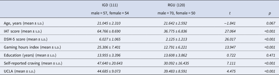
Loneliness was measured using the UCLA Loneliness Scale (Russell, Cutrona, Rose, & Yurko, Reference Russell, Cutrona, Rose and Yurko1984). The scale contains 20 items and quantifies feelings of loneliness. On the day of the experiment, subjects were asked to rate aspects of their loneliness on a 4-point Likert scale ranging from 1 to 4 (‘1 – never’ to ‘4 – often’). Higher scores indicate more loneliness (Cronbach's α = 0.81) (Hueluer et al., Reference Hueluer, Drewelies, Eibich, Duezel, Demuth, Ghisletta and Gerstorf2016). Participant information is shown in Table 1.
Demographic information and group differences

IGD, Internet gaming disorder; RGU, Regular game use; IAT, Internet addiction test; DSM-5, Diagnostic and Statistical Manual of Mental Disorders-5; Gaming playing hours index = (gaming days per week) × (gaming hours per day); Self-reported cravings; UCLA, UCLA Loneliness Scale.
Imaging acquisition
Imaging acquisition was performed using a 3 T Siemens Trio MRI system. For each subject, resting-state functional images were collected by an echo-planar imaging (EPI) pulse sequence with the following parameters: 33 axial slices with a slice thickness = 3 mm, TR = 2000 ms, TE = 30 ms, FOV = 220 × 220 mm2, flip angle = 90°, matrix = 64 × 64, and total volume = 210. High-resolution T1-weighted images were acquired with a three-dimensional spoiled gradient recalled sequence (192 sagittal slices with a slice thickness = 1 mm, TR = 2530 ms, TE = 2.34 ms, inversion time = 1100 ms, FOV = 256 × 256 mm2, flip angle = 7°, matrix = 256 × 256).
Data preprocessing
Resting-state fMRI data were preprocessed by Dpabi_v4.2 (Yan, Wang, Zuo, & Zang, Reference Yan, Wang, Zuo and Zang2016). After removal of the first 10 raw EPI volumes to eliminate non-equilibrium effects of magnetization, slice timing and motion parameters of each brain volume were estimated and corrected. Corrected functional images were registered to the corresponding T1-weighted anatomical image and spatially transformed to the standard MNI space with 3 × 3 × 3 mm3 voxels. The normalized data were then smoothed with a 6 mm FWHM isotropic Gaussian kernel. In addition, to control for motion and physiologic noise effects, six head-motion parameters and averaged signals from the cerebrospinal fluid and white matter were regressed out. Finally, the resultant images were detrended and filtered with a bandpass filter of 0.01–0.08 Hz.
Regions of interest selection
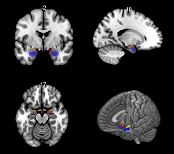
For the subsequent GC, masks of the amygdalar subdivisions were generated by the Anatomy Toolbox_v2.2b (Eickhoff et al., Reference Eickhoff, Stephan, Mohlberg, Grefkes, Fink, Amunts and Zilles2005) based on SPM12 (https://www.fil.ion.ucl.ac.uk/spm/software/spm12/). All masks included only unilateral subregions that were composed of the maximum probability maps of the LBA, CMA, and SFA (Amunts et al., Reference Amunts, Kedo, Kindler, Pieperhoff, Mohlberg, Shah and Zilles2005). The detailed locations of the selected ROIs are shown in Fig. 1.
Amygdala subdivisions. The laterobasal amygdala (LBA) is shown in blue, the centromedial amygdala (CMA) is shown in yellow, and the superficial amygdala (SFA) is shown in red. The bottom right plot shows the three amygdala subdivisions.

Granger causality analysis
The amygdalar-subdivision-based GC was implemented by the DynamicBC toolbox (Liao et al., Reference Liao, Wu, Xu, Ji, Zhang, Zang and Lu2014) and applied separately to 6 ROIs. GC is an exploratory data-driven approach that infers the causal influence between time series through temporal precedence (Roebroeck et al., Reference Roebroeck, Formisano and Goebel2005). Specifically, if the prediction of the current value of time course y can be improved by incorporating past values of x rather than using the past value of y alone, then x Granger causes y. Therefore, GC that uses temporal precedence information to calculate causality can identify voxels that have a causal effect on any selected ROI and does not require the specification of a prior model. In the present study, the time series of the 6 ROIs were defined as the seed time series x, while the time courses of the other voxels within the whole brain were defined as y. A bivariate coefficient GC was performed to examine GC between the amygdalar subdivisions and each voxel of the whole brain. Finally, the values of the GC input and output maps for all subjects were transformed into z-scores using Fisher's r-to-z transformation formula.
Statistical analysis
A two-sample t test was used to compare the z-valued GC maps between IGD subjects and RGUs. To control for multiple comparisons, family-wise error (FWE) correction with Gaussian random field theory was used, in which the voxel threshold was set to p < 0.001 and the cluster threshold was set to p < 0.05. The associations between the neuroimaging findings (i.e. the between-group differences of effective connectivity) and clinical characteristics in IGD (i.e. IAT and UCLA scores) were then evaluated by Pearson correlations. Bonferroni correction was used at this step to control for false-positive results.
Finally, given that the directionality between IAT and loneliness has been controversial in previous mediation tests, here we used the amygdalar subdivision-based effective connection pattern as a possible mediating factor to explore the directional relationship between loneliness and IGD. We tested two mediation models with (1) IAT as the predictor (i.e. IAT – loneliness) and (2) loneliness as the predictor (i.e. loneliness – IAT). The significance of the mediation effect was assessed using PROCESS bootstrapping and bias-corrected 95% confidence intervals (CI) in SPSS 20.0 (Hayes, Reference Hayes2012); a CI that does not contain zero indicates that there is a significant mediation effect for the proposed mediating factor.
Results
Demographics
There were no significant differences in age, sex (χ2 = 2.000, p = 0.157), or education between IGD subjects and RGUs. In contrast, IAT scores (t = 27.064, p < 0.001), DSM scores (t = 26.017, p < 0.001), gaming time (t = 13.947, p < 0.001), and self-reported craving scores (t = 7.111, p < 0.001), were greater for the IGD subjects than for the RGUs. Additionally, as expected, the IGD subjects also exhibited higher UCLA scores (t = 4.475, p < 0.001) than the RGUs, and there was a significant positive correlation between the IAT and UCLA scores (r = 0.291, p < 0.001).
Granger causality differences
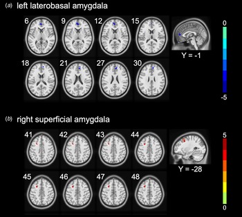
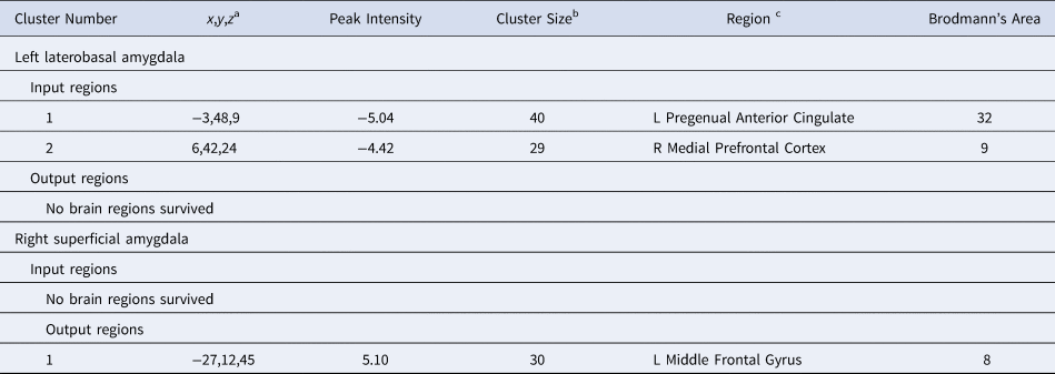
Compared to RGUs, IGD subjects showed a reduced strength in the effective connection from the left pregenual anterior cingulate (pACC) to the left LBA and, from the right medial prefrontal cortex (mPFC) to the left LBA (see Fig. 2). In addition, the IGD subjects showed increased strength in the effective connectivity from the left middle prefrontal gyrus (MFG) to the right SFA compared with RGUs. Generally, positive strength indicates excitement effects and negative strength indicates inhibitory effects. Thus, the current results suggest that inhibitory influences of the effective connections from the left pACC and right mPFC to the right LBA are increased in IGD subjects compared to RGUs, while the connection from the right SFA to the left MFG exhibits more excitatory regulation in IGD subjects than in RGUs. There were no significant between-group differences in the remaining 4 ROI-based effective-connectivity measurements, and detailed information about regional coordinates is given in Table 2.
Amygdala subdivision-based effective connectivity difference between IGD and RGU subjects. IGD subjects showed reduced effective connectivity from the left pregenual anterior cingulate cortex (pACC) and the right medial prefrontal cortex (mPFC) to the LBA and increased effective connectivity from the left middle frontal gyrus (MFG) to the right SFA.

Brain regions showing between-group differences in amygdala-based effective connectivity

a Peak MNI Coordinates.
b Number of voxels. FWE (p < 0.05) with voxel-p < 0.001. Voxel size = 3 × 3 × 3.
c The brain regions were referenced to the software SPM12 (https://www.fil.ion.ucl.ac.uk/spm/software/spm12/) and verified through comparisons with a brain atlas.
Correlations and mediation analysis
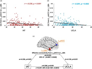
Correlational analyses were conducted between clinical variables (i.e. IAT and UCLA scores) and the 3 amygdalar-subdivision-based effective connections surviving Bonferroni correction [p = 0.008 (0.05/6)] for multiple comparisons. Negative correlations with the IAT score were found in the effective connections from the left pACC (r = −0.238, p < 0.001) and the right mPFC (r = −0.304, p < 0.001) to the left LBA, while the effective connection from the left MFG to the right SFA (r = 0.289, p < 0.001) was positively associated with IAT scores. In addition, only a negative correlation between UCLA scores and the effective connection from the left pACC to the left LBA (r = −0.201, p = 0.002) was found.
Given the finding that the effective connection from the left pACC to the left LBA was correlated with both the IAT and UCLA scores, mediation analyses were performed to test whether this effective connection could mediate the relationship between IGD severity and loneliness. Our results showed that when the IAT was examined as a possible predicting factor, the effective connection from the left pACC to the left LBA (0.0007, 0.0449) was a significant mediator in the relationship between IGD severity and loneliness (see Fig. 3). No mediation effects were detected when loneliness was examined as a possible predicting factor (−0.0314, 0.0922). In addition, we used sex, age, and educational history as covariates to verify the stability of the above findings (see online Supplementary materials).
Correlation and mediation analysis. Plots A and B show that effective connectivity from the left pACC to the LBA is negatively associated with both the IAT and UCLA scores. Plot C shows that the effective connectivity from the left pACC to the LBA significantly mediates the relationship between the IAT scores and the UCLA loneliness scores (0.0007, 0.0449).

Discussion
In the present study, we found changes in specific amygdalar-subdivision-based effective connectivity strengths between IGD subjects and RGUs in the pathways from the left pACC and the right mPFC to the LBA and from the left MFG to the right SFA, which supported our hypotheses and reflected independent connectivity patterns of each subdivision. Importantly, we also demonstrated that the effective connection from the left pACC to the LBA mediated the relationship between the IAT and UCLA scores, which provides novel insights into neural underpinnings of IGD-loneliness associations.
The effective connection from the left pACC to the LBA mediates the relationship between IGD and loneliness
In the current study, greater loneliness was observed in IGD subjects compared to RGUs, as was a positive correlation between IAT and UCLA scores. Individuals with IGD generally display higher levels of loneliness (King et al., Reference King, Delfabbro, Zwaans and Kaptsis2013), while healthy online gaming behaviour may promote social interaction and reduce loneliness (Martoncik & Loksa, Reference Martoncik and Loksa2016; Sundberg, Reference Sundberg2018). Here, we found that the effective connection from left the pACC to the LBA significantly mediated the relationship between the IAT and loneliness scores, suggesting this effective connection could be a promising neurobiological substrate linking IGD and loneliness.
The LBA, as a subdivision of the amygdala, receives inputs from several cortical and subcortical regions (e.g. the PFC, ACC, hippocampus, and thalamus) and facilitates associative emotional conditioning processes (LeDoux, Reference LeDoux2003; Phelps & LeDoux, Reference Phelps and LeDoux2005). Previous studies of emotional disorders have suggested the presence of disrupted neural circuits between the amygdala and ACC (Fonzo et al., Reference Fonzo, Simmons, Thorp, Norman, Paulus and Stein2010; Yuan et al., Reference Yuan, Pantazatos, Zhu, Li, Miller, Rubin-Falcone and Mann2019), which may form the basis for emotional dysregulation. Data suggest that functional impairments and structural deficits of the ACC may contribute to the development and maintenance of IGD (Dong, Shen, Huang, & Du, Reference Dong, Shen, Huang and Du2013; Kuss & Griffiths, Reference Kuss and Griffiths2012); meanwhile, the ACC has been implicated in emotional processing. Similar to the amygdala, it is not a homogeneous structure but rather consists of subdivisions that connect to different regions and contribute to specific functions (Margulies et al., Reference Margulies, Kelly, Uddin, Biswal, Castellanos and Milham2007; Vogt, Reference Vogt2005). Within the ACC, the pACC has been shown to sparsely connect to the amygdala in specific emotional disorders (Wang et al., Reference Wang, Kalmar, He, Jackowski, Chepenik, Edmiston and Blumberg2009) and is implicated in the top-down regulation of emotional stimuli (Ghashghaei, Hilgetag, & Barbas, Reference Ghashghaei, Hilgetag and Barbas2007). In line with these findings, individuals exposed to prolonged internet gaming may have changes in neural pathways from the left pACC to the LBA (Root, Denny, Hen, & Axel, Reference Root, Denny, Hen and Axel2014), which may possibly result in unsuccessful emotional regulation. Such processes may underlie loneliness in IGD.
The effective connection from the right mPFC to the left LBA is associated with craving in IGD
In addition to the pathway from the left pACC to the LBA, we also observed an inhibitory connection from the right mPFC to the left LBA in IGD subjects. This pathway may relate to conflicts between two neural systems in the human brain: an impulsive, amygdala system and a reflective, prefrontal cortex system. Bechara (Reference Bechara2005) noted that addiction may result from an imbalance between two separate, but interacting neural systems. Reduced functional connectivity between the mPFC and amygdala has been observed in people using cocaine (Gu et al., Reference Gu, Salmeron, Ross, Geng, Zhan, Stein and Yang2010). Within the amygdala, interactions between the mPFC and LBA in cue-elicited craving and drug-seeking has been reported (Chefer, Wang, & Shippenberg, Reference Chefer, Wang and Shippenberg2011; Petrovich & Gallagher, Reference Petrovich and Gallagher2003), which may be associated with inhibition of neuropeptide Y1 receptors neurons projecting from the mPFC to the LBA (Robinson et al., Reference Robinson, Marrero, Perez-Heydrich, Sepulveda-Orengo, Reissner and Thiele2019).
Craving may involve interactions between impulses, motivations, and emotions associated with specific addictive rewards. The inhibitory connection from the mPFC to the LBA may reflect imbalance between impulsive and reflective systems in IGD, which further suggests a potential link with craving. The relationship between amygdalar subdivisions and craving should be further examined in the future.
The effective connection from the left MFG to the right SFA reflects the interaction between poor emotional regulation and poor executive control in IGD
In comparison with the RGU subjects, IGD subjects showed a stronger effective connection from the left MFG to the right SFA. The SFA is part of the medial amygdala, generally receiving information from the olfactory system, promoting innate odour-driven behaviours in animals and complex socio-affective information processing in humans (Maras & Petrulis, Reference Maras and Petrulis2008). The MFG is located in the dorsolateral PFC and has been implicated in multiple functions including self-regulation, impulse control, and reward processing (Dong, Li, Wang, & Potenza, Reference Dong, Li, Wang and Potenza2018; Goldstein & Volkow, Reference Goldstein and Volkow2011). The hyperactivation of the bilateral MFG in subjects with IGD has been reported in a meta-analysis, possibly reflecting poor executive control (Meng et al., Reference Meng, Deng, Wang, Guo and Li2015).
In the current study, the right SFA exhibited greater excitatory regulation over the left MFG in IGD relative to RGU subjects, which suggests that poor emotional regulation in individuals with IGD may impair their executive control. The development and maintenance of addictive behaviours has been proposed to depend on interactions between predisposing variables, affective, and cognitive responses to specific rewards, and executive functions (Brand et al., Reference Brand, Wegmann, Stark, Muller, Wolfling, Robbins and Potenza2019). The current findings may have additional insight into affective mechanisms involved in addictions.
Limitations
First, although we recruited 231 subjects, they were mainly college students, and the current findings may not generalize to other populations of different ages, such as adolescents. Second, due to the cross-sectional design, we could not determine whether loneliness came before IGD or vice versa. Third, the current definitions for the IGD and RGU group may be a limitation warranting consideration. Although the DSM-5 proposes criteria for IGD, the criteria are provisional and intended to help research efforts (Petry & O'Brien, Reference Petry and O'Brien2013). The specific criteria and thresholding warrant additional study given that IGD is not currently a formal mental disorder (Petry, Zajac, & Ginley, Reference Petry, Zajac and Ginley2018). In addition, the IAT lacks externally validated thresholding (Laconi, Rodgers, & Chabrol, Reference Laconi, Rodgers and Chabrol2014), and this has been a criticism of this measure, among other concerns. Current uncertainties regarding the conceptualization and measurement of IGD should be considered in interpreting the findings, and future work should aim to establish further criteria and instruments with good psychometric properties.
Fourth, loneliness in the IGD population is often accompanied by other negative emotional states such as depression and anxiety, and common neural features may underlie these states. Given the inclusion/exclusion criteria limiting depression in the current study, future studies should investigate samples with more depression and anxiety to investigate directly their neural underpinnings in IGD.
In addition, our image acquisition parameters generated voxels of 3.4 × 3.4 × 3.2 mm3. Future studies of individual divisions of the amygdala may derive benefit from alternate acquisition parameters (e.g. multi-band or multi-plex) that generate smaller voxels and may offer greater precision for such studies.
Finally, there may be potential concerns about the application of Granger causality to fMRI data, largely because of the temporal aggregation associated with temporal bandpass filtering which may obscure or create spurious temporal dependencies in the data (Deshpande & Hu, Reference Deshpande and Hu2012). Here we applied a 0.01–0.08 Hz bandpass filter, which is an important preprocessing step but one that nonetheless introduces problems of determining temporal precedence due to downsampling. Another issue is Granger causal inferences that may be easily confounded by regional differences in the latencies of the hemodynamic response functions. Although Seth, Chorley, and Barnett (Reference Seth, Chorley and Barnett2013) noted that Granger causal inferences are robust to various changes in hemodynamic response properties, they may still be affected by downsampling and measurement noise. Thus, further studies are needed to better understand and assess this approach.
Conclusions
The current study showed that amygdalar-subdivision-based effective connectivity is associated with IGD severity and loneliness in IGD subjects. These findings may relate to poorly controlled craving and poor emotional regulation in IGD. Importantly, the connectivity from the left pACC to LBA strength mediated the relationship between IAT and UCLA scores, and this connectivity may underlie poor emotional regulation in IGD and contribute to the high prevalence of loneliness. The findings shed new insight into the neural correlations of loneliness and IGD severity and increase understanding of affective features in IGD.
Supplementary material
The supplementary material for this article can be found at https://doi.org/10.1017/S0033291720002366.
Acknowledgements
The current research was supported by the Zhejiang Natural Science Foundation (LY20C090005). Marc Potenza received support from the Connecticut Council on Problem Gambling, the Connecticut Department of Mental Health and Addictions and the National Center for Responsible Gaming. The funding agencies did not have input into the writing of this manuscript.
Author contributions
Min Wang wrote the first draft of the manuscript. Ningning Zeng analyzed the data. Hui Zheng and Xiaoxia Du contributed to fMRI data collection. Guang-heng Dong designed this research and edited the manuscript. Marc Potenza edited the manuscript. All authors contributed to and approved the final manuscript.
Conflict of interest
The authors declare that no competing interests exist. Marc Potenza has consulted for and advised Opiant Pharmaceuticals, Idorsia Pharmaceuticals, AXA, Game Day Data and the Addiction Policy Forum; has received research support from the Mohegan Sun Casino and the National Center for Responsible Gaming; has participated in surveys, mailings or telephone consultations related to drug addiction, impulse control disorders or other health topics; and has consulted for law offices and gambling entities on issues related to impulse control or addictive disorders.