Implications
Supplementing different ranges of camphor, as a novel feed additive, has been evaluated in Japanese quail. Herein, we showed that camphor can effectively improve certain aspects of immunity and health status. Animal feed producers and even poultry farmers may incorporate this drug in their feed to benefit from its modulatory function on immunity, intestinal bacteria and health status in flocks.
Introduction
Microbial resistance to antibiotics and deposits in animal products, which is due to the use of antibiotics, has motivated a worldwide effort to reduce such drugs in animal nutrition, providing harmless animal products for consumers (Jin et al., Reference Jin, Ho, Abdullah and Jalaludin1998). Alternatively, among the potential alternatives to antibiotic growth promoters, the use of phytochemical compounds are considered to be fascinating, with more reliability and suitability among consumers, as harmless and natural additives (Hashemi and Davoodi, Reference Hashemi and Davoodi2011). Under these circumstances, the use of phytochemicals in poultry feed has been closely monitored. Of recent, it is better to investigate the plant material effects in terms of performance, intestinal microbiology, quality and health of carcass meat as the specified criteria (Botsoglou et al., Reference Botsoglou, Florou-Paneri, Christaki, Fletouris and Spais2002), because it is well documented that some plant extracts can affect the secretion of digestive enzymes and act as anti-bacterial, anti-viral and anti-oxidising agents (Ertas et al., Reference Ertas, Güler, Çiftçi, Dalkiliç and Yilmaz2005; Cross et al., Reference Cross, McDevitt, Hillman and Acamovic2007).
Many known phytochemical compounds and their effects on poultry have been investigated; however, the effects of some herbal ingredients are still unknown. Camphor is one of the compounds, yet to be subjected to the nutritional experimental research. It occurs naturally, and is isolated from the wood of the camphor laurel tree (Cinnamomum camphora); it is used as a major active component in the treatment of various disorders (Xu, Reference Xu2005) and has been extensively used for medicinal purposes (Cherneva et al., Reference Cherneva, Pavlovic, Smelcerovic and Yancheva2012). Camphor is also found in a variety of natural products, either alone or in combination with other ingredients. Herbal medicine and essential oils such as rosemary (Brenes and Roura, Reference Brenes and Roura2010), sagebrush (Guenther et al., Reference Guenther, Hewitt, Erickson, Fall, Geron, Graedel and Zimmerman1995), Thymus algeriensis (Ben El Hadj Ali et al., Reference Ben El Hadj Ali, Chaouachi, Bahri, Chaieb, Boussaïd and Harzallah-Skhiri2015), Artemisia annua (Engeu et al., Reference Engeu, Omujal, Agwaya, Kyakulaga and Obua2015) and chicory (Shahabi et al., Reference Shahabi, Jorsaraei, Moghadamnia, Barghi, Zabihi, Amiri, Maliji, Faraji, Boora and Ghazinejad2014) include camphor as an essential component.
The literature on camphor’s effects on animals is limited and shows diverse results. Camphor has been shown to affect the reproductive function in rats (Jamshidzadeh et al., Reference Jamshidzadeh, Sajedianfard, Nekooeian, Tavakoli and Omrani2006) and quails (Sedaghat et al., 2016). Nevertheless, evidence suggests that camphor acts as an insecticidal, anti-microbial, anti-viral, anti-coccidial, analgesic, anti-cancer and anti-tussive agent (Chen et al., Reference Chen, Vermaak and Viljoen2013). It has also been found that camphor extract modulates the immune function in rats (Lee et al., Reference Lee, Hyun, Yoon, Kim, Rhee, Kang, Cho and Yoo2006).
Nevertheless, there are few, if any, research studies to examine whether its inclusion as a feed additive would have a growth-promoting and immune-stimulating effect on live birds. To the best of our knowledge, there is no direct evidence till date for camphor’s effects on poultry performance and health status. Therefore, the present study was conducted to determine the effects of dietary graded levels of camphor on immune functions, microbial populations of gastrointestinal tract (GIT), performance and some blood parameters of Japanese quails.
Material and methods
Birds, diets and experimental procedure
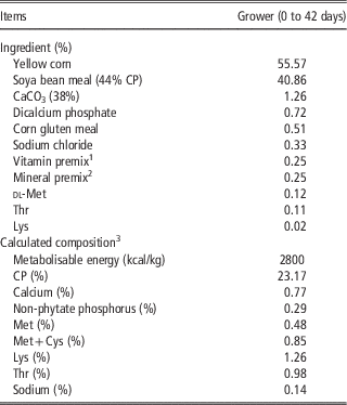
The procedures involved in this study were approved by the Animal Care and Use Committee of Tarbiat Modares University, Tehran, Iran. In all, 280 1-day-old quail chicks were randomly divided into 28 groups of 10 chicks each. Each group was assigned to a cage (50×30×40 cm) with ad libitum access to feed and water. Chicks were brooded with a 100 W bulb throughout the rearing period. They were then divided into seven treatment groups (four cages per treatment) and were fed one of the seven dietary treatments. Diets were: basal diet (control), basal diet+250 ppm camphor, basal diet+500 ppm camphor, basal diet+750 ppm camphor, basal diet+1000 ppm camphor, basal diet+5000 ppm camphor and basal diet+10 000 ppm camphor. The basal diet used in the study was a commercial corn-soya bean diet, formulated to meet the nutrient requirements (NRC, 1994) during the growth (0 to 42 days) period (Table 1). Standard brooding temperatures were preserved for all birds during the rearing period.
Ingredients and nutrient composition of basal diets (as-fed basis)

1 The vitamin premix supplied the following per kilogram of complete feed: vitamin A, 9000 IU (retinyl acetate); D3, 2000 IU; vitamin E, 12.5 mg (dl-α-tocopheryl acetate); vitamin B12, 0.015 mg; menadione, 1.76 mg; calcium d-pantothenate, 6.4 mg; biotin, 0.12 mg; pyridoxine, 1.97 mg; thiamine, 1.2 mg; nicotinic acid, 28 mg; riboflavin, 3.2 mg; choline chloride, 320 mg; cyanocobalamine, 0.01 mg; folic acid, 0.38 mg.
2 The mineral premix supplied the following per kilogram of complete feed: manganese (MnSO4·H2O), 60 mg; zinc (ZnO), 51.74 mg; iron (FeSO4·7H2O), 80 mg; copper (CuSO4·5H2O), 8 mg; selenium (Na2SeO3), 0.2 mg; iodine (iodised NaCl), 0.8 mg.
3 Calculated from NRC (1994).
Performance parameters
Birds were weighed on days 14, 21, 28, 35 and 42 on cage basis to determine the live BW and BW gain (BWG). Consumed feed was recorded daily, unconsumed feed was discarded and average daily feed intake (ADFI) and feed conversion ratio (FCR) were calculated. Mortality was recorded as it occurred to correct feed conversion data.
Internal organs relative weight
On day 42, two birds were selected from each replicate (n=8/treatment) and slaughtered. The birds were dissected, and the weights of the carcass (without giblet), abdominal fat, proventriculus, gizzard, liver, testis, heart, ovaries and oviduct were recorded.
Biochemical analysis
Whole blood samples were randomly collected from eight birds per treatment from the heart into heparinised syringes. The samples were then centrifuged at 1000×g for 20 min at room temperature to obtain the plasma. The plasma samples were kept in Eppendorf tubes at –20°C until the analysis. The concentrations of plasma biochemical factors including uric acid, albumin, total protein, cholesterol, triglycerides, glucose and high-density lipoprotein (HDL) cholesterol were determined spectrophotometrically using a microplate reader (Stat Fax 3200; Awareness Technology Inc., Palm City, FL, USA) and commercial analytical kits provided by Pars Azmun Co. (Tehran, Iran), as described by the manufacturers.
Immune responses
Cell-mediated immune response
The attributes used to evaluate the cell-mediated immune (CMI) activity included the cutaneous basophilic hypersensitivity (CBH) and 2,4-dinitro 1-chlorobenzene (DNCB 10 mg/ml, in a vehicle consisting of an acetone and olive oil (4 : 1) mixture) (Merck; Darmstadt, Germany) response. The CBH response to phytohaemagglutinin-P (PHA-P) was used to evaluate in vivo CMI response on day 40. For the PHA-P challenge, the right wing-web of each bird (8 birds/treatment) was intra-dermally injected with 0.04 ml of PHA-P in sterile phosphate buffered saline (PBS). The left wing-web was injected with 0.04 ml of PBS as a sham control. For CBH response to DNCB, a relatively featherless zone of ~1 cm2 was restricted on the right side for anointing 0.1 mL of DNCB, whereas a similar zone on the left side was chosen for the vehicle application alone, which served as the control. The reactions were assessed for both PHA-P and DNCB by measuring skin thickness via digital calipers, immediately before (0 h) the challenge and 24 h after the challenge. The increase in the mean skin thickness of birds was calculated as follows: ((thickness of right side pre-challenge−thickness of right side post-challenge)−(thickness of left side pre-challenge−thickness of left side post-challenge)/basal skin thickness).
Humoral immune response
On day 28, as a T-cell-dependent antigen, 0.1 ml of 5% sheep red blood cells (SRBC) suspension in PBS was injected into the breast muscle of eight birds per treatment, and the birds were then given a booster injection on day 35 to determine the secondary anti-SRBC antibody response. Each bird’s whole heparinised blood sample was drawn 7 days after the booster injection. The anti-SRBC antibody titer was determined by the micro-haemagglutination test using 96-well microplates. The plasma samples were heat inactivated (56°C for 30 min) and then analysed. In brief, 25 µl of plasma sample and 25 µl of PBS were placed in the first row and incubated at 37°C for 30 min. Moreover, 25 µl of PBS was added to the remaining wells to sort a 2-fold serial dilution of each sample on successive rows. Furthermore, 25 µl of a 1% SRBC suspension was placed in each well, and the plate was re-incubated (37°C for 30 min) and monitored by a haemagglutination test. Antibody titers were reported as log2 of the reciprocal of the highest dilution giving a detectible agglutination.
Newcastle disease virus and avian influenza virus-specific antibody assay
Two birds per replicate (eight birds per treatment) were vaccinated against the Newcastle disease virus (NDV) using an eye dropper (Live B1 strain; Vetrina, Zagreb, Croatia) and avian influenza virus (AIV) via subcutaneous injection, according to the manufacturer’s recommendations, at 33 days of age. Whole blood samples were collected from the heart of each bird at 42 days of age to determine the antibody titers against NDV and AIV. Antibody titers were performed using haemagglutination inhibition (HI) assay on the obtained sera. Initially, four haemagglutination (HA) units for NDV and eight HA units for AIV were added (25 µl/well), and one HA unit of the virus was determined as the highest dilution of virus, which causes the agglutination of freshly prepared (1 : 100) chicken red blood cell (RBC) suspension under standard conditions of pH (7.2) and temperature (25°C). Moreover, 25 µl of plasma was added to a microplate containing PBS (25 µl/well) to create 2-fold serial dilutions. A 1% chicken RBC suspension (25 µl) was then added to each well and incubated at 37°C for 45 min. The HI titer was expressed as the log2 of the reciprocal of the highest plasma dilution, which inhibited the HA activity.
Lymphoid organs
Eight birds from each treatment were humanly decapitated for determining the weight of spleen and bursa of Fabricius, and the values were adjusted for BW.
Heterophil-to-lymphocyte ratio
On day 42, the whole blood sample was collected to prepare smears for the heterophil-to-lymphocyte (H:L) ratio. Blood smears were air-dried and then stained with Wright’s-Giemsa (Saikin Kagaku Institute Co. Ltd, Sendai, Japan). The heterophil and lymphocyte cells were then enumerated to a total of 100 white blood cells per slide with a light microscope (standard 20; Carl Zeiss, Göttingen, Germany) at 1000× magnification.
Bacteriology
On day 42, eight quails from each treatment were slaughtered and the small intestine (from the distal end of the jejunum to the ileocecal junction) was removed from each bird to determine the number of total aerobic bacteria, coliform bacteria, lactic acid bacteria and spore forming bacteria. The small intestine was immediately opened longitudinally, the mucosal surface and digesta were scraped with a sterile knife and then the ileal contents were transferred under aseptic conditions into the sterile tubes. A quantity of 1 g of the homogenised ileal content was serially diluted from 101 to 108 in PBS. The selective agar media including plate count agar (Merck, Darmstadt, Germany), MRS agar (Merck, Darmstadt, Germany) and Mac Conkey agar (Merck) were used for the enumeration of total aerobic bacteria and spore forming bacteria, lactic acid bacteria and coliform bacteria, respectively. Each duplicate sample dilution was inoculated on media and enumerated after incubation at 37°C for 24 h. For spore forming bacteria, in order to inactivate the heat sensible bacteria, samples were submerged in the water bath at 80°C for 30 min before inoculation. Following enumeration data were expressed as log10 colony forming units per gram of ileum content (log10 CFU/g).
Statistical analysis
This study was carried out with a randomised complete block design. Data were subjected to the ANOVA using the GLM procedure of SAS software (SAS, 2014). Duncan’s multiple range test was used to compare the statistical significance among the means at a 95% significance level. All data analyses were performed using SAS 9.4 for Windows. The original data of bacterial counts were transformed to log10 CFU/g of the intestinal content. Polynomial orthogonal contrasts were directed for camphor levels in order to consider the linear and quadratic trends.
Results
Performance parameters
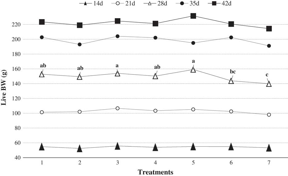
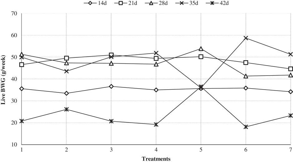
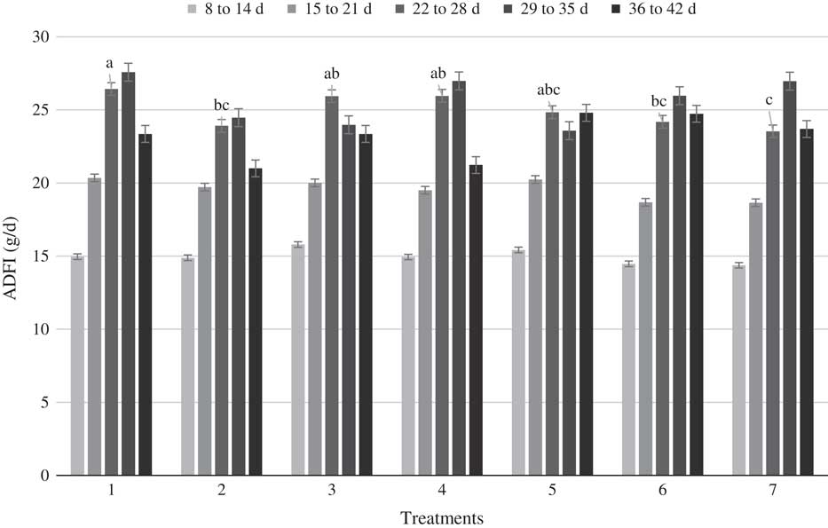
Camphor treatment did not affect the live BW, with an exception on day 28 (P=0.003), which linearly tended to reduction (P=0.0003) with 5000 and 10 000 ppm of camphor in the diet (Figure 1). Moreover, contrast comparison expressed the linear (P=0.01) affection of BW on day 21. The results showed that the quail BWG was not affected by camphor supplementation (Figure 2) but had significant linear differences on days 21 (P=0.009) and 28 (P=0.01). The ADFI was monitored daily throughout the trial. The mean ADFI is presented in Figure 3. Overall, the weekly average for ADFI among treatments was not statistically significant; however, from 22 to 28 days of age interval, ADFI was significantly (P=0.02) greater in control and lower dosage of camphor groups, that is, camphor caused slight linear reductions from 8 to 14, 15 to 21 and 22 to 28 days of age intervals; in particular, camphor treatment had a reducing effect on ADFI in juvenile quail chicks until 28 days of age (Figure 3). There was no significant difference in FCR for quails fed on diets containing camphor during the whole experimental period. Contrast comparisons showed that different levels of camphor supplementation had a linear (P=0.03) and quadratic (P=0.04) effect on FCR from 8 to 14 day-old and 15 to 21 day-old quails, respectively (data not shown).
Effect of dietary camphor levels on live BW (g) in growing Japanese quail at different age intervals that given control diet (0 camphor) or diet supplemented with camphor at 250, 500, 750, 1000, 5000 or 10 000 ppm. Treatments 1, 2, 3, 4, 5, 6 and 7 stand for 0, 250, 500, 750, 1000, 5000 and 10 000 ppm camphor, respectively. a,b,cValues with different superscripts differed significantly (P<0.05).

Effect of dietary camphor levels on live BW gain (BWG) (g/week) in growing Japanese quail at different age intervals that given control diet (0 camphor) or diet supplemented with camphor at 250, 500, 750, 1000, 5000 or 10 000 ppm. Treatments 1, 2, 3, 4, 5, 6 and 7 stand for 0, 250, 500, 750, 1000, 5000 and 10 000 ppm camphor, respectively.

Effect of dietary camphor levels on average daily feed intake (ADFI) (g/d) in growing Japanese quail at different age intervals that given control diet (0 camphor) or diet supplemented with camphor at 250, 500, 750, 1000, 5000 or 10 000 ppm. Treatments 1, 2, 3, 4, 5, 6 and 7 stand for 0, 250, 500, 750, 1000, 5000 and 10 000 ppm camphor, respectively. a,b,cValues with different superscripts differed significantly (P<0.05).

Organ relative weight
The effect of dietary camphor levels on different internal organ weights as percentages of BW revealed that no significant effects of camphor supplementation were observed on organ weights during the experimental period (data not shown).
Immune response
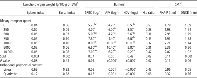
The effects of dietary camphor on lymphoid organ weights (spleen and bursa of Fabricius), humoral (SRBC, AIV, NDV and H:L ratio) and CMI responses (PHA-P and DNCB) are given in Table 2. In this study, camphor showed no significant effect on spleen and bursa indices. Nevertheless, humoral immune response was affected by camphor supplementation in which the greatest secondary responses to SRBC were seen in 750 and 1000 ppm of camphor than other groups (P<0.05). In addition, the greatest AIV antibody titers were found in 1000 and 5000 ppm of camphor-treated groups (P<0.05), and the values of NDV antibody titers increased with the increase in camphor consumption (P<0.05). In this trial, the H:L ratio was also unaffected by the camphor treatment. Data on CBH response to PHA-P and DNCB indicated that neither PHA-P nor DNCB tests influenced the response to the skin challenge.
Effect of dietary camphor levels on relative weight of the lymphoid organs, cell-mediated and humoral immune responses in growing Japanese quail

CMI=cell-mediated immune; SRBC=sheep red blood cell; AIV=avian influenza virus; NDV=Newcastle disease virus; H:L=heterophil-to-lymphocyte ratio; PHA-P=phytohemagglutinin-P; DNCB=2,4-Dinitro 1-chlorobenzene.
a,b,cMeans with different letters within the same column differ significantly (P<0.05).
1 The organ index is expressed as lymphoid organ weight (g) per BW (g).
2 Cell-mediated immunity – Cutaneous basophil hypersensitivity test – as measured by increase in skin thickness after PHA-P and DNCB injection.
Intestinal microbiota
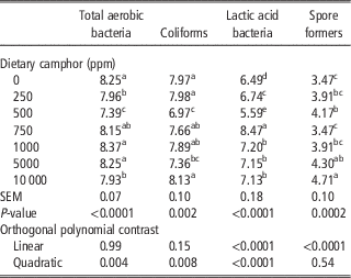
Total aerobic bacteria, coliforms, lactic acid bacteria and spore forming bacterial populations of quail’s gut at 42 days of age, fed on diets with camphor levels are summarised in Table 3. The treatment with 1000 and 5000 ppm camphor and control groups produced significantly greater populations of total aerobic bacteria (P<0.05) than the other groups. As shown in Table 3, in birds consuming the diet containing 500 ppm of camphor, coliform population positively declined. A significant (P<0.05) increase in lactic acid bacteria was also observed at 42 days of age in quails fed on the diet containing 750 ppm camphor, when compared with birds in the other treatment groups. The results of the effect of camphor on spore forming bacteria indicated a linear increase with an increase in camphor dosage, thereby 10 000 ppm of camphor group exhibited the greatest population of spore forming bacteria.
Effect of dietary camphor levels on intestinal microbiota in growing Japanese quail at day 42Footnote 1

a,b,c,d,eMeans with different letters within the same column differ significantly (P<0.05).
1 The number of colonies was counted and all the data were expressed as log10 CFU/g.
Biochemical blood parameters
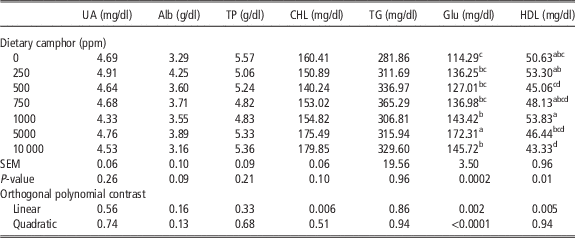
To assess biochemical blood plasma characteristic, urea acid, albumin, total protein, cholesterol, triglyceride, glucose and HDL were measured in eight 42-day-old quails per treatment. Urea acid, albumin, total protein and triglyceride were not affected by camphor treatments (Table 4). In addition, cholesterol concentration was significantly different in the linear manner (P=0.006) by which 500 ppm of camphor resulted in a lower level of cholesterol. Alternatively, camphor supplementation positively affected glucose and HDL concentrations, which exhibited greater concentrations by 5000 and 1000 ppm of camphor, respectively, when compared with groups receiving lower doses of camphor as well as the control group, which showed the lowest values of such parameters (Table 4).
Effect of dietary camphor on some blood characteristics in growing Japanese quail

UA=uric acid; Alb=albumin; TP=total protein; CHL=cholesterol; TG=triglycerides; Glu=glucose; HDL=high-density lipoprotein.
a,b,c,dMeans with different letters within the same column differ significantly (P<0.05).
Discussion
This paper presented a novel additive in poultry feed industry. The main purpose of the experiment was to draw attention to camphor supplementation and its effects on performance and related parameters. As live BW did not change throughout the trial, it can be concluded that camphor cannot be classified as a growth-promoting substance in Japanese quails. Nevertheless, the treatment with 1000 ppm of camphor produced a significantly greater BW on day 28. These results are in good agreement with those reporting no significant change in BW by the use of camphor derivatives in rats (Carou et al., Reference Carou, Deguiz, Reynoso, Szwarcfarb, Carbone, Moguilevsky, Scacchi and Ponzo2009) and camphor (Eucalyptus globules) in quails (Abu Taleb et al., Reference Abu Taleb, Salah, Ezzat and Barkouky2003). Alternatively, this result is in contrast with the findings of researchers who observed the beneficial effect of camphor derivatives on BW in rats (Schlumpf et al., Reference Schlumpf, Jarry, Wuttke, Ma and Lichtensteiger2004a), and significantly greater BW in chickens fed with diet containing 119 ppm of A. annua, which included camphor as the active ingredient (Allen et al., Reference Allen, Lydon and Danforth1997).
The data (Figure 3) show that as dietary camphor levels increased, ADFI significantly decreased from 22 to 28 days of age. These results were in agreement with those of previous studies, which demonstrated a significantly decreased feed intake in rats and rabbits (Herzog and Leuschner, Reference Herzog and Leuschner1997). Similar to ADFI, during the whole experimental period, FCR was not significantly different after the treatment. The decrease in ADFI can be contributed to the reduction in γ-aminobutyric acid (GABA), aspartate and glutamate, which clearly demonstrated that using 4-methylbenzyldene camphor (4-MBC) can suppress feed intake in rats (Carou et al., Reference Carou, Deguiz, Reynoso, Szwarcfarb, Carbone, Moguilevsky, Scacchi and Ponzo2009). The aforementioned amino acids are neurotransmitters in the central nervous system and can, in turn, act as feed intake regulators. Studies reported that feed intake was stimulated by GABA induction in chickens and turkeys (Denbow, Reference Denbow1991; Jonaidi et al., Reference Jonaidi, Babapour and Denbow2002) and glutamate induction in broilers (Zendehdel et al., Reference Zeni, Seidler, De Carvalho, Freitas, Marino-Neto and Paschoalini2009) and pigeons (Zeni et al., Reference Zeni, Seidler, De Carvalho, Freitas, Marino-Neto and Paschoalini2000). In addition, in a previous report (Michiels et al., Reference Michiels, Missotten, Ovyn, Dierick, Fremaut and De Smet2012), feeding piglets a diet with 200 mg/kg of BW camphor supplementation reduced palatability. Moreover, some studies show that camphor stimulates the secretion of leptin (Johari et al., Reference Johari, Mahjoor, Fallahi, Kargar Jahromi, Abedini, Poor Danesh and Zamani2013); according to the known effect of leptin in reducing feed intake (Denbow et al., Reference Denbow, Meade, Robertson, McMurtry, Richards and Ashwell2000; Dridi et al., Reference Dridi, Swennen, Decuypere and Buyse2005), it seems that stimulating leptin secretion by dietary camphor could be attributable to the reduction of feed intake in the present experiment.
In the present study, no measured internal relative organ weight had significant differences (P<0.05). In contrast to our results, a greater weight in the ovaries of female and testis of male rats was achieved using 4-MBC in the pre-pubertal period (Schlumpf et al., Reference Schlumpf, Schmid, Durrer, Conscience, Maerkel, Henseler, Gruetter, Herzog, Reolon, Ceccatelli, Faass, Stutz, Jarry, Wuttke and Lichtensteiger2004b). These inconsistencies in results may be due to the dose-dependent effects of camphor-containing products (Schlumpf et al., Reference Schlumpf, Jarry, Wuttke, Ma and Lichtensteiger2004a). Notably, pure camphor was used in the present study rather than camphor-containing products used in other studies.
This is the first study to describe an enhanced secondary anti-SRBC antibody response, as well as greater responses against AIV and NDV in poultry produced from quail fed on diets supplemented with camphor (Table 2). In this case, our experimental findings concur with the in vitro results of Lee et al. (Reference Lee, Hyun, Yoon, Kim, Rhee, Kang, Cho and Yoo2006), which revealed an improvement in the cell immune functions. The C. camphora has long been used in traditional medicine (Lee et al., Reference Lee, Hyun, Yoon, Kim, Rhee, Kang, Cho and Yoo2006) for the treatment of inflammation-related diseases. Lee et al. (Reference Lee, Hyun, Yoon, Kim, Rhee, Kang, Cho and Yoo2006) demonstrated that camphor could inhibit pro-inflammatory cytokines produced through the challenge with lipopolysaccharides (LPS) and suppress nitric oxide and prostaglandin E2 production stimulated by LPS. Nevertheless, this issue in poultry clearly requires elucidation by future research.
The anti-microbial properties of camphor in the present investigation are supposed to favour a healthy gut, which in turn could be related to the enhancement of immune functions. It is frequently suggested that camphor-containing essential oils can exhibit a strong anti-microbial activity (Hammerschmidt et al., Reference Hammerschmidt, Clark, Soliman, El-Kashoury, Abd El-Kawy and El-Fishawy1993; Magiatis et al., Reference Magiatis, Skaltsounis, Chinou and Haroutounian2002; Kotan et al., Reference Kotan, Kordali, Cakir, Kesdek, Kaya and Kilic2008). Indeed, it was revealed that such products display an activity against Enterococcus hirae, Candida albicans and Saccharomyces cerevisiae (Juteau et al., Reference Juteau, Masotti, Bessière, Dherbomez and Viano2002). Some studies have reported a poor anti-microbial activity for pure camphor alone (not in combination with essential oils and/or extracts) (Setzer, Reference Setzer2004); despite this, higher lactic acid bacteria and lower coliform counts were achieved in quails, which were fed diets with 750 and 500 ppm of camphor, respectively. This result is in agreement with the trial in which a potentially reducing effect of camphor (E. globules) was observed on salmonella in quail GIT (Abu Taleb et al., Reference Abu Taleb, Salah, Ezzat and Barkouky2003). Notably, other camphor anti-microbial activities were well reviewed by Chen et al. (Reference Chen, Vermaak and Viljoen2013). Due to their lipophilic property, terpenoids and phenylpropanoids can be attracted via bacteria and can easily penetrate the cell membrane (Helander et al., Reference Helander, Alakomi, Latva-Kala, Mattila-Sandholm, Pol, Smid, Gorris and von Wright1998). Likewise, the anti-microbial activity of essential oils is attributed to their cyclic compound and specific functional groups. Therefore, one can deduce the cyclic isoprene existing in the camphor molecule that can be attributed to its anti-microbial property in the GIT.
Considering the results of plasma biochemical profile, it is demonstrated that camphor has positive effects on cholesterol and glucose metabolism, by which cholesterol linearly diminished up to 500 ppm of camphor, and HDL was increased by 1000 ppm of camphor when compared with the other groups. Contrary to the findings of Abdullah-Al-Mamun et al. (Reference Abdullah-Al-Mamun, Huque, Biswas, Bhuiyan and Rashid2014), which demonstrated a significant modification in total protein, albumin and triglyceride concentrations in albino rats, an insignificant change was observed in such characteristics in this study; however, uric acid was in agreement with the mentioned study. The mechanism by which dietary camphor supplement affects the concentrations of plasma glucose and cholesterol is not fully understood. Nevertheless, this result may be due to the inhibiting effect of camphor on 3-hydroxy-3-methylglutaryl-CoA reductase, which is the key enzyme for de novo cholesterol biosynthesis leading to an improvement in the liver activity and kidney function, thereby affecting lipid metabolism (Abdullah-Al-Mamun et al., Reference Abdullah-Al-Mamun, Huque, Biswas, Bhuiyan and Rashid2014).
Conclusion
Summing up the results, it can be concluded that camphor, which is a multi-purpose molecule with a most diverse range of applications in animals and humans, can be used as an additive in diets for Japanese quails to achieve modulation in gut microbial populations, higher glucose and HDL and immune functions. Overall, lower levels of camphor supplementation (500, 750 and 1000 ppm) had more beneficial effects on performance and health-related parameters, which revealed the dose-dependence of this substance. In addition, due to its effect on the immune response to SRBC, AIV and NDV, evaluation of its effect on other important viruses in the poultry industry, in vivo and in vitro, can be recommended to achieve its vigour for anti-viral activities. Camphor also may act in a synergistic manner with other herbal constituents possessing anti-microbial activity. Moreover, it is useful in improving the immune functions of broiler production. These results are non-conclusive, and the present study is a pioneering work exploring effects of camphor on poultry. Further studies are needed to focus on the effects of diverse levels of C. camphora (pure substance) alone and in combination with other terpenoids.
Acknowledgements
The authors wish to thank Tarbiat Modares University Research Council for financial support.