INTRODUCTION
Role of chemotherapy and radiotherapy has increased enormously in effective management of various types of cancer in recent past. This success story has other side also and includes the severe form of side effects of these therapies. The important being the disruption in the functions and integrity of the mouth.Reference Worthington, Clarkson and Eden1Candida albicans is a normal inhabitant and frequently colonizes in the oral cavity in a large population of normal individuals and does not cause disease under normal circumstances. The changes in systemic as well as local factors may lead to clinical oral fungal infections. The factors responsible for oral fungal infection include immunosupression, changes in normal oral microbial flora and local tissue damage. Cancer patients receiving radiation or chemotherapy are prone for all these risk factors.Reference Lalla, Latortue, Hong, Ariyawardana, D’Amato-Palumbo, Fischer, Martof, Nicolatou-Galitis, Patton, Elting, Spijkervet and Brennan2–Reference Schelenz, Abdallah, Gray, Stubbings, Gow, Baker and Hunter3 Radiotherapy-induced hyposalivation encourages oral Candida colonization that often leads to oral and/or pharyngeal candidiasis. Compromised salivary function secondary to destruction of glandular tissue by radiation is thought to be a major factor leading to Candida infection.Reference Koc and Aktas4
Oral candidiasis can present as various clinical presentations, such as pseudomembranous candidiasis (thrush), chronic hyperplastic candidiasis, erythematous candidiasis, and angular cheilitis. Whatever the presentation, oral candidiasis have a significant impact on quality of life and can impair nutritional status. As oral candidiasis cause burning sensation while eating and impairs significantly taste sensations, it may adversely affect the food intake by the patients. In patients suffering with cancer, maintenance of nutritional intake is very important.
If neglected, the colonized Candida can spread to other organs and may result in systemic effects or septicemia. Identification of oral candidiasis forms an important part of management of cancer patients. Determination of the epidemiology of Candida isolates in patients receiving head and neck radiation has traditionally involved taking individual cultures and identifying yeasts to the species level, as well as using techniques to identify specific yeast strains with serotyping and biotyping. In the past, Candida albicans has been by far the most predominant organism isolated.Reference Redding, Zellars, Kirkpatrick, McAtee, Caceres, Fothergill, Lopez-Ribot, Bailey, Rinaldi and Patterson5
Recently published studies have shown that many other Candida species, such as Candida glabrata, Candida tropicalis, Candida krusei, Candida parapsilosis, Candida kefyr and Candida dubliniensis have been found to be responsible for oral infections.Reference Lalla, Latortue, Hong, Ariyawardana, D’Amato-Palumbo, Fischer, Martof, Nicolatou-Galitis, Patton, Elting, Spijkervet and Brennan2,Reference Belazi, Velegraki, Eremondi, Andreadis, Hini, Arsenis, Eliopoulou, Destouni and Antoniades6
Fluconazole is the predominant medication used to treat oropharyngeal candidiasis caused by Candida albicans.Reference Redding, Zellars, Kirkpatrick, McAtee, Caceres, Fothergill, Lopez-Ribot, Bailey, Rinaldi and Patterson5 Some studies have reported an increase in Candida resistance to first-line antifungal agents such as fluconazole. Hence early recognition of oral candidiasis and the subsequent speciation and susceptibility testing is important.Reference Schelenz, Abdallah, Gray, Stubbings, Gow, Baker and Hunter3
Oral candidiasis can be easily treated particularly in earlier stages, so it is important to identify and treat or take preventive measures in the high-risk cancer patients.Reference Schelenz, Abdallah, Gray, Stubbings, Gow, Baker and Hunter3
The objective of this study was to identify the Candida species causing oropharyngeal candidiasis in patients suffering with oral cavity cancers undergoing radiotherapy and to find out antifungal susceptibility pattern.
MATERIAL AND METHODS
Patients undergoing radiation therapy for oral cavity cancers were included in this study.
Informed and written consent was taken from each patient before the sample collection. The ethics committee of Government Medical College, Aurangabad gave approval for this study. The study was carried over a period of 1 year in the Department of Microbiology, Government Medical College and Hospital, Aurangabad, Maharashtra, India. This is a tertiary care hospital and training centre providing facility of radiotherapy and/or chemotherapy to the patients suffering from cancers.
Swabs were collected from 60 patients who developed lesions suggestive of oral candidiasis such as erythematous, hyperplastic, or pseudomembranous patches. At the end of 1st week of radiation therapy, two swabs were collected from each patient with sterile cotton swabs. One swab was used for Gram staining, and the other was used for culture on Sabouraud’s dextrose agar.
In the case of positive culture, yeast identification was done by conventional methods like germ tube test and corn meal agar was used to study detail morphological features such as presence of chlamydospores, pseudohyphae, true mycelium and blastospore arrangement. For further characterization, each isolate was subjected to carbohydrate assimilation and fermentation tests. To differentiate Candida albicans from C. dubliniensis, all C. albicans isolates were subjected to growth at 45°C. Infection was defined in case of growth on culture media and clinical signs of oral candidiasis. Antifungal susceptibility of each of the isolated species was done using disc diffusion method following CLSI guidelines. Antifungal discs (HiMedia Laboratories Limited) used were Nystatin (100 units/disc), Amphotericin B (100 units/disc), Fluconazole (10 μgm/disc), Clotrimazole (10 μgm/disc). ATCC C.kefyr-8614 was used as a control strain.
RESULTS
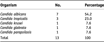
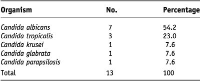
Majority of patients were men in the age group of 51–70 years. On Gram stain, 8 swabs showed presence of yeast cells and pseudohyphae and 13 swabs showed growth on Sabouraud’s dextrose agar. Out of 13 isolates, 7 were Germ tube test positive.
The species isolated are shown in Table 1.
Table 1 shows Candida species isolated
DISCUSSION
Cancer and cancer treatment profoundly impairs oral health. Oral complications presenting as mucositis, xerostomia, bleeding and infections are commonly seen. The clinical diagnosis of oral infections may be difficult due to reduced inflammatory responses in the immunocompromised host. It has been found that oropharyngeal colonization of Candida species may increase the risk of systemic infection especially when oral ulcers develop during the neutropenic episodes. Risk factors for systemic fungal infection also include the use of broad-spectrum antibiotics and steroids.Reference Alberth, Majoros, Kovalecz, Borbás, Szegedi, Márton and Kiss7
Chemotherapy or radiotherapy induced local tissue damage leads to oral mucositis. Damage to salivary glands results in hypofunctioning of salivary glands ultimately compromising the oral hygiene badly.Reference Lalla, Latortue, Hong, Ariyawardana, D’Amato-Palumbo, Fischer, Martof, Nicolatou-Galitis, Patton, Elting, Spijkervet and Brennan2 These factors probably play a major role in causation of oral candidiasis. The epidemiology of C. albicans and other yeasts from the oropharynx of patients receiving radiation for head and neck cancer is quite varied. C. albicans is the predominant organism associated with symptomatic infection.Reference Redding, Zellars, Kirkpatrick, McAtee, Caceres, Fothergill, Lopez-Ribot, Bailey, Rinaldi and Patterson5 In recent studies, C. glabrata has emerged as a notable pathogenic agent in oropharyngeal candidiasis in patients receiving radiotherapy.Reference Dongari-Bagtzoglou, Dwivedi, Ioannidou, Shaqman, Hull and Burleson8
The incidence of colonization and infection of Candida has been reported from 43–90% and 13–52%, respectively.Reference Koc and Aktas4–Reference Belazi, Velegraki, Eremondi, Andreadis, Hini, Arsenis, Eliopoulou, Destouni and Antoniades6,Reference Galitis, Velegraki, Sotiropoulou-Lontou, Dardoufas, Kouloulias, Kyprianou, Kolitsi, Skarleas, Pissakas, Papanicolaou and Kouvaris9–Reference Epstein, Freilich and Le10 In our study, we found the fungal infection in 21.6% of cases, corresponding with the other studies.
Still Candida albicans is the dominant species causing the oropharyngeal candidiasis in immunocompromised patients, dental wearers, systemic malignancies or patients undergoing radiation therapy. The emergence of other species can be attributed to prophylactic use of antifungal medications.Reference Schelenz, Abdallah, Gray, Stubbings, Gow, Baker and Hunter3–Reference Dongari-Bagtzoglou, Dwivedi, Ioannidou, Shaqman, Hull and Burleson8,Reference Jain, Veerabhadrudu, Kulkarni, Ajantha, Shubhada and Amruthkishan11–Reference Baradkar and Kumar15
The identification of species causing candidiasis is important and crucial for both clinical treatment and epidemiological studies. As pathogenicity and antifungal susceptibility varies among various species, an essential prerequisite for the laboratories is a suitable culture medium which facilitates the recovery and differentiation of Candida species. The conventional identification of pathogenic fungi in the clinical microbiology laboratory involves the examination of colony morphology, microscopic morphology and the assessment of various biochemical reactions. The presence of more than one Candida species in the oral cavity of the same host is not uncommon. By conventional methods, identification of yeasts may take several days, and, in presence of mixed cultures, it is difficult to isolate and identify the organisms. For differentiation of respective species of Candida, various phenotypic methods have been reported such as formation of chlamydospores; the pattern of carbohydrate assimilation, D-glucosidase activity, Staib agar, Niger agar, Tobacco agar and growth in Sabouraud agar at temperatures between 42 and 45°C.Reference Chavasco, Paula, Hirata, Aleva, Melo, Gambale, Ruiz Lda and Franco16 There are new CHROM medias which are being advocated in identifying the Candida species. New chromogenic media can complement traditional identification methods for identifying clinical yeast isolates.Reference Eraso, Moragues, Villar-Vidal, Sahand, González-Gómez, Pontón and Quindós17–Reference Abraham18 Recently, DNA-based methods such as DNA–DNA reassociation, DNA fingerprinting and southern hybridization with appropriate DNA probes have been described to identify Candida species in culture and in clinical materials. Currently, there are other molecular techniques apart from DNA-based methods such as pulsed field electrophoresis and PCR-based methods.Reference Chavasco, Paula, Hirata, Aleva, Melo, Gambale, Ruiz Lda and Franco16 However, these genotypic methods have the disadvantage of being laborious and time- consuming, and besides they require specialized equipments.Reference Liguori, Lucariello, Colella, De Luca and Marinelli19–Reference Das, Brown, Kellar, Holloway and Morrison20 These state-of-the-art techniques are not readily available to the majority of laboratories in India. Hence, identification of organisms largely rests on traditional culture and biochemical studies in Indian setup.
Chakrabarti et al.Reference Chakrabarti, Ghosh, Kanta and Kumar21 expressed that the various methods done for detection of antifungal susceptibility pattern are having very desperate outcomes, and the results are often noncomparable. The disc diffusion method for study of antifungal susceptibility pattern is simple and easy to perform. Reliable, reproducible antifungal susceptibility tests depends on standardization of several factors like pH, inoculum size, liquid versus solid medium, medium formulation, time and temperature of inoculation. They observed that 2–5% Candida strains were resistant to Amphotericin B and 10–22% strains were intermediate. In case of Fluconazole, resistance was observed in 13–20% strains. These simple disc diffusion methods can suitably be standardized in the laboratories as it showed a good correlation with the reproducible broth dilution method. However, when the results are equivocal, especially for azoles, the broth dilution test should be used.Reference Chakrabarti, Ghosh, Kanta and Kumar21
In our study, few strains of Candida showed resistance to the drugs used; (Table 2) however, the number studied is too low to make any decisive comment. Topical agents are considered preferable to systemic agents due to lower risk of side effects and drug interactions. The previously published studies have used fluconazole as tropical agent, and it had shown to be very effective in the prevention of clinical oral fungal infection and in reducing oral fungal colonization in patients receiving cancer therapy. Lately, studies are being published showing resistance to FluconazoleReference Schelenz, Abdallah, Gray, Stubbings, Gow, Baker and Hunter3 as in our study. A close monitoring is required for frequency of different non-albicans Candida species isolated from patients with systemic infection and of antifungal susceptibility pattern because of recent increased use of Fluconazole as prophylactic agent against oral candidiasis.
Table 2 shows antifungal susceptibility pattern.

The pathogenic agents colonizing the oral cavity may induce symptomatic infections, often systemic ones, and may promote the development of infections with drug-resistant strains. Regular dental check-up examinations, oral microbial surveillance and application of professional oral hygienic measures in patients with oral cancer may decrease the incidence, duration and severity of infectious complications. Considering the high prevalence of oral candidiasis in cancer patients, identification of more effective topical antifungal agents to avoid the potential side effects of systemic agents would be beneficial.
LIMITATIONS OF THE STUDY
The number of positive culture is very small to make confirmatory remarks about the antifungal susceptibility pattern of the Candida species.