Introduction
It has been over a decade since the birth of Dolly, the first somatic cell nuclear transfer (SCNT) animal to be produced, which is a revolution both in theory and in methodology (Wilmut et al., Reference Wilmut, Schnieke, McWhir, Kind and Campbell1997). Since then, viable offspring have been produced in many other species using similar techniques (Wakayama et al., Reference Wakayama, Perry, Zuccotti, Johnson and Yanagimachi1998; Baguisi et al., Reference Baguisi, Behboodi, Melican, Pollock, Destrempes, Cammuso, Williams, Nims, Porter, Midura, Palacios, Ayres, Denniston, Hayes, Ziomek, Meade, Godke, Gavin, Overstrom and Echelard1999; Li et al., Reference Li, Sun, Chen, Liu, Wisely, Zhou, Renard, Leno and Engelhardt2006). However, the efficiencies of development of healthy offspring derived from this standard nuclear transfer (SNT) technique remain low, being estimated at less than 5% of oocytes used. Furthermore, this technique contains complex procedures that require considerable technical skills. The procedures of enucleation and donor nucleus transfer mainly rely on micromanipulation, which is technically demanding, time consuming, and clearly damages cytoplast spatial organization. Several approaches, including handmade cloning (Vajta et al., Reference Vajta, Lewis, Hyttel, Thouas and Trounson2001), reverse-order nuclear transfer (Peura, Reference Peura2003) and induced-enucleated nuclear transfer (INT) (Ibáñez et al., Reference Ibáñez, Albertini and Overstrom2003), have been attempted to simplify the procedures and improve the developmental competence of nuclear transfer (NT) embryos. Among these recently developed nuclear transfer techniques, INT can produce larger numbers of recipient cytoplasts in a shorter time and with reduced labour intensity and is, therefore, a very attractive prospect. A number of reagents have been utilized to achieve complete enucleation, including etoposide (Elsheikh et al., Reference Elsheikh, Takahashi, Katagiri and Kanagawa1998), a combination of etoposide and cycloheximide (Fulka & Moor, Reference Fulka and Moor1993), or ethanol and demecolcine (DC) (Ibáñez et al., Reference Ibáñez, Albertini and Overstrom2003). Although viable offspring have been obtained after transfer of cloned embryos produced by DC-induced enucleation in the mouse (Baguisi & Overström, Reference Baguisi and Overström2000; Gasparrini et al., Reference Gasparrini, Gao, Ainslie, Fletcher, McGarry, Ritchie, Springbett, Overstrom, Wilmut and De Sousa2003), the developmental competence of reconstructed embryos derived from DC induced-enucleated cytoplasts is impaired compared with those enucleated by conventional mechanical method. In INT, recipient cytoplasts used for NT were pre-activated in order to induced enucleation (IE) and this process usually occurred in telophase (TII) prior to the donor nuclei transfer. However, metaphase II (MII) cytoplasts and delayed activation were thought to be important for the successful cloning of mice and other species by allowing an increased opportunity for restoration or recruitment of some chromatin-associated factors that were important for nuclear reprogramming.
To ensure the exposure of donor nuclei to MII cytoplasts rather than activated cytoplasts in INT, in this study, we modified the INT procedures by transferring donor nuclei into recipient cytoplasts prior to activation and induced enucleation. The dynamic changes of maternal and transferred donor nuclei in the reverse-order and induced-enucleated nuclear transfer (RINT) oocytes were monitored to evaluate the feasibility of this new NT technique by timed immunofluorescence. In vitro development of embryos derived from RINT, INT and SNT was compared in order to assess the efficiency of RINT.
Materials and Methods
All chemicals used in the present study were purchased from the Sigma Chemical Company unless otherwise noted. Disposable, sterile plasticware was purchased from Nunclon.
Animals
ICR female mice, aged 8 to 10 weeks, were used as oocyte donors. All animals used in this study were maintained according to the guidelines of the experimental laboratory animal centre of Xi'an Medical University. The mice were kept under controlled temperature (23–25°C).
Isolation of MII oocytes
ICR female mice were induced to superovulate by consecutive treatment of PMSG (10 IU) and hCG (10 IU) 48 h apart. Cumulus–oocyte complexes (COCs) were collected from oviducts 20 h after treatment with hCG. The cumulus cells were dispersed by treatment with 0.1% bovine testicular hyaluronidase in phosphate-buffered saline (PBS).
Culture of donor cells for SCNT
Primary culture was obtained by mincing 14-day ICR fetal tissue with sterile scissors in a 35 mm Petri dish. The small tissue pieces (approximately 1 mm in diameter) were cultivated for 1–2 weeks in DMEM/F12 containing 20% fetal calf serum (FCS), 1 mM sodium pyruvate, 100 IU/ml penicillin and 100 mg/ml streptomycin. Once cells had reached 90% confluence, they were trypsinized and recultivated at a concentration of 1 × 10⁶ cells/ml. The second to fifth passages were utilized as nucleus donors. Cells were induced into G0/G1 phase of cell cycle by serum deprivation (0.5% FCS, 2 days) for subsequent use in SCNT experiments.
Standard nuclear transfer, activation and embryo culture
For enucleation, 15% of the circumference of the zona pellucida of the MII oocyte was dissected with a fine glass needle directly above the nucleus in a micromanipulation chamber in PBS containing5 μg/ml cytochalasin B and 20% FBS. A blunt polished pipette with an inside diameter of 15 μm was inserted through the slit and the nucleus was aspirated into the enucleation pipette. For nuclear transfer, cultured fetal fibroblast cells, which were induced into G0/G1 phase of cell cycle by serum deprivation, were transferred into the perivitelline space of enucleated oocytes and the oocyte-cell couplets were electrically fused using two direct current pulses of 120 V/mm for 20 μs in 0.28 M mannitol supplemented with 0.1 mM MgSO4 and 0.1% polyvinyl alcohol (PVA). Successfully reconstructed embryos were kept in potassium simplex optimized medium (KSOM) containing 5 μg/ml cytochalasin B for 2 h until activation. Reconstructed embryos were activated by incubation in calcium-free KSOM supplemented with 10 mM SrCl2 and 5 μg/ml cytochalasin B for 6 h and were incubated further for 120 h in 50 μl droplets of KSOM under mineral oil at 37°C and 5% CO2 in air.
Induced-enucleated nuclear transfer
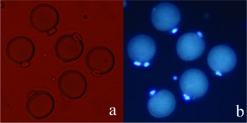
For INT, MII oocytes were first activated by exposure to 7% ethanol in PBS for 7 min. Then, they were incubated in KSOM containing 0.4 μg/ml DC for 90 min immediately after activation. The success of IE was assessed by staining with 10 μg/ml Hoechst 33342. Those oocytes that exhibited all chromatin material in expelled polar bodies (Pb) and the obvious border between Pb and cytoplast were scored as IE (Fig. 1). The oocytes scored as IE were selected for Pb aspiration by using a glass micropipette of 15 μm in diameter. Nuclear transfer, fusion, activation and culture of reconstructed embryos were performed as described in SNT.
Mouse oocytes following demecolcine induced enucleation. Obvious border between Pb and cytoplasts was observed (a). Induced enucleated oocytes were assessed under an inverted microscope using ultraviolet light. The oocytes exhibited that all chromatin materials were expelled with the second Pb (b). Original magnification × 200.

Reverse-order and DC induced-enucleated nuclear transfer
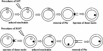
For RINT, the donor cells were transferred into recipient oocytes prior to enucleation. The transfer of donor cells into recipient oocytes was performed as described in SNT. Immediately after stimulation by electrical pulse for fusion, reconstructed embryos were cultured in KSOM containing 0.4 μg/ml DC. Fusion results were examined under a stereomicroscope 30 min later. The successfully reconstructed oocytes were activated by 7% ethanol for 5 min and then cultured in KSOM containing 0.4 μg/ml DC for IE. An outline of the INT and RINT techniques was illustrated in Fig. 2.
Outline of the procedures of RINT and INT techniques. See text for details.

Immunocytochemistry analysis of the dynamic changes of maternal and transferred donor nuclei in the RINT oocytes
In order to evaluate the feasibility of the RINT technique, and provide a basis for selection of successfully reconstructed embryos for culturing, the dynamic changes of maternal and transferred donor nuclei in the RINT oocytes were monitored by timed immunofluorescence. All reagents for fixation, washing and blocking were purchased from Beyotime Institute of Biotechnology (Jiangsu, China). RINT oocytes were fixed for 1 h at 20, 40, 60 min post-activation and DC treatment according to the experimental design, then permeabilized with 0.1% Triton X-100 for 4 min, and blocked overnight. Embryos were incubated in α-tubulin primary antibodies (Abcam) for 3 h. Then, they were washed extensively and incubated with FITC-conjugated secondary antibody for 1 h. DNA was stained with 10 μg/ml propidium iodide (PI) for 30 min, and the embryos were mounted on slides for confocal microscopy. Fluorescence was detected under Zeiss LSM 510 laser confocal scanning system (Carl Zeiss).
Selection, activation and culture of RINT embryos
Successfully reconstructed RINT embryos were selected according to the indicator of Immunocytochemistry. After DC treatment for 60 min, RINT oocytes were stained with 10 μg/ml Hoechst 33342 for 10 min and observed under ultra-violet light. The oocytes exhibiting a distinct border between Pb and cytoplast, and containing only one part of chromatin material in the cytoplasts were scored as RINT success. Activation and culture of reconstructed embryos were performed as described in SNT.
Count of the total cell number of blastocysts
The total cell number of day 5 blastocysts was determined. Blastocysts were fixed in buffered formal saline (containing 5% formaldehyde), mounted on slides under coverslips, and subsequently stained with Hoechst 33342. The number of nuclei in the blastocysts was then recorded under fluorescence microscope.
Statistical analysis
Four replicates for each experiment were conducted. All percentage data were pooled and then tested by chi-squared analysis using the DPS Data Processing System 6.01 software (Refine Information Tech. Co. Ltd). Differences between experimental groups were considered significant at p < 0.05. Cell numbers of individual groups were compared using the general linear model (SAS Genmod procedure), where p < 0.05 was considered significant between experimental groups.
Results
Immunocytochemistry analysis of the dynamic changes of maternal and transferred donor nuclei and meiotic progression in the RINT oocytes after activation and DC treatment
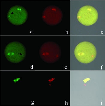
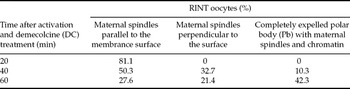
As shown in Fig. 3 and Table 1, the effects of RINT on the dynamic changes of maternal and transferred donor nuclei and meiotic progression in the RINT oocytes were evaluated at three intervals, from 20 min to 60 min after activation. Because the spindle was disrupted by DC during the RINT procedures and the structure of the spindle could not be clearly observed, it was difficult to determine the exact meiotic resumption phase of the RINT oocytes. We evaluated the process of meiotic resumption by the relative location (parallel or perpendicular) of spindle and the surface of the oocyte membrane or the extrusion of the second Pb. At 20 min post-activation, 81.1% of the observed maternal spindles were located parallel to the surface of the reconstructed oocytes and microtubules were observed around the transferred donor nuclei in all the RINT oocytes. At 40 min post-activation, the maternal spindles in 50.3% of the observed oocytes were still located parallel to the surface of the reconstructed oocytes; only 32.7% of the observed oocytes processed to maternal spindles were located perpendicular to the surface and 10.3% oocytes showed completely expelled Pb that contained all maternal spindles and chromatins. Although α-tubulin was observed around most of the donor nuclei (76.6%, n = 58), the intensity of the fluorescence was obviously weaker compared with that observed at 20 min post-activation. Alpha-tubulin was not observed around the transferred donor nuclei and about half of the observed oocytes showed one or two completely extruded Pb (42.2%, n = 83) at 60 min post-treatment. In all observed oocytes, none showed extrusion of the donor nucleus with the second Pb, while 42.2% of oocytes at 60 min post-treatment showed completely extrusion of all maternal chromatin with Pb. These results provided a basis for selection of successfully reconstructed RINT embryos for further culture.
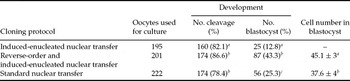
Effects of NT technique on the in vitro developmental capacity of reconstructed embryos.

a–cWithin the same column, values with different superscripts are different (p < 0.01).
Confocal observation of the dynamic changes of maternal and transferred donor nuclei in the RINT embryos. Oocytes were fixed at 20 min (a–c), 40 min (d–f) and 60 min (g–i) after activation and demecolcine treatment. Microtubules were stained by indirect immunofluorescence with antibodies to α-tubulin (green) and DNA was counterstained with propidium iodide (red). For each oocyte, anti-tubulin staining patterns were shown on the left, propidium iodide staining patterns were shown in the middle and merged detailed images were shown on the right. Arrows in (a) and (d) indicated α-tubulin around the transferred donor nuclei. Microtubules were not observed around the transferred donor nuclei at 60 min (g). Original magnification ×200.

Effects of nuclear transfer techniques on IE and in vitro developmental competence of cloned embryos
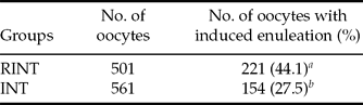
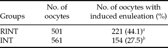
As shown in Tables 2 and 3, although there was no difference in cleavage rate, the rates of successful IE and blastocyst development were significantly increased in RINT compared with INT. Compared with SNT, there was no difference in cleavage rate, but the blastocyst developmental rate was significantly increased in RINT group. Blastocysts derived from RINT have a much higher total cell number than those from SNT.
Meiotic progressin of reverse-order and induced-enucleated nuclear transfer (RINT) oocytes.

Effects of reverse-order and induced-enucleated nuclear transfer (RINT) and induced-enucleated nuclear transfer (INT) on induced enucleation efficiency in mouse oocytes.
a,bWithin the same column, values with different superscripts are different (p < 0.01)
Discussion
In the present study, we modified the INT technique by transferring donor nuclei into recipient cytoplasts prior to IE and developed a new NT technique, which we call RINT. We found this new NT technique was feasible because none of the observed RINT oocytes showed extrusion of transferred donor nuclei, while 42.2% of the oocytes showed extrusion of all maternal chromosomes and spindles with the second Pb after DC treatment. Compared with INT, the NT efficiencies were significantly improved in RINT in both of IE and blastocyst rates.
As the major cytoskeleton component, microtubules play an important role in the modulatio of the movement of chromosomes during meiosis and cell division. Disruption of spindle microtubules by microtubule specific inhibitors, such as etoposide and DC, has been shown to suppress the extent and completion of spindle rotation and extrusion of Pb (Russell et al., Reference Russell, Ibanez, Albertini and Overstrom2005; Hou et al., Reference Hou, Lei, Liu, Cui, An and Chen2006). Although the exact mechanism underlying complete extrusion of all maternal chromatin after DC treatment remains unclear, previous studies indicated that timely disruption of spindle microtubules in some, but not complete extent was necessary for success of IE (Gasparrini et al., Reference Gasparrini, Gao, Ainslie, Fletcher, McGarry, Ritchie, Springbett, Overstrom, Wilmut and De Sousa2003; Ibáñez et al., Reference Ibáñez, Albertini and Overstrom2003). Our studies demonstrated that none of the RINT oocytes showed extrusion of donor nuclei with the second Pb, while about half of them completely extruded all maternal chromatin after DC treatment. In the present study, spindle microtubules can be observed at 80 min after DC treatment, which is consistent with previous study (Gasparrini et al., Reference Gasparrini, Gao, Ainslie, Fletcher, McGarry, Ritchie, Springbett, Overstrom, Wilmut and De Sousa2003). However, microtubules around the transferred donor nuclei were completely disrupted and were not observed under confocal microscope at 40 min after DC treatment. These results indicated that the disruption of microtubules by DC might be affected by the patterns of microtubule organization. The complete disruption of microtubules around donor nuclei might explain why donor nuclei were not extruded in RINT procedures.
The RINT technique described here differs from INT technique in several aspects. The most significant difference is the exposure of donor nuclei to factors in MII cytoplasm. In all successful SCNT cloning experiments, oocytes in MII phase are confirmed to be the most suitable recipient. Various factors in the MII cytoplasm have been reported to facilitate the remodelling and reprogramming of somatic cell nuclei, and such factors are lost or insufficient following fertilization or artificial activation within 1 to 4 h (Szollosi et al., Reference Szollosi, Czolowska, Soltynska and Tarkowski1986; Szollosi et al., Reference Szollosi, Czolowska, Szollosi and Tarkowski1988; Latham, Reference Latham1999; Kim et al., Reference Kim, Ogura, Nagata and Aoki2002; Gao et al., Reference Gao, Chung, Parseghian, King, Adashi and Latham2004). In INT procedures, oocytes are activated and cultured in DC for about 90 min to induce enucleation before donor nuclei were transferred, so at least two hours had passed and the recipient cytoplasts had entered TII phase when the donor nuclei were exposed to recipient cytoplasts. Another difference of RINT technique is that the donor nuclei were co-incubated with the maternal chromosome complements for more than 1 h. It has been reported that numbers of important cytoplasmic factors, such as AKAP95 and active MPF, are localized near metaphase chromosomes (Casas et al., Reference Casas, Betancourt, Bonilla, Duculomb, Zayas and Trejo1999; Eide et al., Reference Eide, Coghlan, Orstavik, Holsve, Solberg, Skalhegg, Lamb, Langeberg, Fernandez, Scott, Jahnsen and Tasken1998). The presence of maternal chromatin associated factors was reported to be beneficial for nuclear reprogramming (Kang et al., Reference Kang, Koo, Park, Choi, Lee and Han2001; Latham, Reference Latham2005). Unfortunately, in SNT and INT technique, most of the cytoplasmic factors localized near metaphase chromosomes are lost with enucleation. So the advantage of RINT in this study can be summarized as follows: (i) MII cytoplasts as recipient cytoplasts; (ii) factors existing in or surround oocyte nucleus; and (iii) minimal loss of the cytoplasmic volume and protein/molecular content.
In this study, our aim was to improve the developmental competence of cloned embryos derived from INT technique. Interestingly, IE rate in RINT was also significantly improved. Low efficiency of IE was another reason that hampered the wide application of the INT protocol. The reported IE rate ranged from 26 to 57% (Baguisi & Overström, Reference Baguisi and Overström2000; Gasparrini et al., Reference Gasparrini, Gao, Ainslie, Fletcher, McGarry, Ritchie, Springbett, Overstrom, Wilmut and De Sousa2003). The genetic background of the oocyte and the concentration and duration of DC treatment on IE rate have been studied extensively. However, to the best of our knowledge, the effect of activation protocol on IE rate has not been studied. In our study, most oocytes with a completely expelled Pb were IE after DC treatment. The low rate of IE was due to the failure of completed second Pb extrusion (Fischer et al., Reference Fischer, Ibanez, Cibelli, Albertini and Overstrom2002; Ibáñez et al., Reference Ibáñez, Sanfins, Combelles, Albertini and Overström2002). Therefore, we hypothesized that the process in oocytes activation was altered after DC treatment. The current activation protocol was less efficient for induced enucleation. There were dual activation stimulations during the DC treatment in RINT. To fuse the donor cell with the recipient oocyte, electrical pulse was performed. Following this event, the fused karyoplast–cytoplast couplets underwent activation stimulation again by 7% ethanol to induce enucleation. High IE rate in the present study may be due to the two different activation stimulations, which activate oocytes by different activation ways and may contribute to the full activation of the oocyte and the second Pb extrusion.
Blastocyst quality analysis revealed that cloned embryos derived from SNT have a much lower total cell number than embryos derived from the in vivo group. This inferior blastocyst quality can be overcome by application of RINT, which gave comparable total cell number between SNT and the in vivo group. A total of 200 and 160 cloned embryos derived from SNT and RINT were transfer to 10 ICR recipients respectively. However, no living fetus was obtained (data not shown). The successful development to term of a cloned mouse was strictly dependent on the genetic background of the oocyte. Most of the living cloned mice were derived from B6D2F1 (Ogura et al., Reference Ogura, Inoue, Takano, Wakayama and Yanagimachi2000; Wakayama & Yanagimachi, Reference Wakayama and Yanagimachi2001). Therefore, to evaluate developmental competence of cloned mice derived from RINT protocol, mouse strains such as B6D2F1 should be studied in the future.
In conclusion, the RINT technique described here is feasible and efficient, and might offer another choice of methodology for NT research. Plans are being undertaken to simplify this new technique by combining it with a zona-free cloning technique.
Acknowledgements
We thank Guoliang Pei for the assistance with the confocal microscopy. This work was supported by the 863 Programme from the Ministry of Science and Technology of China (No. 2004AA213072).