INTRODUCTION
The genus Plasmodium (Haemosporida: Plasmodiidae) is a cosmopolitan group of heteroxen protists composed of more than 100 described species in different vertebrate hosts such as mammals, reptiles and birds (Garnham, Reference Garnham1966; Valkiūnas, Reference Valkiūnas2005). Plasmodium species are known to be transmitted by several species of mosquitoes (Culicidae) from different genera (Garnham, Reference Garnham1966; Atkinson and van Riper, Reference Atkinson, Van Riper, Loye and Zuk1991; Lourenço-de-Oliveira et al. Reference Lourenço-de-Oliveira and Castro1991; Valkiūnas, Reference Valkiūnas2005; Kimura et al. Reference Kimura, Darbro and Harrington2010; Ejiri et al. Reference Ejiri, Sato, Kim, Tsuda, Murata, Saito, Watanabe, Shimura and Yukawa2011; Njabo et al. Reference Njabo, Cornel, Bonneaud, Toffelmier, Sehgal, Valkiūnas, Russell and Smith2011; Lyimo et al. Reference Lyimo, Keegan, Ranford-Cartwright and Ferguson2012; Santiago-Alarcon et al. Reference Santiago-Alarcon, Havelka, Schaefer and Segelbacher2012). Plasmodium can also be found parasitizing wild birds and domestic fowls (Garnham, Reference Garnham1966; Valkiūnas, Reference Valkiūnas2005). Recently, a large number of studies have been conducted to assess the prevalence and molecular and phylogenetic relationships of Plasmodium spp. from wild birds in different habitats and continents (Sehgal, Reference Sehgal2010; Belo et al. Reference Belo, Pinheiro, Reis, Ricklefs and Braga2011, Reference Belo, Rodríguez-Ferraro, Braga and Ricklefs2012; Marzal et al. Reference Marzal, Ricklefs, Valkiūnas, Albayrak, Arriero, Bonneaud, Czirják, Ewen, Hellgren, Hořáková, Iezhova, Jensen, Križanauskienė, Lima, de Lope, Magnussen, Martin, Møller, Palinauskas, Pap, Pérez-Tris, Sehgal, Soler, Szöllosi, Westerdahl, Zetindjiev and Bensch2011; Asghar et al. Reference Asghar, Westerdahl, Zehtindjiev, Ilieva, Hasselquist and Bensch2012; Lapointe et al. Reference Lapointe, Atkinson and Samuel2012; Lacorte et al. Reference Lacorte, Félix, Pinheiro, Chaves, Almeida-Neto, Neves, Leite, Santos and Braga2013). Despite the relevance of malaria to domestic fowl due to its potential economic importance, few studies in this field have been conducted.
There are two main known Plasmodium species that naturally infect chickens: Plasmodium gallinaceum and Plasmodium juxtanucleare (Valkiūnas, Reference Valkiūnas2005). Plasmodium gallinaceum has a restricted global distribution, and its parasite–host interactions can be used as an excellent model in many types of studies, including those related to pathogenesis, cellular interactions and immunological responses (Frevert et al. Reference Frevert, Späth and Yee2008; Nagao et al. Reference Nagao, Arie, Dorward, Fairhurst and Dvorak2008; Macchi et al. Reference Macchi, Quaresma, Herculano, Crespo-López, DaMatta and Nascimento2010). By contrast, P. juxtanucleare has been found to infect chickens worldwide; nevertheless, this infection has been poorly studied (Versiani and Gomes Reference Versiani and Gomes1941; Al-Dabagh, Reference Al Dabagh1961; Bennett and Warren Reference Bennett and Warren1966a, Reference Bennett and Warrenb; Elisei et al. Reference Elisei, Fernandes, Forlano, Madureira, Scofield, Yotoko, Soares, Araújo and Massard2007; Vashist et al. Reference Vashist, Silveira, Cabral, Amaral, Soares and D'Agosto2008; Silveira et al. Reference Silveira, Damatta and D'Agosto2009a). Malaria caused by P. juxtanucleare in chickens generally has a benign evolution (Bennett and Warren, Reference Bennett and Warren1966a, Reference Bennett and Warrenb; Silveira et al. Reference Silveira, Vashist, Cabral, Amaral, Soares and D'Agosto2009a), frequently consisting of an asymptomatic chronic phase evidenced by low parasitaemia in the peripheral blood. However, the infection may be significantly pathogenic during the acute phase, as previously demonstrated (Al Dababh, Reference Al Dabagh1960, Reference Al Dabagh1961; Krettli, Reference Krettli1972; Silveira et al. Reference Silveira, Vashist, Cabral, Amaral, Soares and D'Agosto2009a). It is noteworthy that despite having low levels of bloodstream circulating parasites, fowls experiencing chronic infections could represent an excellent source of gametocytes to susceptible vectors (Santos-Prezoto et al. Reference Santos-Prezoto, D'Agosto and Daemom2004), spreading the malarial infection to susceptible hosts. The susceptibility and pathogenicity of avian hosts to P. juxtanucleare infection are variable, but high mortality has been reported in experimentally infected chicks (Al-Dabagh, Reference Al Dabagh1961). In addition, studies related to natural infection have demonstrated that infected white-eared pheasants (Crossoptilon crossoptilon) and penguins (Spheniscus demersus) exhibited both clinical signs and moderate to high levels of parasitaemia (Grim et al. Reference Grim, van der Merwe, Sullivan, Parsons, Mc Cutchan and Cranfield2003; Murata et al. Reference Murata, Nii, Sasaki, Ishikawa, Sato, Sawabe, Tsuda, Matsumoto, Suda and Ueda2008). The clinical signs of infection caused by P. juxtanucleare are non-specific, but depression, apathy, weight loss and anaemia may be observed (Grim et al. Reference Grim, van der Merwe, Sullivan, Parsons, Mc Cutchan and Cranfield2003; Valkiūnas, Reference Valkiūnas2005; Murata et al. Reference Murata, Nii, Sasaki, Ishikawa, Sato, Sawabe, Tsuda, Matsumoto, Suda and Ueda2008). Necropsy of clinically affected penguins may reveal pulmonary oedema and hydropericardium (Grim et al. Reference Grim, van der Merwe, Sullivan, Parsons, Mc Cutchan and Cranfield2003).
The potential pathogenic effect of simultaneous infection with other organisms on the outcome of avian malarial infection is poorly understood. The interaction of different pathogens in a specific host may have synergistic or antagonistic effects. Coinfection of birds by Plasmodium relictum and Plasmodium ashfordi, two generalist avian malaria parasites, has been demonstrated to have a synergistic effect in siskins (Spinus spinus) and crossbills (Loxia curvirostra) (Palinauskas et al. Reference Palinauskas, Valkiūnas, Bolshakov and Bensch2011). However, an antagonistic effect was observed in chickens infected by P. gallinaceum and the nematode Ascaridia galli; the resulting avian malaria caused a significant reduction in the establishment of intestinal parasites (Permin and Juhl, Reference Juhl and Permin2002). Although P. gallinaceum is considered one of the best models for experimental infection, chickens infected by P. gallinaceum frequently die due to the effects of severe infection, interrupting the parasitic cycle and precluding continuity in experimental studies. Thus, the P. juxtanucleare–chicken host model could provide substantial information, allowing measurement of the impacts caused by different pathogens during chronic malarial infection. In this context, coinfection with this plasmodia species and the chicken anaemia virus (CAV), a widespread infection (Nogueira et al. Reference Nogueira, Piantino, Martins, Luiz, Lazzarin and Brentano2007; Craig et al. Reference Craig, Rimondi, Delamer, Sansalone, König, Vagnozzi and Pereda2009; Eltahir et al. Reference Eltahir, Qian, Jin, Wang and Qin2011; Snoeck et al. Reference Snoeck, Komoyo, Mbee, Nakouné, Le Faou, Okwen and Muller2012; Marin et al. Reference Marin, Barrios, Rios, Resende, Resende, Santos and Martins2013) that is potentially immunosuppressive (Ragland et al. Reference Ragland, Mazija, Cvelic-Cabrilo, Savic, Novak and Pogacnik1998; Kuscu and Gurel, Reference Kuscu and Gurel2008; Schat, Reference Schat2009; Haridy et al. Reference Haridy, Sasaki, Ikezawa, Okada and Goryo2012; Gallardo et al. Reference Gallardo, van Santen and Toro2012), could be considered a useful model in determining possible synergistic or antagonistic effects.
The aim of this present study was to assess infection caused by P. juxtanucleare in a model using chicks previously infected by a potentially immunosuppressive virus and to determine whether the pathogenic effect on the host is aggravated during coinfection. The experimental model established here, based on the infection of specific-pathogen-free (SPF) chickens with P. juxtanucleare after CAV immunosuppression, simulates the natural transmission conditions of both pathogens and could be useful in developing various animal management strategies.
MATERIALS AND METHODS
Animals
All of the experimental protocols were approved by the Universidade Federal de Minas Gerais Animal Ethics Committee (protocol number 166/2010).
Eighty SPF eggs (Laboratório Biovet, Vargem Grande Paulista, SP, Brazil) were maintained in brooders (Premium Ecological, Belo Horizonte, MG, Brazil) under controlled temperature and humidity (37·5 °C; 60%). The hatchlings were selected based on complete umbilical healing and transferred randomly to mosquito-free screened cages with water and food provided ad libitum in an experimental infection facility at the Veterinary College of Universidade Federal de Minas Gerais, Brazil.
Plasmodium juxtanucleare and CAV
Plasmodium juxtanucleare was obtained from a naturally infected chicken from a small farm in Minas Gerais state, Brazil (Itabirito, MG – 20°16′03.17″S; 43°49′19.06″W) and was used directly for all experimental inoculations. The strain was named the P. juxtanucleare FMV strain. The infected blood of this donor chicken was cryopreserved and maintained in Laboratório de Malaria, UFMG, Brazil, for future experiments.
The CAV inoculum was prepared from a live attenuated vaccine (Cuxhaven-1 strain®, Thymovac, AvilPro, Lohmann Animal Health, Germany) containing 105·5 TCID50, and was administered via intraperitoneal injection (ip), as previously described (Hoop, Reference Hoop1992). The administration route of the inoculum and the dose used did not follow the recommendations of the manufacturer, which suggest oral vaccine administration at a dose of 104·5 TCID50 for chickens older than 8 weeks.
Plasmodium juxtanucleare infection was detected in stained blood smears (procedure described below) and subsequently confirmed by molecular diagnosis and sequencing (Hellgren et al. Reference Hellgren, Waldenström and Bensch2004).
Methylprednisolone acetate (26 mg kg−1) was given intramuscularly to the chicken donor to cause immunosuppression and to increase parasitaemia (Krettli, Reference Krettli1972). After 10 days, the chicken exhibited 1% parasitaemia and a total erythrocyte count of 2·4 × 106μL; at that time, blood was collected and used as P. juxtanucleare inoculum for other chicks.
The donor chicken was negative for CAV DNA as determined by PCR and for CAV-specific antibodies as determined by ELISA (FlockCheck®, Idexx Laboratories, Maine, USA). ELISAs were performed using serum diluted to 1 : 100 according to the manufacturer's specifications. The PCR for CAV detection was performed as described below.
To verify the absence of CAV infection before the chicks were subjected to experimental inoculations, six individuals were euthanased, and the blood, bursa of Fabricius, spleen, thymus and gonads were collected for CAV detection by PCR. In addition, an ELISA was performed to detect CAV-specific antibodies.
Molecular CAV detection
The CAV diagnosis was confirmed by PCR using DNA extracted from different tissues and by the silica adsorption method (Boom et al. Reference Boom, Sol, Salimans, Jansen, Wertheim-van and van der Noordaa1990), in the presence of NaI (chaotropic agent) (Vogelstein and Gillespie, Reference Vogelstein and Gillespie1979) according to the method adapted by Marin et al. (Reference Marin, Barrios, Rios, Resende, Resende, Santos and Martins2013), using peripheral blood from the chicks. The genomic region encoding the VP1 protein of CAV was amplified, using previously described primers (Todd et al. Reference Todd, Mawhinney and McNulty1992): CAV1-F 5′ACTGTTAAGATGGCAAGACGAGCT-3′141 and CAV2R-5′GGCTGAAGGATCCCTCCATTC-3′. For the amplification reaction, 300 ng of DNA, 1 μL of 1X buffer (200 mm Tris-HCl pH 8·4, 500 mm KCl), 2 mm of each dNTP, 1 μL of 1·5 mm MgCl2, 1 μL of each primer at a concentration of 10 pmol, 1 unit of Taq polymerase (Platinum Taq DNA Polymerase, Invitrogen, São Paulo, Brazil) and 50 μL of ultra-pure water were used. The PCR amplification consisted of an initial phase at 94 °C for 5 min, followed by 35 cycles at 94 °C for 30 s, 54 °C at 60 s and 72 °C for 90 s, and a final extension at 72 °C for 10 min. DNA extracted from Cuxhaven-1 strain® (Thymovac, AvilPro, Lohmann, Animal Health, Germany) was used as positive control.
Morphological analyses and parasitaemia determination in chicks infected by P. juxtanucleare
The morphological stages of P. juxtanucleare were identified according to the original description (Versiani and Gomes, Reference Versiani and Gomes1941). Blood smears were air-dried, fixed in absolute methanol and stained with Giemsa 10%. A minimum of 100 fields were examined at high magnification (1000×, Olympus® CX31 microscope, Tokyo, Japan). Parasitaemia was calculated by counting the number of infected erythrocytes per 10 000 erythrocytes in blood smears (Godfrey et al. Reference Godfrey, Fedynich and Pence1987).
Sequencing and phylogeny of P. juxtanucleare FMV field strain
Blood from the donor chicken (20 μL) was used for DNA extraction (Wizard Genomic DNA Purification Kit, PROMEGA®, Madison, Wisconsin, USA) according to the manufacturer's protocol. A nested-PCR protocol designed to amplify a 479 bp fragment from the mitochondrial cytochrome b (cyt b) gene was used (Hellgren et al. Reference Hellgren, Waldenström and Bensch2004); the primers HaemNFI 5′-CATATATTAAGAGAAITATGGAG-3′ and HaemR3 5′ ATAGAAAGATAAGAAATACCATTC-3′ were used in the first reaction. For the second PCR reaction, the primers HaemF 5′-ATGGTGCTTTCGATATATGCATG-3 and HaemR2 5′-GCATTATCTGGATGTGATAATGGT-3′ were used. PCR products were purified from 1·5% agarose gels using the QIAquick gel extraction kit (Qiagen®, Valencia, USA). Bi-directional sequencing with dye-terminator fluorescent labelling was performed in an ABI Prism 3100 automated sequencer (Applied Biosystems, Inc. Bedford, MA, USA). Genomic DNA from P. gallinaceum (obtained from experimentally infected chickens and kindly provided by Dr Paulo F. Pimenta – CPqRR-Fiocruz/MG, Belo Horizonte, Minas Gerais, Brazil) was used as a positive control. DNA from recently hatched non-infected SPF chicks (Laboratório Biovet, Vargem Grande Paulista, SP, Brazil) that were hatched at the Veterinary College, UFMG, was used as negative control.
Phylogenetic relationships among the cyt b lineages of P. juxtanucleare, including the cyt b sequences of P. juxtanucleare described in previous studies, and related haemosporidian parasites, were inferred using cytochrome b gene sequences from the MalAvi database (Bensch et al. Reference Bensch, Hellgren and Pérez-Tris2009). The phylogenetic tree was constructed using the Bayesian inference method implemented in MrBayes v3.0b4 (Huelsenbeck and Ronquist, Reference Huelsenbeck and Ronquist2001). Bayesian inference was performed with two Markov chains that ran simultaneously for 3 000 000 generations; trees were sampled every 100 generations for a total of 30 000 trees. The first 7500 trees were considered ‘burn-in’ steps and discarded, and the remaining trees were used to calculate the posterior probabilities.
Experimental design
The 2-day-old SPF chicks were divided randomly into four groups of 20 individuals: (i) chicks infected exclusively with P. juxtanucleare (Malaria group); (ii) chicks infected exclusively with CAV (CAV group); (iii) chicks infected with both P. juxtanucleare and CAV (Coinfected group) and (iv) uninfected chicks (Control group). The weight of the chicks before inoculation was similar across the groups. Three pilot experiments were conducted before the main experiment described here was performed.
The chicks were experimentally infected with the CAV strain Cuxhaven-1 7 days before P. juxtanucleare inoculation to establish an immunosuppressive model of coinfection. The animals from the CAV and Coinfected groups were inoculated intraperitoneally with 300 μL of CAV inoculum. In one laboratory room, the two groups that received CAV inoculum were isolated from the Control and P. juxtanucleare-infected and were maintained in another laboratory room.
The FMV P. juxtanucleare strain inoculations were performed using heparinized blood collected from the ulnar vein of the infected donor chicken; 300 μL was promptly injected into the peritoneal cavity of the chicks. Control chicks were intraperitoneally inoculated with 300 μL of 0·8% sterile saline solution. To confirm infection in the CAV and Coinfected groups, infected chicks were tested 15 days after CAV inoculation by PCR and at 30 days after inoculation by ELISA. Experimental infection by P. juxtanucleare in the chicks was monitored by evaluating parasitaemia, as determined using peripheral blood smears prepared three times per week for 30 days. PCR for Plasmodium DNA detection was also performed on peripheral blood to confirm malarial infection 15 days post inoculation (dpi).
Haematological and clinical parameters
Twelve chicks were used to evaluate haematological, clinical and pathological parameters in each group. Haematocrit, haemoglobin and erythrocyte (RBC) counts were determined in six chicks from each group 4, 6, 8, 11, 13, 15, 18, 21, 23, 26, 28 and 30 dpi after P. juxtanucleare infection. To avoid errors caused by frequent blood sampling, an extra set of six chicks from each group was maintained under the same conditions and used for blood collection. The values obtained for the Control group were used as the reference standard in this study.
Blood was collected from the ulnar vein in heparinized capillary tubes and centrifuged at 13 000 rpm for 5 min in a microhaematocrit centrifuge (Fanem®, São Paulo, Brazil) to determine haematocrit levels. The haematocrit values were expressed as a percentage of the total blood volume. To determine the RBC counts and haemoglobin concentration, approximately 1 mL of blood was collected into EDTA tubes (MiniCollect®, BD, Microtainer). Erythrocytes were counted in a Neubauer chamber using Natt–Herrick's solution at a 1 : 100 dilution (Natt and Herrick, Reference Natt and Herrick1952). Haemoglobin values were determined via the cianometahaemoglobin reaction method using Drabkin solution at a 1 : 250 dilution (Campbell, Reference Campbell1995), and the samples were centrifuged (2000 g for 5 min) prior to reading.
The temperature and body weight of the chicks were measured at 4, 6, 11, 13, 21, 23, 26 and 28 dpi after infection by P. juxtanucleare.
Necropsy and histological examination
Two individuals from each group were euthanased weekly by cervical dislocation and examined for lesions in the heart, spleen, liver, thymus and bursa of Fabricius. Formalin-fixed paraffin-embedded 5 μ m tissue sections were obtained, stained with haematoxylin and eosin and examined under a light microscope.
Statistical analyses
The one-sample D'Agostino–Pearson test was used to determine whether the variables followed a normal distribution pattern. Non-parametric tests (Mann–Whitney U test and Kruskal–Wallis followed by Dunn's post-test) were used because the statistical assumptions of the parametric tests were violated. A P value <0·05 was considered significant. Survival analysis was performed using the Log-Rank test. All analyses were carried out using Prisma 5.0 for Windows (GraphPad Software, Inc.).
RESULTS
Sequencing and phylogeny of P. juxtanucleare from donor chicken
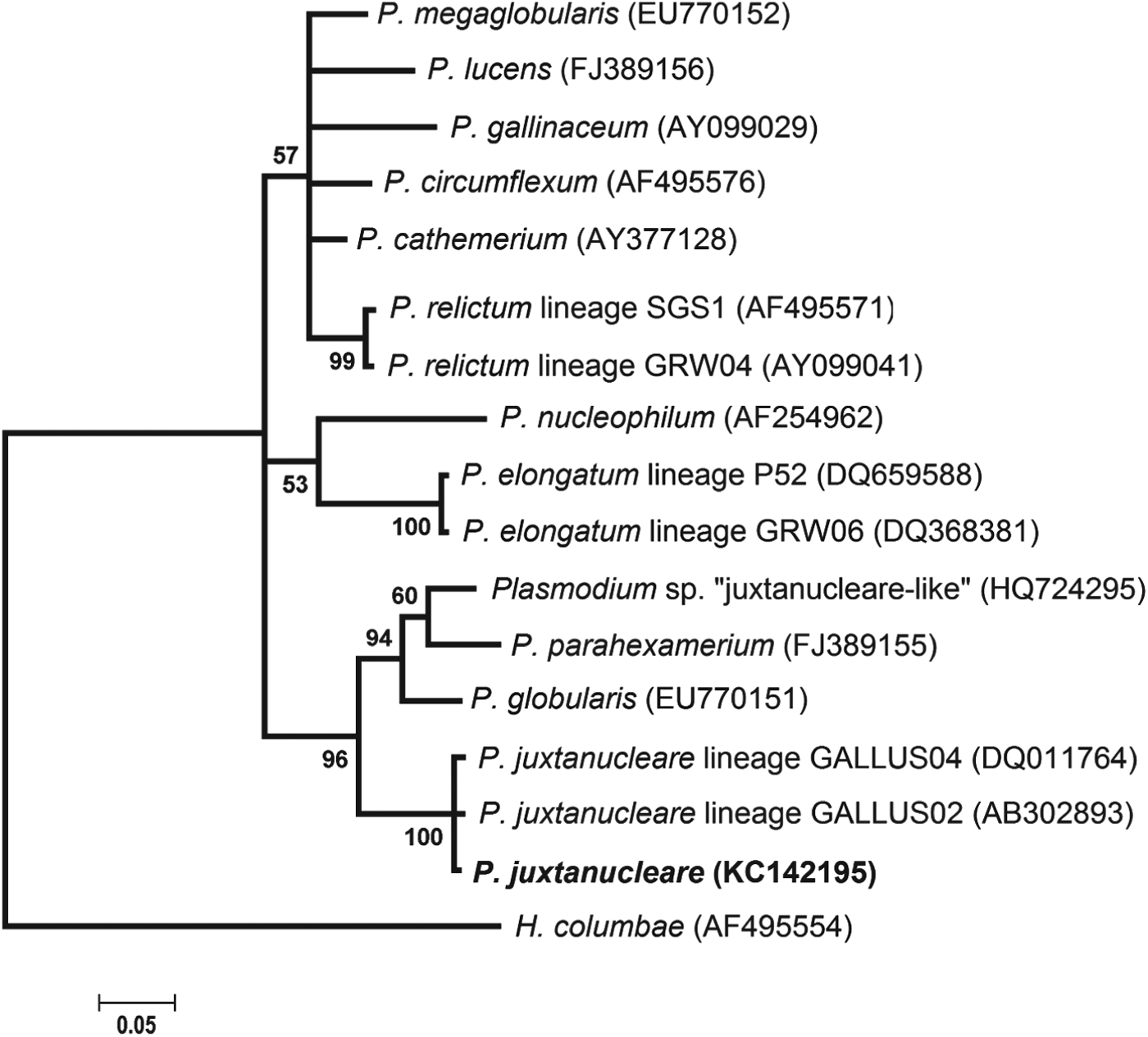
The Bayesian phylogenetic tree showed that the sequence recovered represented one lineage of P. juxtanucleare (strain FMV) and constituted a well-supported clade with other P. juxtanucleare sequences available in the GenBank and MalAvi databases, as shown in Fig. 1. The P. gallinaceum lineage GALLUS01, which is also commonly found to infect chickens, was placed in a clade distinct from the P. juxtanucleare lineages, supporting their classification in two different Plasmodium subgenera. The cyt b lineage found in this study is different from the P. juxtanucleare lineages GALLUS02 (reported in chickens and pheasant from Japan) and GALLUS04 (also reported in chickens from Brazil) by only one nucleotide at distinct sites. The lineage reported by Pacheco et al. (Reference Pacheco, Escalante, Garner, Bradley and Aguilar2011) in two Masked bobwhite quails and a Common raven and identified as ‘P. juxtanucleare-like’ (accession numbers HQ724295 and HQ724297) appeared in a distinct clade from the P. juxtanucleare sequences and showed a genetic divergence of 5·6% from the P. juxtanucleare sequence described here.
Phylogenetic relationships among the P. juxtanucleare cyt-b lineage described in this study (FMV strain) and the cyt b lineages of Plasmodium morphospecies available in MalAvi and Genbank. The Cyt b Plasmodium lineage described by Pacheco et al. (Reference Pacheco, Escalante, Garner, Bradley and Aguilar2011) is also included. GenBank accession numbers are shown in parentheses after the species/lineage names. The P. juxtanucleare lineage recovered in this study is shown in bold. Values near the nodes correspond to Bayesian probabilities (in per cent values).

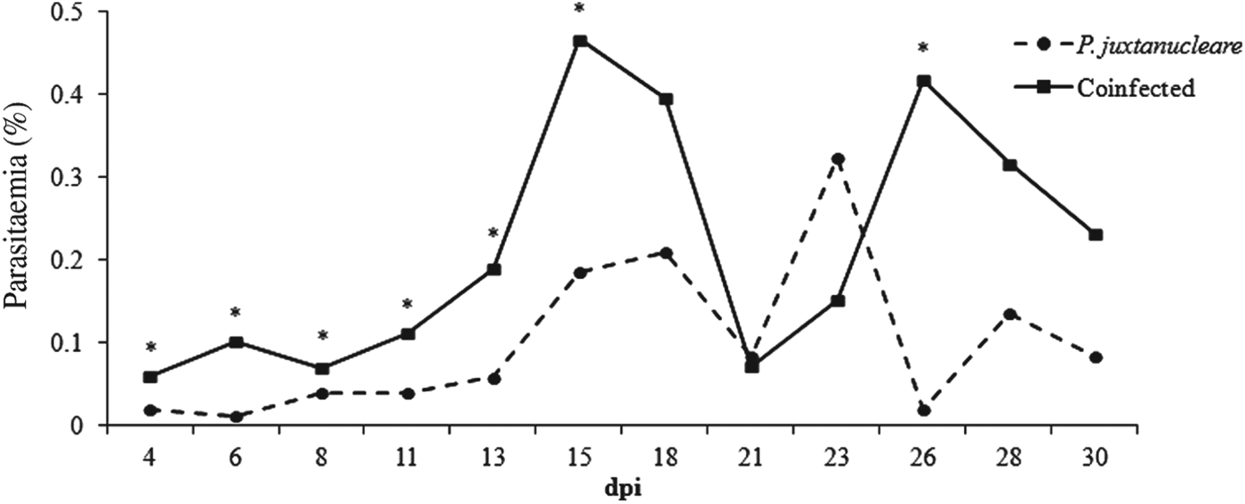
Parasitaemia (%) observed in coinfected chicks (CAV and Plasmodium juxtanucleare – Coinfected group) and in chicks exclusively infected with P. juxtanucleare (Malaria group). The asterisk indicates a significant difference according to a two-tailed Mann–Whitney U test (N = 6).
Parasitaemia in infected chicks
It is important to note that the three pilot experiments performed before the main experiment provided evidence that the proposed model had the potential to identify different clinical and pathological features associated to CAV-P. juxtanucleare coinfection.
All of the erythrocytic stages (trophozoits, schizonts and gametocytes) were observed in Giemsa stained blood smears from chicks in the Malaria and Coinfected groups. The trophozoite was the predominant stage (90%), followed by schizont (10%); gametocytes were rarely observed. Poliparasitism and infection of immature erythrocytes were observed in chicks from the Coinfected group only. In general, the chicks from the Coinfected and Malaria groups exhibited low parasitaemia (Fig. 2). In addition to low mean values, parasitaemia was significantly higher (P<0·05) in the chicks of the Coinfected group in comparison with chicks of the Malaria group at 4, 6, 8, 11, 13, 15 and 26 dpi. In the Coinfected group, two peaks of parasitaemia were observed. The first was at 15 dpi, with a mean parasitaemia of 0·47% (±0·3%), and 2% was the highest individual value. The second peak occurred at 26 dpi, with a mean parasitaemia of 0·42% (±0·7%). In the Malaria group, the highest (P<0·05) mean parasitaemia value was detected at 23 dpi (0·33±0·16%), and the highest (P<0·05) individual value observed was 1%.
Haematological analyses in infected chicks
CAV infection in the CAV and Coinfected groups was confirmed using PCR and ELISA and was not confirmed in the Malaria and Control groups. Plasmodium infection was also confirmed by PCR in the Coinfected and Malaria groups and was not confirmed in the CAV and Control groups.
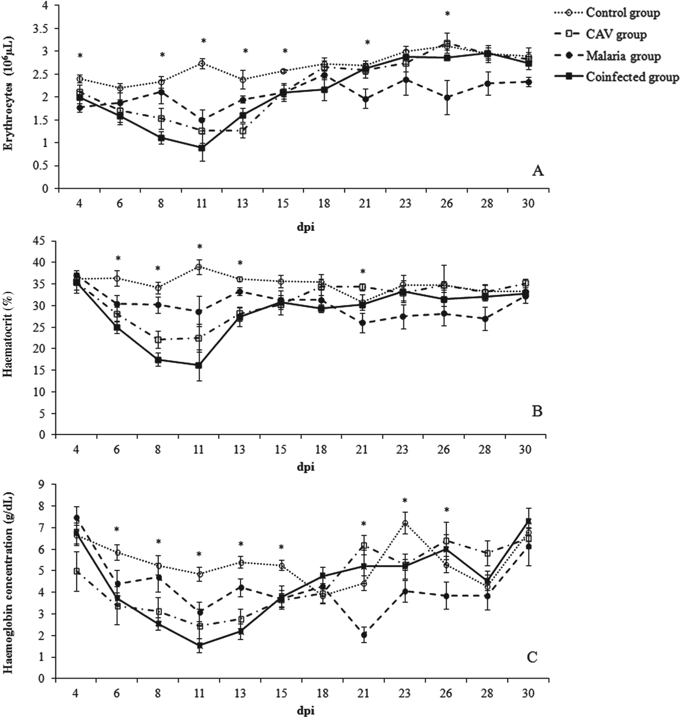
Haematological parameters such as the RBC, haematocrit and haemoglobin values were clearly lower (P<0·05) among chicks from the Coinfected and CAV groups in comparison with chicks from the Control group at the initial phase (8 and 11 dpi; Fig. 3). In addition, all of the haematological parameters had the lowest values in the Coinfected group (P<0·05) compared with values observed in the other groups, mainly during the early phase of the infection. The Malaria group showed lower RBC levels (P<0·05) compared with values measured in the Control group at 11 and 21 dpi; the haematocrit and haemoglobin levels were also significantly lower at 21 dpi when compared with the Control group (P<0·05).
Changes in erythrocyte, haematocrit and haemoglobin counts in CAV and Plasmodium juxtanucleare-coinfected (Coinfected group), CAV-infected (CAV group), P. juxtanucleare-infected (Malaria group) and uninfected chicks (Control group). Bars indicate the standard error. The asterisk indicates a significant difference according to a Kruskal–Wallis test, followed a Dunn's post test (N = 6).
The coinfected chicks showed the lowest RBC mean values, mainly at 8 and 11 dpi (1·1±0·3 and 0·9±0·2×106μL, respectively) (Fig. 3A). The means values were significantly (P<0·05) lower than the values observed for the Control group (2·3±0·3×106 and 2·7±0·2×106μL, respectively) at the same day post inoculation. Moreover, the RBC for the Coinfected group was significantly lower (P<0·05) at 8 dpi when compared with that for the Malaria group (1·1±0·3×106 and 2·1±0·2×106μL, respectively). No significant differences (P<0·05) were observed in the RBC counts between the Coinfected group and the CAV group.
The lowest (P<0·05) haematocrit levels were observed for the Coinfected group chicks at 8 and 11 dpi (17± 3·7 and 16±6·7%, respectively; Fig. 3B). The haematocrit in the Coinfected group was lower (P<0·05) compared with that in both the Control (34±3·2 and 39±4·3%) and Malaria groups (30±4·8 and 28±4%) (Fig. 3B). The haematocrit measured in the Coinfected group was significantly lower (P<0·02) than that observed for the CAV group at 11 dpi (22±5%).
The haemoglobin concentration in the Coinfected group decreased at 6 dpi, reaching its lowest value at 11 dpi (Fig. 3C). Haemoglobin values were lower in the Coinfected group compared with the CAV group at 8 and 11 dpi, although no statistical difference was observed (P>0·05).
Body weight and temperature of infected chicks
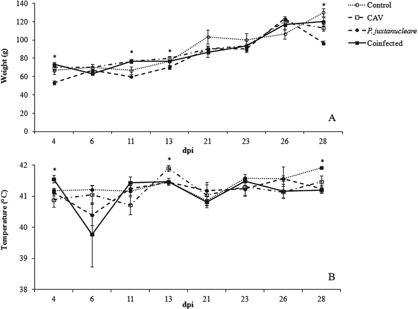
The mean of body weight and temperature of the chicks in all of the experimental groups were compared with those of the Control group chicks (Fig. 4A and B, respectively).
Changes in body weight and temperature in CAV and Plasmodium juxtanucleare-coinfected (Coinfected group), CAV (CAV group), P. juxtanucleare (Malaria group) and uninfected chicks (Control group). Bars indicate the standard error of body temperature expressed in °C and body weight expressed in g for each group. The asterisk indicates a significant difference according to a Kruskal–Wallis test, followed by Dunn's post test (N = 6).
The chicks from the Coinfected and CAV groups did not show significant weight decreases when compared with the Control group (P>0·05; Fig. 4A). However, the Malaria group showed a lower mean weight in comparison with the Control group at 4, 11 and 28 dpi (P<0·05).
The mean body temperature of the Coinfected chicks (39·5±1·5 °C) at 6 dpi was lower compared with that of chicks from the Control group (41·2±0·7 °C, P<0·05). However, the Malaria and CAV groups did not show significant changes (Fig. 4B).
Additionally, in the Coinfected and Malaria groups, we observed ruffled feathers and diarrhoea. Moreover, in the Coinfected group, severe signs of prostration and marked pallor were observed.
Mortality, macroscopic and microscopic tissue changes
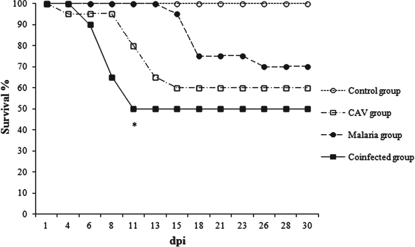
The cumulative mortality was higher for chicks from the Coinfected group at 11 dpi (Fig. 5; P<0·05). For chicks in the CAV and Malaria groups, mortality was only detected after 11 dpi.
Mortality of CAV and Plasmodium juxtanucleare-coinfected (Coinfected group), CAV (CAV group), P. juxtanucleare (Malaria group) and uninfected chicks (Control group). The asterisk indicates a significant difference between the Coinfected and other groups according to a Log-Rank test (N = 20). The chicks were infected with P. juxtanucleare 7 days after CAV infection.
The three experimentally infected groups presented paleness compared with the non-infected Control group. However, more severe paleness was observed in the Coinfected and CAV groups. A moderate increase was observed in the sizes of the liver and of spleen in the Coinfected group. The size decrease of the thymus and bursa of Fabricius in this group was moderate compared with chicks in the CAV and Control groups at 7 and 21 dpi. Suffusions and petechiae were observed in the heart of all coinfected chicks in the Coinfected group (see supplementary material – in Online version only). Moreover, this group exhibited haemorrhage in the bursa of Fabricius and liver at 14 and 28 dpi, respectively. Hepatomegaly and splenomegaly were more severe in the Malaria group compared with the Control and Coinfected groups. Furthermore, the size increase in these organs in the Malaria group was observed in all necropsies at 7 to 28 dpi. Cardiomegaly was observed in the Malaria group, particularly at 21 and 28 dpi. In addition, haemorrhagic foci and hydropericardium were also observed in the heart of chicks from the Malaria group. A mild-to-moderate decrease in the size of bursa de Fabricius and thymus were observed on necropsy in this group. The chicks of the CAV group presented severe atrophy of the lymphoid organs (spleen, bursa of Fabricius and thymus), particularly at 14 dpi. Suffusions and petechiae in the heart were also observed but at a lower intensity compared with chicks from the Coinfected group. Moreover, haemorrhagic foci were observed in the liver and in skeletal muscle in CAV group.
Changes in the infected chicks were detected via histopathology (Fig. 6). Splenic red pulp hyperplasia, with brown pigments, was detected in chicks of the Malaria and Coinfected groups (Fig. 6B and D). Lymphoid depletion in the spleen was observed in the Coinfected and CAV-infected chicks (Fig. 6C and D). Lymphoid depletion in the bursa of Fabricius and thymus was observed in animals from the three infected groups, although the depletion was more evident in the CAV group. A moderate reduction in the number of follicles with depletion in lymphocytes was observed in chicks from the Coinfected group (Fig. 7A). Mild-to-moderate multifocal lymphocytic hepatitis and pericarditis were present in chicks from all infected groups but was more evident in the Malaria and Coinfected groups (Fig. 7B and C).
Histopathology of the spleen. A. Control group normal spleen. B. Malaria group. Increased cellularity of the red pulp (RP) with accumulation of brown pigment (arrow). C. CAV group. Decreased cellularity of the white pulp (WP) due to marked lymphoid depletion. D. Coinfected group. Mildly increased cellularity of the red pulp and moderate lymphoid depletion. HE, 200×.
Histopathology of CAV and Plasmodium juxtanucleare coinfected chick organs. A. Bursa of Fabricius atrophy. Moderate reduction in follicles with depletion of lymphocytes (asterisk). HE, 400×. B. Multifocal moderate hepatitis. Moderate portal mononuclear infiltrate (large arrow) associated random areas of necrosis (N). HE, 400×. C. Multifocal moderate lymphoplasmacytic pericarditis (narrow arrow). HE, 200×.
DISCUSSION
The effects of simultaneous infection by P. juxtanucleare and CAV on SPF chicks are described for the first time herein. For this study, we choose two pathogens that are widely distributed in Brazil and that are not included in programmes that have obligatory health control strategies (Brentano et al. Reference Brentano, Silva, Sayd and Flores2000; Simionatto et al. Reference Simionatto, Lima-Rosa, Binneck, Ravazzolo and Canal2006; Barrios et al. Reference Barrios, Marín, Resende, Rios, Resende, Horta, Costa and Martins2009; MAPA, 2009). Therefore, CAV and P. juxtanucleare are two pathogens that could potentially be found naturally in chickens. In Brazil, the vaccination of chickens against CAV is not mandatory, leaving breeders to decide whether to immunize their chickens (MAPA, 2009). The presence of P. juxtanucleare has not been studied in poultry farms on the grounds that it is a parasite that has a low pathogenic effect on the host. In fact, during the chronic infection caused by this parasite, there is low pathogenic effect. However, in cases of coinfection, the effects of infection are unknown. In the present study, we observed that although coinfected chicks had higher pathogenicity compared with the chicks with single infections, exclusive P. juxtanucleare infection also has harmful effects on susceptible hosts, as discussed below.
Experimental coinfection with avian malaria parasites has been investigated previously (Permin and Juhl, Reference Juhl and Permin2002; Wright et al. Reference Wright, Nayar and Forrester2005; Marzal et al. Reference Marzal, Bensch, Reviriego, Balbontin and De Lope2008; Zehtindjiev et al. Reference Zehtindjiev, Ilieva, Westerdahl, Hansson, Valkiūnas and Bensch2008; Palinauskas et al. Reference Palinauskas, Valkiūnas, Bolshakov and Bensch2011). Although Plasmodium may negatively affect host health, it is still unclear whether and how the simultaneous presence of a pathogen and/or immunosuppression influence the course of infection. Multiple infections associated with different pathogens, especially immunosuppressive viruses, may have more severe effects on chicks than do single infections (Loving et al. Reference Loving, Brockmeier, Vincent, Palmer, Sacco and Nicholson2010; Tan et al. Reference Tan, Zhu, Wang, Wang, Wei, Sun and Sheng2011; Liang et al. Reference Liang, Zhao, Liu, Yang, Zuo, Cui, Zhong, Sun, Liu and Zhu2013). Indeed, coinfection upregulated plasmodia parasitaemia, most likely due to the immunosuppressive effect of CAV, which favoured the development of the P. juxtanucleare asexual erythrocytic stages. It is known that CAV may infect chicks after hatching, leading to temporary or permanent immune dysfunction, which increases the likelihood of opportunistic infections. Studies of human malaria have shown a synergistic pathological effect during coinfection with HIV. Humans infected with HIV exhibit higher P. falciparum parasitaemia and develop more severe clinical signs compared with patients experiencing single infection (Khasnis and Karnad, Reference Khasnis and Karnad2003; Chalwe et al. Reference Chalwe, Van Geertruyden, Mukwamataba, Menten, Kamalamba, Mulenga and D'Alessandro2009). On the other hand, in murine malaria, an antagonistic interaction was reported during coinfection by a lethal (Plasmodium berghei NK65) and a non-lethal parasite (Plasmodium yoelii 17X or P. berghei XAT). In this case, coinfection by low pathogenic Plasmodium strains suppressed the high levels of parasitaemia and severe pathogenesis caused by lethal P. berghei NK65 (Niikura et al. Reference Niikura, Kim, Hunt, Burnside, Morgan, Dodgson and Cheng2007). The early age at which the chicks were infected is a factor that was not investigated in this study but that could contribute to increased pathogenicity. Wright et al. (Reference Wright, Nayar and Forrester2005) observed that 1-week-old turkeys concomitantly infected by turkeypox virus and Plasmodium hermani or exclusively infected by one of these pathogens showed more severe signs of infection compared with turkeys infected under the same conditions at 10 weeks of age. In the present study, we attempted to establish a new experimental model based on malarial infection post virus acquisition by newborn chicks. The fact that CAV transmission principally occurs by a vertical route supports the validity of the experimental design presented here. In nature, we expect that chicks are born with the virus or are infected very early through the fecal–oral route. Therefore, it seems that under natural conditions, infection by P. juxtanucleare occurs after CAV infection, reinforcing the value of the new model established.
More severe anaemia (reduced haematocrit, RBC and haemoglobin levels) was observed in the Coinfected group compared with the Control and Malaria groups. More severe anaemia (reduced haematocrit, RBC or haemoglobin levels) was observed in the Coinfected group compared with the Control and Malaria groups. The mechanisms by which anaemia develops could be different for the two pathogens (P. juxtanucleare and CAV). CAV is an agent that has immunosuppressive ability and that also causes severe anaemia in infected individuals, especially the very young (Rosenberger and Cloud, Reference Rosenberger and Cloud1998). Anaemia in CAV-infected chicks is mainly detected at approximately 15 dpi, as observed in the present study. It is known that the host immune response and clinical features during infection with viruses such as CAV are associated with several factors, including anaemia (Rosenberger and Cloud, Reference Rosenberger and Cloud1998). Plasmodium juxtanucleare has low pathogenicity during the chronic phase of the infection, and anaemia is rare. However, previous studies showed that the experimental acute phase in chicks caused anaemia, increases in leukocytes, and inflammatory damage in several tissues (Krettli, Reference Krettli1972; Silveira et al. Reference Silveira, Damatta and D'Agosto2009a, Reference Silveira, Vashist, Cabral, Amaral, Soares and D'Agostob, Vashist et al. Reference Vashist, Falqueto, Lustrino, Tunholi, Tunholi-Alves, dos Santos, D'Agosto, Massard and Pinheiro2011). In those studies, it was also observed that P. juxtanucleare generally caused low parasitaemia and that parasitaemia is not always associated with anaemia or other changes. Experimental infection with P. juxtanucleare was not previously evaluated in SPF chicks. The possibility that SPF chicks may exhibit greater susceptibility to malaria compared with regular chicks may not be excluded. SPF animals have been shown to have lower numbers of specific immune system cells compared with animals kept in regular environments (Umesaki et al. Reference Umesaki, Setoyama, Matsumoto and Okada1993; Beirão et al. Reference Beirão, Fávaro, Nakao, Caron, Zanata and Mercadante2012). Indeed, there is evidence that infection by P. juxtanucleare may increase pathogenicity in more susceptible hosts. For example, the infection caused by P. juxtanucleare resulted in death among penguins (S. demersus) and white-eared pheasants (C. crossoptilon) (Grim et al. Reference Grim, van der Merwe, Sullivan, Parsons, Mc Cutchan and Cranfield2003; Murata et al. Reference Murata, Nii, Sasaki, Ishikawa, Sato, Sawabe, Tsuda, Matsumoto, Suda and Ueda2008).
The unlimited availability of food ‘ad libitum’ may have equilibrated weight and favoured body mass similarity among the experimental groups. Various studies have reported no weight change during single or double Plasmodium infections (Zehtindjiev et al. Reference Zehtindjiev, Ilieva, Westerdahl, Hansson, Valkiūnas and Bensch2008; Palinauskas et al. Reference Palinauskas, Valkiūnas, Krizanauskiene, Bensch and Bolshakov2009, Reference Palinauskas, Valkiūnas, Bolshakov and Bensch2011; Silveira et al. 2009a). In free-range or field conditions, where access to feed is limited, more severe infections, such as in the coinfected birds, could result in lower weight (Marzal et al. Reference Marzal, Bensch, Reviriego, Balbontin and De Lope2008). Only coinfected chicks showed hypothermia during early infection, which was associated with severe anaemia and deteriorating health conditions.
Marked lymphoid cell depletion was detected in the Coinfected and CAV groups, particularly in the thymus, but also in the spleen and bursa of Fabricius. Lymphoid organ atrophy was detected in the CAV and Coinfected groups, in accordance with other experimental CAV infection studies, which identified atrophy based on depletion of lymphoid cells in addition to haemorrhagic lesions (Goryo et al. Reference Goryo, Hayashi, Yoshizawa, Umemura, Itakura and Yamashiro1989, McNulty et al. Reference McNulty, McIlroy, Bruce and Todd1991; Pope, Reference Pope1991; Smyth et al. Reference Smyth, Moffett, McNulty, Todd and Mackie1993; Rosenberger and Cloud, Reference Rosenberger and Cloud1998; Peters et al. Reference Peters, Crabb, Tivendale and Browning2007). It is important to note that in the present study, cardiomegaly and hepato-splenomegaly were observed among animals from the Malaria group. Spleen and liver enlargement were observed in earlier studies related to P. juxtanucleare infection in chickens (Vashist et al. Reference Vashist, Falqueto, Lustrino, Tunholi, Tunholi-Alves, dos Santos, D'Agosto, Massard and Pinheiro2011). However, cardiomegaly and hydropericardium by P. juxtanucleare in experimentally infected SPF chicks are reported herein for the first time.
We conclude that a P. juxtanucleare challenge 7 days after CAV infection leads to more severe malarial infection, possibly due to CAV immunosuppression and other potential synergistic effects during coinfection. The relationship among the immunological mechanisms associated with these clinical, haematological and histopathological changes must be clarified in future studies. In Brazil, it may be particularly important to investigate the infection by P. juxtanucleare due to its high prevalence, especially in domestic chicken (Santos-Prezoto et al. Reference Santos-Prezoto, D'Agosto and Daemom2004). The relevance of P. juxtanucleare infection in chickens to the poultry industry is largely unknown, and future studies should evaluate its potential impacts on performance, productivity and economic factors.
ACKNOWLEDGEMENTS
The authors are grateful to Francisco C. Ferreira Junior, Rodrigo S. Horta and Daniel A. R. Vilela (Veterinary College, UFMG), for assistance in laboratory.
FINANCIAL SUPPORT
This study was financially supported by CNPq (Conselho Nacional de Desenvolvimento Cientifico e tecnologico) FAPEMIG (Fundação de Desenvolvimento da Pesquisa do Estado de Minas Gerais) and CAPES (Coordenação de Pessoal de Nível Superior) with grants and scholarships.
SUPPLEMENTARY MATERIAL
To view supplementary material for this article, please visit http://dx.doi.org/10.1017/S0031182013001170.