Introduction
The inhaled sharp foreign body is usually amenable to bronchoscopic extraction. When this fails, the rational management hierarchy needs to consider the potential benefits and complications of each form of treatment, and even no treatment, in an otherwise asymptomatic patient. We present a case which highlights the difficulties faced, and we examine our clinical decisions in light of the currently available evidence.
Case report
A 12-year-old girl had been biting on her lapel, to which a remembrance poppy had been pinned, when a friend had made her laugh, causing her to inhale suddenly. The pin holding the poppy had disappeared, and she had felt a scratch in her throat. This had been followed by a single episode of minimal haemoptysis.
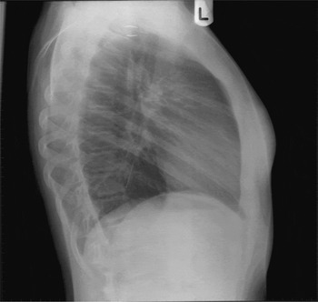
On presentation to the accident and emergency service several hours later, a chest radiograph showed the pin to be present in the left lung (Figures 1 and 2). The patient was otherwise asymptomatic.
Postero-anterior chest radiograph showing pin in left lower lobe.

Lateral chest radiograph showing pin in left lower lobe chest.

An otolaryngological review was performed. Due to the distal location of the pin, a cardiothoracic referral was also made.
Consent was obtained to perform bronchoscopy and extraction of the pin, and, if this failed, to proceed to thoracotomy and extraction.
Following general anaesthesia, rigid bronchoscopy was attempted; however, the pin could not be located. Rigid bronchoscopy was repeated with a magnet at the tip of the bronchoscope, with no success. Flexible paediatric bronchoscopy was also attempted in the hope of accessing the more distal airways, and was repeated with radiological screening. This showed the pin to be lodged 3 cm distal to the tip of the bronchoscope.
After these multiple attempts, a decision was made to proceed with a posterolateral thoracotomy. The pin was located by digital palpation through the lung tissue, within the medial segment of the anteromedial basal segment of the left lung, and was manipulated through the parenchyma.
Post-operatively, the patient made an uneventful recovery.
Discussion
The sharp foreign body in the bronchial tree is a unique challenge. Although foreign body inhalation is in general commoner in infants and young children, inhalation of pins is commoner in young girls. The literature contains several reports of turban pin inhalation in young Islamic girls, who are in the habit holding the scarf pin between their teeth to keep both hands free while wearing or adjusting their head scarves.Reference Murthy, Ingle, George, Ramakrishna and Shah1 It is common for the inhaled pin to lodge in the airways with its point turned upwards; this is because the point tends to lodge in the mucosal wall, causing the pin to tumble and the point to trail.Reference Holinger2
Typically, these patients lack significant symptoms.Reference Murthy, Ingle, George, Ramakrishna and Shah1, Reference Kaptanoglu, Dogan, Onen and Kunt3 The large majority of foreign bodies are organic, e.g. nuts and seeds. In contrast, pins are conveniently radiopaque, allowing easy radiological location and diagnosis. Pins are less inflammatory than organic foreign bodies. However, in a series of 105 patients reported recently, one patient who presented with high fever was found to have infective secretions surrounding the aspirated pin, confirming the potential of such aspiration to cause infection even as a relatively uncommon occurrence.Reference Kaptanoglu, Dogan, Onen and Kunt3 Furthermore, lung atelectasis may also occur and may be seen on the radiograph.Reference Rafanan, Mehta, Wang, Mehta and Turner4
The overriding concern regarding sharp foreign bodies, however, is their potential to migrate, and this has been reported in the literature. In the aforementioned series, 11 pins migrated distally and one migrated proximally and was actually spontaneously expectorated.Reference Kaptanoglu, Dogan, Onen and Kunt3 In a series of five patients who had inhaled pins, a delay of up to 10 days before intervention appeared to be safe.Reference Shabb, Taha, Hamada and Kanj5 However, in a larger series of 124 patients who underwent bronchoscopy for inhaled pins, complications were seen in 44 (35.48 per cent), and these complications occurred significantly more frequently in patients whose treatment was delayed.Reference Sersar, Elshazli, Abdel-Hakam and Mahdy6 This, then, would justify early rather than late intervention.
Rigid bronchoscopy under general anaesthesia is the general method of removing aspirated pins in children. Distally located pins can sometimes be accessed by flexible bronchoscopy. Some authors, however, prefer fibre-optic bronchoscopy in the first instance.Reference Shabb, Taha, Hamada and Kanj5 During pin extraction, it is important to ensheath the pointed end within the bronchoscope to protect the bronchial mucosa from damage. Two methods have been described, one using extraction forceps and the other using postural drainage to assist with suction, without forceps (the Sersar or Egyptian technique).Reference Sersar, Hamza, AbdelHameed, AbdulMaaty, Gowaeli and Moussa7 When the pin point is impacted within the mucosa, pushing the pin distally or bending it in the middle may assist dislodgement. The use of laser photocoagulation to vaporise the granulation tissue around the point of the pin has been described in some centres.Reference Rafanan, Mehta, Wang, Mehta and Turner4
Sersar et al. reported a series of 124 patients who underwent bronchoscopy for removal of inhaled pins, and noted laryngeal oedema in 10, of which three needed temporary tracheostomy.Reference Sersar, Elshazli, Abdel-Hakam and Mahdy6 Therefore, it seems preferable to repeat the bronchoscopy at a later date rather than unnecessarily prolong the procedure. These authors recommended thoracotomy for pin removal when three trials of bronchoscopy had failed, in patients with or without symptoms, in order to prevent inflammation.
• An inhaled pin needs removal even in the absence of symptoms, as migration and infection can occur
• Up to three attempts at repeat bronchoscopy on separate occasions may be needed, after which it may be necessary to proceed to open surgery
• In case the foreign body is found to be beyond the reach of the bronchoscope, it is useful to have obtained consent to proceed to thoracotomy in the first instance
Radiological screening is invaluable during bronchoscopy, in order to enable a peri-operative decision to proceed to thoracotomy if the pin is obviously out of reach of the bronchoscope. Such screening could also assist location of the foreign body during open surgery. Even in an open procedure, the difficulty of locating a pin within the lung parenchyma, and the role of careful digital palpation, should not be underestimated. The use of lung resection has been described to remove destroyed lung segments arising as a complication of an aspirated foreign body, rather than to remove the foreign body itself.
Conclusion
This case highlights important aspects of decision-making when dealing with a patient with an inhaled pin. The authors suggest that the patient be informed that the pin needs removal even in the absence of symptoms, as migration and infection can occur. The patient should be warned that up to three attempts at repeat bronchoscopy may be needed, on separate occasions, after which it may be necessary to proceed to open surgery. In case the foreign body is found to be obviously beyond the reach of the bronchoscope, it is useful to have obtained consent to proceed to thoracotomy in the first instance. Good cooperation within the multidisciplinary team is invaluable in dealing with this unexpected challenge.