Introduction
Phenotype-based circumscriptions of genera have repeatedly been challenged in different groups of lichenized fungi. In the hyperdiverse family Parmeliaceae, many genera that were chiefly separated based on vegetative traits have not been supported as monophyletic clades in molecular phylogenetic reconstructions (reviewed in Lumbsch Reference Lumbsch2007; Printzen Reference Printzen2010; Crespo et al. Reference Crespo, Divakar and Hawksworth2011; Thell et al. Reference Thell, Crespo, Divakar, Kärnefelt, Leavitt, Lumbsch and Seaward2012; Divakar & Crespo Reference Divakar and Crespo2015). The frequent incongruence between traditional circumscriptions of genera in Parmeliaceae and monophyletic evolutionary lineages highlights the necessity to carefully evaluate generic circumscriptions within an evolutionary context.
Currently in Parmeliaceae c. 80 genera are accepted based on phenotypic features and analyses of multilocus sequence data (Thell et al. Reference Thell, Crespo, Divakar, Kärnefelt, Leavitt, Lumbsch and Seaward2012; Divakar et al. Reference Divakar, Crespo, Wedin, Leavitt, Hawksworth, Myllys, McCune, Randlane, Bjerke and Ohmura2015). The largest group within the family is the parmelioid core, to which the genera Relicina (Hale & Kurok.) Hale and Relicinopsis Elix & Verdon belong (Crespo et al. Reference Crespo, Kauff, Divakar, Amo, Arguello, Blanco, Roca-Valiente, Núñez-Zapata, Cubas and Argüello2010; Divakar et al. Reference Divakar, Crespo, Wedin, Leavitt, Hawksworth, Myllys, McCune, Randlane, Bjerke and Ohmura2015). The evolutionary relationships of these two genera have only been partially explored. The genus Relicinopsis was segregated from Pseudoparmelia Lynge based on morphological features, such as the presence of simple marginal cilia, fusiform conidia and usnic acid as a cortical extrolite (Elix et al. Reference Elix, Johnston and Verdon1986). This genus includes a total of five species, which are widely distributed in South-East Asia and Australasia (Hale Reference Hale1976; Elix Reference Elix1993, Reference Elix1994; Divakar & Upreti Reference Divakar and Upreti2005). The genus Relicina was segregated from Parmelia Ach. s. lat. (Hale Reference Hale1974) based on having bulbate marginal cilia and bifusiform conidia, and containing usnic acid in the upper cortex. This genus includes c. 54 species (Thell et al. Reference Thell, Crespo, Divakar, Kärnefelt, Leavitt, Lumbsch and Seaward2012) with a centre of distribution in South-East Asia and Australasia (Hale Reference Hale1975; Elix Reference Elix1993). In a recent study, Relicina and Relicinopsis formed a well-supported sister-group relationship, although the taxon sampling was limited and monophyly was not supported in an mtSSU single locus phylogeny (Buaruang et al. Reference Buaruang, Scharnagl, Divakar, Leavitt, Crespo, Nash, Manoch, Lücking and Lumbsch2015). Moreover, the distinction of the two genera was supported in the ‘1GENE’ data analysis by Crespo et al. (Reference Crespo, Kauff, Divakar, Amo, Arguello, Blanco, Roca-Valiente, Núñez-Zapata, Cubas and Argüello2010). In the present study we used an extended taxon sampling to 1) examine the monophyly of Relicina and Relicinopsis and 2) evaluate the taxonomic significance of phenotypic features in these two genera.
Materials and Methods
Taxon sampling
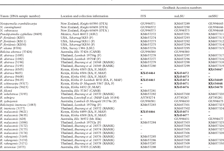
Data matrices of 36 samples including four of the five described species of Relicinopsis and six species of Relicina were analyzed, including eight new samples of these genera collected from East Africa. We assembled a multilocus DNA matrix comprised of nuLSU, ITS and mtSSU rDNA to infer evolutionary relationships. The multilocus data set included 74 sequences from a previous study (Buaruang et al. Reference Buaruang, Scharnagl, Divakar, Leavitt, Crespo, Nash, Manoch, Lücking and Lumbsch2015) and 14 sequences generated for this study. Three species of Notoparmelia were used as the outgroup since the genus has been shown to be closely related to Relicina (Crespo et al. Reference Crespo, Kauff, Divakar, Amo, Arguello, Blanco, Roca-Valiente, Núñez-Zapata, Cubas and Argüello2010; Buaruang et al. Reference Buaruang, Scharnagl, Divakar, Leavitt, Crespo, Nash, Manoch, Lücking and Lumbsch2015). Information on material studied, including GenBank Accession numbers, is reported in Table 1.
Specimens used in this study together with location, reference collection detail and GenBank Accession numbers. Newly obtained sequences for this study are in bold and missing data are indicated with a dash (–)
DNA extraction and PCR amplification
Total genomic DNA was extracted from small pieces of thallus devoid of any visible damage or contamination using the USB PrepEase Genomic DNA Isolation Kit (USB, Cleveland, OH, USA) in accordance with the manufacturer’s instructions. We generated sequence data from nuclear ribosomal markers, the ITS region and a fragment of the nuLSU, in addition to a fragment of the mtSSU. Polymerase chain reaction (PCR) amplifications were performed using Ready-To-Go PCR Beads (GE Healthcare, Pittsburgh, PA, USA) using dilutions of total DNA. Fungal ITS rDNA was amplified using primers ITS1F (Gardes & Bruns Reference Gardes and Bruns1993), ITS4 and ITS4A (White et al. Reference White, Bruns, Lee and Taylor1990; Larena et al. Reference Larena, Salazar, González, Julián and Rubio1999); nuLSU rDNA was amplified using LR0R and LR5 (Vilgalys & Hester Reference Vilgalys and Hester1990); and mtSSU rDNA was amplified using the primers mrSSU1, mrSSU3R and mrSSU2R (Zoller et al. Reference Zoller, Scheidegger and Sperisen1999). PCR products were visualized on 1% agarose gel and cleaned using ExoSAP-IT (USB, Cleveland, OH, USA). Cycle sequencing of complementary strands was performed using BigDye v3.1 (Applied Biosystems, Foster City, CA, USA) and the same primers used for PCR amplifications. Sequenced PCR products were run on an ABI 3730 automated sequencer (Applied Biosystems) at the Pritzker Laboratory for Molecular Systematics and Evolution at the Field Museum, Chicago, USA.
Sequence editing and alignment
New sequences were assembled and edited using Geneious v8.1.7 (Biomatters Ltd 2005–2015). Multiple sequence alignments for each locus were performed using the program MAFFT v7 (Katoh et al. Reference Katoh, Kuma, Toh and Miyata2005; Katoh & Toh Reference Katoh and Toh2008). For the ITS and nuLSU sequences we used the G-INS-i alignment algorithm and ‘20PAM/K=2’ scoring matrix with an offset value of 0·3 and the remaining parameters set to default values. We used the E-INS-i alignment algorithm and ‘20PAM/ K=2’ scoring matrix, with the remaining parameters set to default values, for the mtSSU sequences. The program Gblocks v0.91b (Talavera & Castresana Reference Talavera and Castresana2007) was used to delimit and remove ambiguous alignment nucleotide positions from the final alignments using the online web server (http://molevol.cmima.csic.es/castresana/Gblocks_server.html), implementing the options for a less stringent selection of ambiguous nucleotide positions including “Allow smaller final blocks”, “Allow gap positions within the final blocks” and “Allow less strict flanking positions” options.
Phylogenetic analyses
Phylogenetic relationships were inferred using maximum likelihood (ML) and Bayesian inference (BI). Exploratory phylogenetic analyses of individual gene topologies showed no evidence of well-supported (≥70% bootstrap values) topological conflict, thus relationships were estimated from a concatenated, three-locus (ITS, nuLSU, mtSSU) data matrix using a total-evidence approach (Wiens Reference Wiens1998). We used the program RAxML v8.1.11 (Stamatakis Reference Stamatakis2006; Stamatakis et al. Reference Stamatakis, Hoover and Rougemont2008) to reconstruct the concatenated ML gene tree using the CIPRES Science Gateway server (http://www.phylo.org/portal2/). We implemented the ‘GTRGAMMA’ model, with locus-specific model partitions treating all loci as separate partitions, and evaluated nodal support using 1000 bootstrap pseudoreplicates. Exploratory analyses using alternative partitioning schemes resulted in identical topologies and highly similar bootstrap support values. We also reconstructed phylogenetic relationships from the concatenated multilocus data matrix under BI using the program BEAST v1.8.2 (Drummond & Rambaut Reference Drummond and Rambaut2007). We ran two independent Markov chain Monte Carlo (MCMC) chains for 20 million generations, implementing a relaxed lognormal clock, a birth-death speciation process prior. The most appropriate model of DNA sequence evolution was selected for each marker using the program PartitionFinder v1.1.1 (Lanfear et al. Reference Lanfear, Calcott, Ho and Guindon2012) treating the ITS1, 5.8S, ITS2, nuLSU, and mtSSU as separate partitions. The first 2 million generations were discarded as burn-in. Chain mixing and convergence were evaluated in Tracer v1.5 (Rambaut & Drummond Reference Rambaut and Drummond2009) considering effective sample size (ESS) values >200 as a good indicator. Posterior trees from the two independent runs were combined using the program LogCombiner v1.8.0 (Drummond et al. Reference Drummond, Suchard, Xie and Rambaut2012) and the final maximum clade credibility (MCC) tree was estimated from the combined posterior distribution of trees.
Alternative hypothesis testing
Since the results of the phylogenetic analyses did not support the monophyly of Relicina as currently circumscribed, we tested whether our data were sufficient to reject the monophyly of that genus. For the hypothesis testing two different methods were employed: 1) Shimodaira-Hasegawa (SH) test (Shimodaira & Hasegawa Reference Shimodaira and Hasegawa1999) and 2) expected likelihood weight (ELW) test (Strimmer & Rambaut Reference Strimmer and Rambaut2002). The SH and ELW tests were performed using TREE-PUZZLE 5.2 (Schmidt et al. Reference Schmidt, Strimmer, Vingron and von Haeseler2002) with the combined data set on a sample of the best trees agreeing with the null hypotheses and the unconstrained ML tree. These trees were inferred in TREE-PUZZLE employing the GTR+I+G nucleotide substitution model.
Morphological and chemical studies
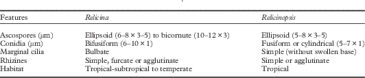
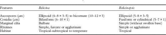
Morphological characters, including lobe shape, size and width, and cilia and rhizines were studied using a Leica Wild M8 dissecting microscope. Key morphological and chemical features used to segregate Relicina and Relicinopsis are listed in Table 2.
Main morphological and chemical features used to distinguish Relicina and Relicinopsis
Chemical constituents were identified by high performance thin-layer chromatography using standard methods (Arup et al. Reference Arup, Ekman, Lindblom and Mattsson1993; Lumbsch Reference Lumbsch2001) with a Camag horizontal developing chamber (Oleico Laboratory, Stockholm) using solvent system A.
Results and Discussion
Molecular phylogeny and phenotypic features
The aligned matrix contained 455 unambiguously aligned nucleotide positions in the ITS, 808 in the nuLSU and 735 in the mtSSU rDNA data sets. The final alignment of the concatenated data set was 1999 positions in length, with 548 variable characters. The ITS PCR product obtained ranged between 600–800 bp. Differences in size were due to the presence or absence of a group I intron of c. 200 bp at the 3' end of the 18S rDNA (Gutierrez et al. Reference Gutierrez, Blanco, Divakar, Lumbsch and Crespo2007). Introns from the ribosomal gene (18S) were removed from the analysis. GTR+I+G for ITS1, K80+I+G for 5.8S rDNA, TrN+G for ITS2, TrN+I+G for nuLSU rDNA and GTR+G for mtSSU rDNA were estimated as best fit models of evolution for each partition. All the newly generated sequences for this study were deposited in GenBank under Accession numbers KX434464–KX434477 (Table 1).
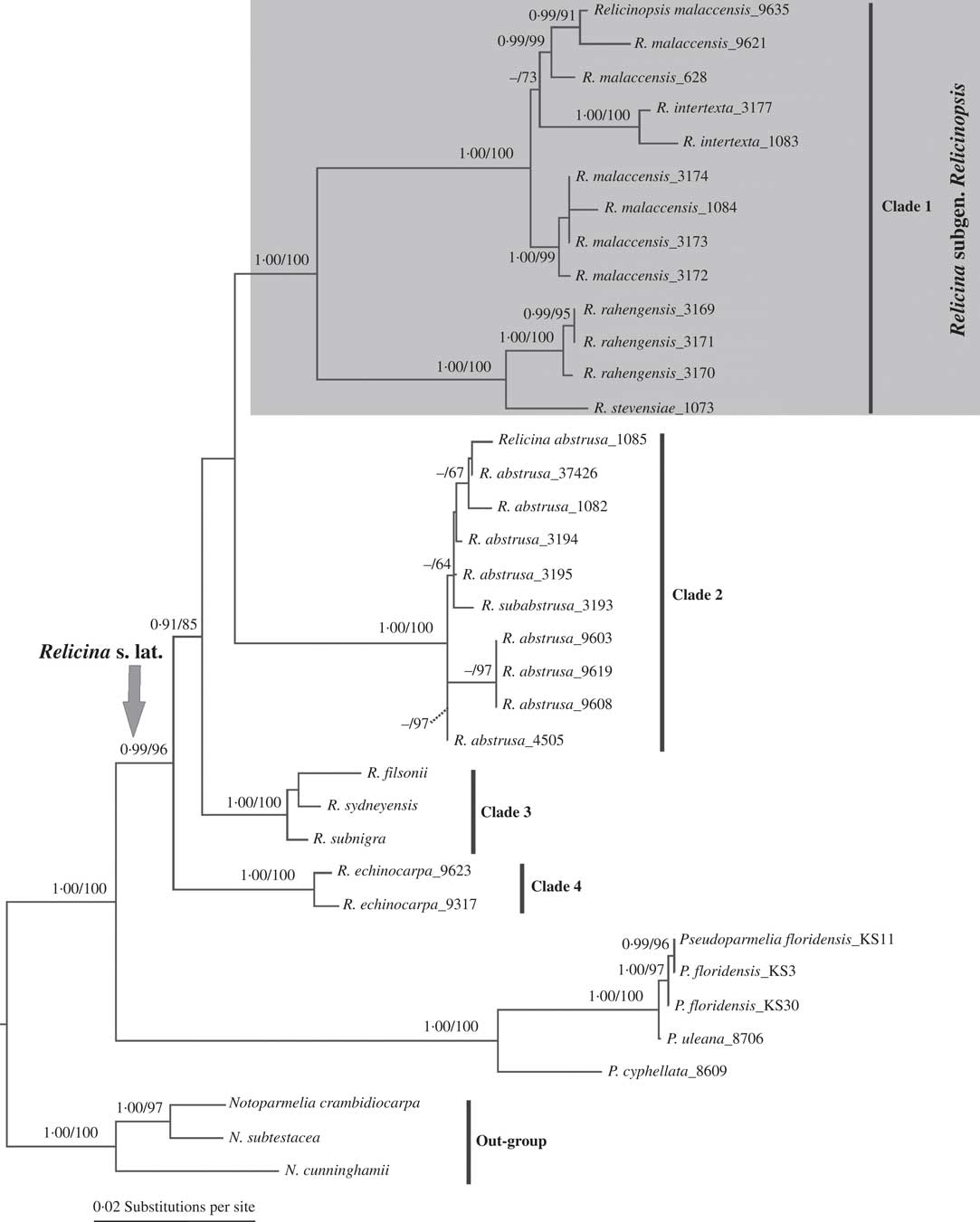
Tests for topological incongruence showed no supported conflicts (results not shown). The partitioned ML analysis of the concatenated data matrix yielded an optimal tree with ln likelihood value=−8048·95 (Fig. 1). In the Bayesian analysis, ESS values of all estimated parameters were well above 200 indicating that convergence among parallel runs was reached. ML and Bayesian topologies were largely similar and did not show well-supported conflict (e.g. PP≥0·95 and ML bootstrap≥70%) and thus the ML tree topology is shown here with the Bayesian posterior probabilities added (Fig. 1).
Phylogenetic relationships of the genera Relicina and Relicinopsis based on maximum likelihood (ML) and Bayesian analyses of a concatenated three locus data set (ITS, nuLSU & mtSSU rDNA.) The ML tree obtained with RAxML is shown here. Posterior probabilities≥0·95 from the Bayesian analysis (before the slash) and ML bootstrap values≥70% (after the slash) are given above branches. Three species of Notoparmelia (N. crambidiocarpa, N. cunninghamii and N. subtestacea) were used as the outgroup.
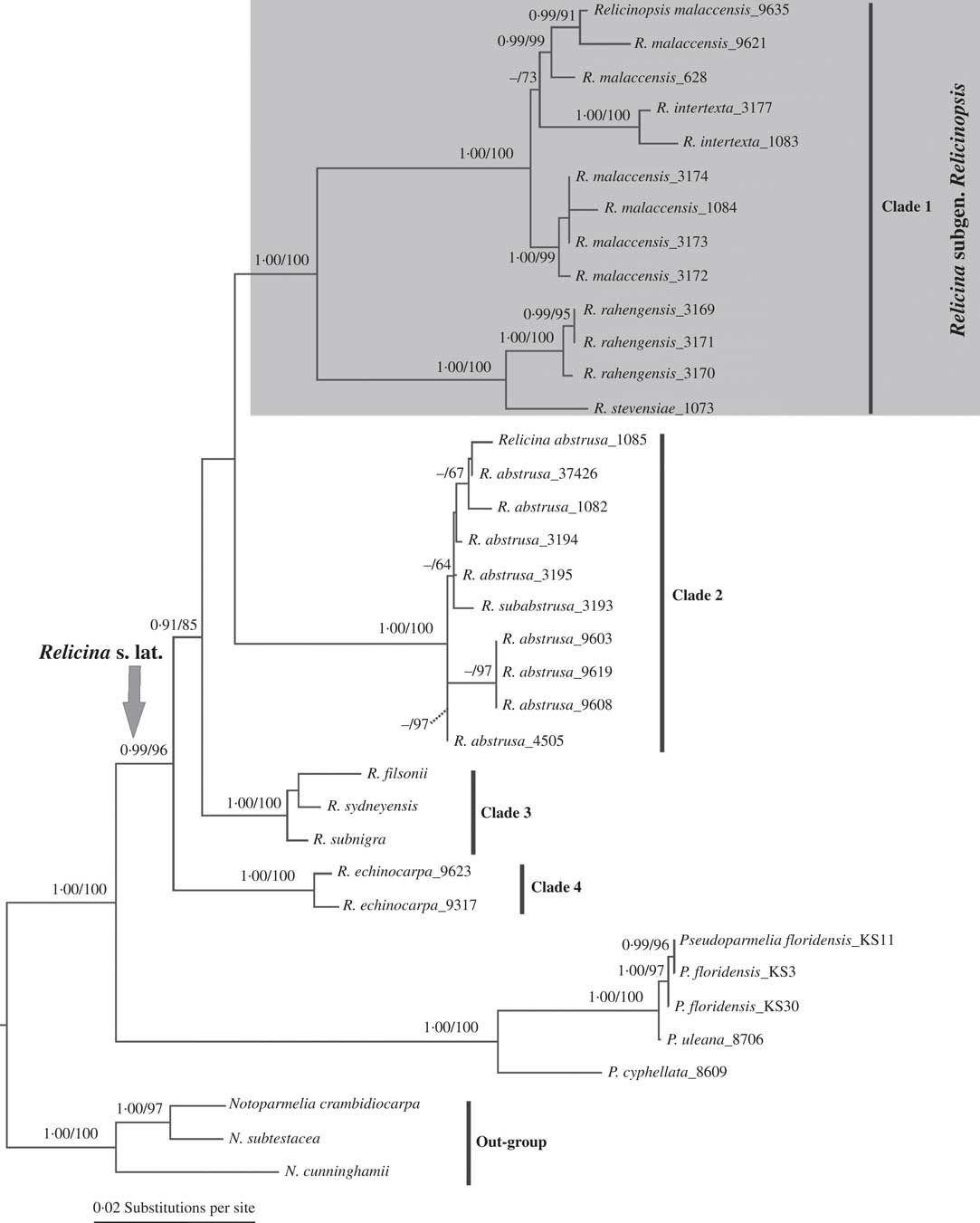
Results of the multilocus phylogeny showed that species of the genus Relicinopsis did not cluster with Pseudoparmelia in which they had previously been classified based on morphology (Hale Reference Hale1975; Swinscow & Krog Reference Swinscow and Krog1988). In agreement with previous molecular studies (Buaruang et al. Reference Buaruang, Scharnagl, Divakar, Leavitt, Crespo, Nash, Manoch, Lücking and Lumbsch2015; Divakar et al. Reference Divakar, Crespo, Wedin, Leavitt, Hawksworth, Myllys, McCune, Randlane, Bjerke and Ohmura2015), however, they grouped with Relicina species. Morphological similarities between Relicinopsis and Relicina species have been discussed previously (Elix et al. Reference Elix, Johnston and Verdon1986; Elix Reference Elix1993; Divakar & Upreti Reference Divakar and Upreti2005). In this study, Relicina was recovered as paraphyletic with Relicinopsis nested within it (Fig. 1). Both the SH and ELW tests significantly rejected monophyly of Relicina as currently circumscribed (P≤0·005). These data clearly indicate that the current phenotype-based generic circumscription (Hale Reference Hale1975) does not reflect evolutionary relationships. Genus-level paraphyly has been found in other groups of parmelioid lichens, including Hypotrachyna (Vain.) Hale (Divakar et al. Reference Divakar, Crespo, Núñez-Zapata, Flakus, Sipman, Elix and Lumbsch2013) and Bulbothrix Hale (Divakar et al. Reference Divakar, Ferencova, Del Prado, Lumbsch and Crespo2010), and similar patterns have been found in other groups of lichen-forming fungi (reviewed in Lumbsch Reference Lumbsch2007; Printzen Reference Printzen2010).
All species of Relicinopsis were recovered in a well-supported (PP=1·00 and ML bootstrap=100%) monophyletic clade (clade 1), nested within Relicina (Fig. 1). Clade 1 of Relicinopsis included four of the five species currently known in this genus, including the type species R. intertexta (Mont. & Bosch.) Elix & Verdon. Species of Relicina were grouped in three well-supported monophyletic clades (clades 2, 3 and 4). However, the sister-group relationship of clade 2 and clade 1 recovered in the ML tree lacked support (Fig 1). Furthermore, in the Bayesian tree, clade 2 formed a well-supported (PP=0·95) sister-group relationship with clade 3 (data not shown). Clade 2 included samples of Relicina abstrusa (Vain.) Hale and R. subabstrusa (Gyeln.) Hale from Australia, Kenya and Thailand, whereas clade 3 consisted of three species, viz. R. filsonii Elix & J. Johnston, R. sydneyensis (Gyeln.) Hale and R. subnigra Elix & J. Johnston occurring in Australasia and South-East Asia. Clade 4 included two samples of R. echinocarpa (Kurok.) Hale from Kenya. This relationship was strongly supported in the ML analysis (ML bootstrap=85%) but received weak support in the Bayesian tree reconstruction (PP=0·91). Our results showed that the relationships among these clades remain unresolved suggesting that additional sampling is necessary to better understand the evolutionary relationships among the clades within the Relicina-Relicinopsis clade. In fact, although all but one of the described Relicinopsis species were studied, only 15 samples collected from Africa, Australia and South-East Asia, representing only six of 54 described Relicina species, were sampled here.
Relicina was initially thought to be closely related to Bulbothrix (Hale Reference Hale1975, Reference Hale1976; Elix Reference Elix1993) since both genera are characterized by the presence of bulbate cilia. However, molecular data showed that the two genera were only distantly related with Bulbothrix belonging to the Parmelina clade and Relicina belonging to the Parmelia clade, closely related to Relicinopsis (Crespo et al. Reference Crespo, Kauff, Divakar, Amo, Arguello, Blanco, Roca-Valiente, Núñez-Zapata, Cubas and Argüello2010; Divakar et al. Reference Divakar, Crespo, Wedin, Leavitt, Hawksworth, Myllys, McCune, Randlane, Bjerke and Ohmura2015). The key phenotypic features used to delineate the genera Relicina and Relicinopsis are summarized in Table 2. Both genera differ in the morphology of the marginal cilia (simple in Relicinopsis vs. bulbate in Relicina) and the type of conidia (fusiform or cylindrical in Relicinopsis vs. bifusiform in Relicina). Other characters, such as ascospore form and size, and rhizine morphology overlap (Table 2). Different types of conidia can be found in a number of currently accepted genera in Parmeliaceae such as Hypotrachyna, Melanelixia O. Blanco et al., Melanohalea O. Blanco et al., Myelochroa (Asahina) Elix & Hale, Parmotrema A. Massal., Punctelia Krog and Xanthoparmelia (Vain.) Hale (Crespo et al. Reference Crespo, Kauff, Divakar, Amo, Arguello, Blanco, Roca-Valiente, Núñez-Zapata, Cubas and Argüello2010); thus this feature can be variable within genera in this family. Consequently, Relicinopsis is reduced here to synonymy with Relicina. However, given that Relicinopsis species formed a well-supported monophyletic clade (clade 1) and are distinguished by conidium morphology, we propose recognising the clade at subgeneric rank. Consequently, the subgenus Relicina is paraphyletic with Relicinopsis nested within it. However, following Divakar et al. (Reference Divakar, Crespo, Núñez-Zapata, Flakus, Sipman, Elix and Lumbsch2013) we consider recognition of the monophyletic clade at the subgeneric level preferable since no paraphyletic taxa at generic level are produced (Hörandl & Stuessy Reference Hörandl and Stuessy2010).
While most of the traditionally circumscribed species in Relicina s. lat. sampled for this study were recovered in monophyletic clusters, a few species did not form monophyletic groups, such as Relicinopsis malaccensis (clade 1) and Relicina abstrusa (clade 2). Additional studies are necessary to evaluate species boundaries in these nominal taxa.
Taxonomic Treatment
Relicina subgen. Relicinopsis (Elix & Verdon) Kirika, Divakar & Lumbsch comb. et stat. nov.
MycoBank No.: MB 817621
Relicinopsis Elix & Verdon, in Elix et al., Mycotaxon 27: 281 (1986); type species: Relicina intertexta (Mont. & Bosch) Kirika, Divakar & Lumbsch, Lichenologist XX: XX (2016).
Parmelia intertexta Mont. & Bosch, in Miquel, Pl. Jungh. 4: 445 (1855).— Pseudoparmelia intertexta (Mont. & Bosch) Hale, Phytologia 29: 190 (1974).— Relicinopsis intertexta (Mont. & Bosch) Elix & Verdon, in Elix et al., Mycotaxon 27: 281 (1986).
A subgenus in the genus Relicina, corresponding to clade 1 in Fig. 1, including all species currently placed in Relicinopsis (Elix et al. Reference Elix, Johnston and Verdon1986; Elix Reference Elix1993).
New Combinations
Relicina dahlii (Hale) Kirika, Divakar & Lumbsch comb. nov.
MycoBank No.: MB 817622
Pseudoparmelia dahlii Hale, Smithson. Contr. Bot. 31: 28 (1976).—Relicinopsis dahlii (Hale) Elix & Verdon, in Elix et al., Mycotaxon 27: 281 (1986).
Relicina intertexta (Mont. & Bosch) Kirika, Divakar & Lumbsch comb. nov.
MycoBank No.: MB 817624
Parmelia intertexta Mont. & Bosch, in Miquel, Pl. Jungh. 4: 445 (1855).— Pseudoparmelia intertexta (Mont. & Bosch) Hale, Phytologia 29: 190 (1974).— Relicinopsis intertexta (Mont. & Bosch) Elix & Verdon, in Elix et al., Mycotaxon 27: 281 (1986).
Relicina malaccensis (Nyl.) Kirika, Divakar & Lumbsch comb. nov.
MycoBank No.: MB 817623
Parmelia malaccensis Nyl., J. Linn. Soc., Bot. 20: 52 (1883).—Pseudoparmelia malaccensis (Nyl.) Hale, Phytologia 29: 190 (1974).—Relicinopsis malaccensis (Nyl.) Elix & Verdon, in Elix et al., Mycotaxon 27: 282 (1986).
Relicina rahengensis (Vain.) Kirika, Divakar & Lumbsch comb. nov.
MycoBank No.: MB 817625
Parmelia rahengensis Vain., Ann. Bot. Soc. Zool.-Bot. Fenn. Vanamo 1: 39 (1923).—Pseudoparmelia rahengensis (Vain.) Hale, Phytologia 29: 191 (1974).—Relicinopsis rahengensis (Vain.) Elix & Verdon, in Elix et al., Mycotaxon 27: 282 (1986).
Relicina stevensiae (Elix & J. Johnst.) Kirika, Divakar & Lumbsch comb. nov.
MycoBank No.: MB 817626
Relicinopsis stevensiae Elix & J. Johnst., Mycotaxon 31: 504 (1988).
Newly obtained DNA sequences were generated in the Pritzker Laboratory for Molecular Systematics and Evolution at the Field Museum and at the Molecular Laboratory, Department of Biology, Faculty of Pharmacy, Complutense University of Madrid. This study was supported by a grant from the IDP/The Field Museum Africa Training Fund and the Spanish Ministerio de Ciencia e Innovación (CGL2013-42498-P).