Introduction
Mammalian preimplantation development, as well as early embryonic development, in non-mammalian species is a very specific period of life. The cell cycle is much shorter, G1 and G2 phases are almost missing and the embryo lives only from maternal stores. Until the event called embryonic genome activation (EGA) or maternal-to-zygotic transition (MZT) the embryo is transcriptionally silent and only maternally derived mRNAs and proteins are present. As both these types of molecules have usually short lifetimes, their processing has to be altered so that they could be stored from oocyte maturation through fertilization to EGA and in some proteins even further. It has been shown that some maternal proteins are stored after EGA (Svarcova et al., Reference Svarcova, Laurincik, Avery, Mlyncek, Niemann and Maddox-Hyttel2007; Ohsugi et al., Reference Ohsugi, Zheng, Baibakov, Li and Dean2008; Toralova et al., Reference Toralova, Benesova, Vodickova Kepkova, Vodicka, Susor and Kanka2012). This prolonged stability may be ensured by some posttranslational modification or complex formation. Such complexes of maternal proteins are formed to persist to preimplantation development and are then involved in driving embryogenesis (reviewed in Lei et al., Reference Lei, Xukun and Jurrien2013). This represents primarily proteins like zona pellucida proteins or the SCMC (subcortical maternal complex). However it is possible that maternal proteins form complexes or are modified in order to be preserved for preimplantation development. Such masking or modification was found in oocytes in protein CENPE (Duesbery et al., Reference Duesbery, Choi, Brown, Wood, Resau, Fukasawa, Cleveland and Vande Woude1997). Moreover, the SCMC has a molecular weight much larger than is expected by the total mass of all participating proteins (669–2000 kDa vs. expected 325 kDa) (Li et al., Reference Li, Baibakov and Dean2008).
Material and methods
In vitro fertilization and embryo culture
Bovine cumulus–oocytes complexes were obtained from abattoir-derived ovaries. The cattle had been slaughtered (Slaughterhouse Rosovice) for publicly edible meat. Those ovaries were discarded without any utilization. Hence, an ethics statement in our paper was not required. The isolated oocytes were subjected to in vitro maturation and subsequent fertilization (Toralova et al., Reference Toralova, Benesova, Vodickova Kepkova, Vodicka, Susor and Kanka2012). The embryos were collected after an appropriate time of cultivation (Benesova et al., Reference Benesova, Kinterova, Kanka and Toralova2016).
Western blotting
Unless otherwise indicated, chemicals were purchased from Sigma. Unlike the anti-p27 and anti-IκBα antibodies (in which was necessary to use 20 embryos per line), anti-Skp1 antibody gave a really intensive signal and therefore we used just six embryos per line. Embryos were lysed in 15 μl of Blue Loading Buffer (772, Cell Signaling Technology, Danvers, MA, USA) with dithiothreitol, boiled for 5 min and subjected to 12% SDS-PAGE. Proteins were transferred from gels to an Immobilon P membrane (Millipore Biosciences, Billerica, MA, USA) using a semidry blotting system (Whatman Biometra GmbH, Hoettingen, Germany) for 28 min at 5 mA/cm2. Blocking of the membrane was performed in 5% BSA in TBS-Tween buffer (TBS-T, 20 mM Tris, pH 7.4, 137 mM NaCl and 0.5% Tween 20) for IκBα, and in 5% non-fat milk in TBS-T for Skp1 and p27, for 1 h and incubated overnight with following antibodies: IκBα - IκBα Antibody (Cell Signaling Technology 9242, Leiden, The Netherlands) 1:1000 in 5% BSA/TBS-T, p27 - Anti-p27 KIP 1 antibody (Abcam ab32034, Cambridge, UK) 1:1000 in 5% non-fat milk/TBS-T or Skp1–SKP1A monoclonal antibody, clone 1H8 (Abnova M01, Heidelberg, Germany) 1:1000 in 5% non-fat milk/TBS-T. After washing in TBS-T, the membranes were incubated with HRP-conjugated donkey anti-rabbit or donkey anti-mouse IgG antibody (both 1:7500; Jackson Immuno Research, Suffolk, UK) in 5% non-fat milk/TBS-T or in 5 % BSA/TBS-T for 1 h at room temperature. Proteins were visualized with Luminata Crescendo Western HRP (Merck Millipore, Darmstadt, Germany) or ECL (Amersham, GE Healthcare Life Science, UK). Precision Plus Protein™ Dual Color Standards (161-0374, Bio-Rad spol s.r.o., Czech Republic) were used for molecular weight estimation.
Results and discussion
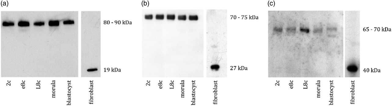
Both embryos and fibroblasts were processed in the same way according to the protocol used in Toralova et al. (Reference Toralova, Benesova, Vodickova Kepkova, Vodicka, Susor and Kanka2012). All experiments were performed at least three times. When performing the western blot analysis, we found the existence of bands of higher molecular weight than expected, in all stages of bovine preimplantation development from the 2-cell stage until the blastocyst stage (representative blot in Fig. 1).
Western blot analysis of bovine preimplantation embryos and bovine fibroblasts. Representative images of western blots of embryos from 2-cell to blastocyst stage: (a) Skp1: 80–90 kDa vs. 19 kDa (six embryos per line). (b) p27: 70–75 kDa vs. 27 kDa (20 embryos per line). (c) IκBα: 65–70 kDa vs. 40 kDa (20 embryos per line). The expected band size responds to the band size in bovine fibroblasts. All experiments were repeated at least three times. 2c, 2-cell stage; e8c, early 8-cell stage, L8c, late 8-cell stage embryos.

These bands were not possible to be dissolved using dithiothreitol or high temperature (5 min boiling). Simultaneously, analysis of bovine fibroblast cells was performed and the bands emerged at the expected molecular weight. This shows that the bands of higher molecular weight might be specific and typical for preimplantation embryos. However, we cannot exclude that this phenomenon does not exist in another cells, especially non-differentiated, rapidly dividing cells. The described proteins play distinct roles in cell functioning, however all of these have connection to E3-ubiquitin ligase SCF complex (Skp1–Cullin1–F-box protein complex). Skp1 is an invariant member of this complex and is involved in its activation/deactivation control (Bai et al., Reference Bai, Sen, Hofmann, Ma, Goebl, Harper and Elledge1996; Zheng et al., Reference Zheng, Yang, Harrell, Ryzhikov, Shim, Lykke-Andersen, Wei, Sun, Kobayashi and Zhang2002). Besides participation in the ubiquitin-proteasome pathway, it is necessary for correct chromosome segregation and euploidy maintenance in mice (Piva et al., Reference Piva, Liu, Chiarle, Podda, Pagano and Inghirami2002). It is supposed to play an important role during mammalian preimplantation development (Benesova et al., Reference Benesova, Kinterova, Kanka and Toralova2016). Incorrect Skp1 expression is involved in development of malignancy (Piva et al., Reference Piva, Liu, Chiarle, Podda, Pagano and Inghirami2002). P27 is a cell cycle regulator especially involved in G1 arrest and in regulation of transcription. Its decreased expression is involved in tumorigenesis and poor prognosis of disease progression (Slingerland & Pagano, Reference Slingerland and Pagano2000). IκBα is involved in NF-κB inhibition by masking its nuclear localization signal and dissociation of NFκB from DNA. Both p27 and IκBα are substrates of the SCF complex, in which Skp1 is incorporated. However, it does not seem that the higher band incidence is related in these three proteins, as the bands are in different heights. It is known that the proteins in early embryos often have multiple isoforms (Tay et al., Reference Tay, Lin, Seow, Tan, Hew and Gong2006), however the large difference in molecular weights does not support this explanation. Moreover, the band height is not a multiple of the expected value, so it is not consequence of polymerization.
The reason for the existence of bands of higher molecular weight remains to be elucidated. However, the finding of them in these three proteins speaks for its importance and common occurrence in preimplantation embryos. We suppose that these bands are complex of proteins that arise to preserve them for further stages of preimplantation and may be (as the higher molecular weight bands are present in all preimplantation stages, including blastocysts) to some period of postimplantation development. We assume that similar results can be found also in many other proteins in preimplantation embryos.
Acknowledgements
We would like to thank J. Tyleckova, J. Cervenka, R. Sucha, M. Zizkova and J. Novak for their helpful comments.
Financial support
The work was supported by grant GACR 13-24730P and CIGA 20172013.
Ethical standards
Not applicable.
Statement of interest
None.