Major aortopulmonary collateral arteries typically arise from the aorta in patients with pulmonary atresia with a ventricular septal defect. Infrequently, major aortopulmonary collateral arteries can arise from coronary arteries and this entity is described as a coronary-pulmonary artery fistula. The incidence of coronary-pulmonary fistulas in pulmonary atresia with ventricular septal defect is reported to be as high as 10%.Reference Alkhushi, Al‐Radi, Ajlan, Abdelmohsen and Attia 1
Patients with pulmonary atresia, ventricular septal defect, and major aortopulmonary collateral arteries can have very intricate anatomy. Unifocalisation of major aortopulmonary collateral arteries is often required as part of the initial surgical palliation and necessitates a complete understanding of the pulmonary blood supply.Reference Alkhushi, Al‐Radi, Ajlan, Abdelmohsen and Attia 1 Pre-surgical planning often depends upon cardiac catheterisation and cross-sectional imaging in these cases. Recently, three-dimensional printed models were reported to be useful for pulmonary atresia with ventricular septal defect and major aortopulmonary collateral arteries.Reference Anwar, Rockefeller, Raptis, Woodard and Eghtesady 2 We present a case of pulmonary atresia with ventricular septal defect and major aortopulmonary collateral arteries involving a coronary-pulmonary artery fistula from the left main coronary artery, in which we used a life-sized, three-dimensional printed model to plan surgical management.
Case report
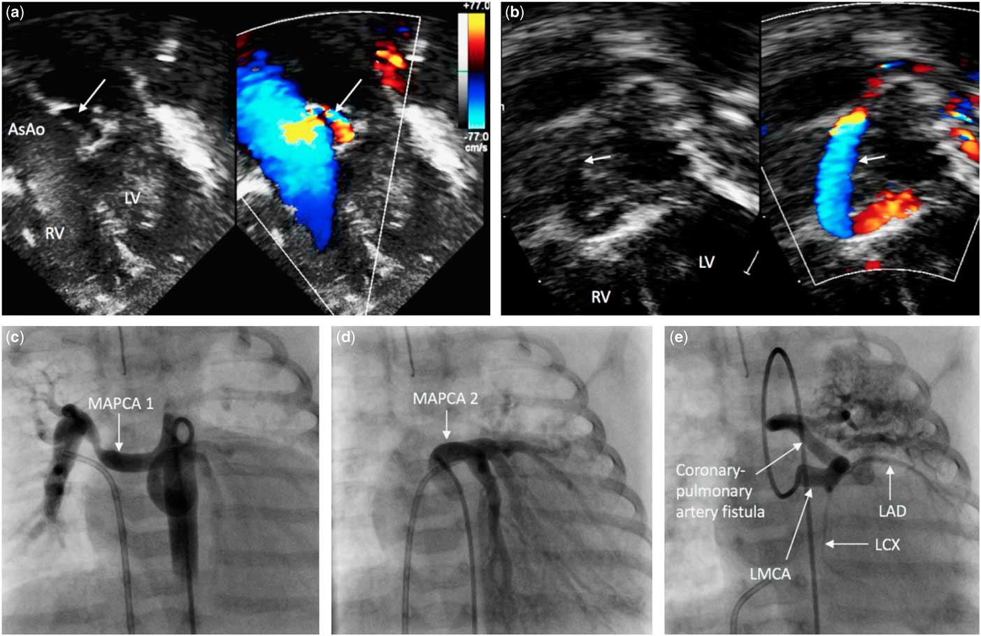
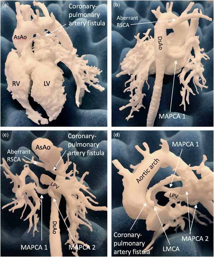
A female neonate with a prenatal diagnosis of pulmonary atresia with ventricular septal defect and chromosome 22q11.2 microdeletion was born at 36 weeks gestation with a birth weight of 1.9 kg. Postnatal echocardiography confirmed the diagnosis of pulmonary atresia with ventricular septal defect and non-confluent pulmonary arteries with two major aortopulmonary collateral arteries arising from the descending aorta. Another unusual vessel appeared to arise from the proximal left coronary artery (Fig 1a and b). The presence of a coronary-pulmonary artery fistula was suspected. To delineate this complex anatomy, cardiac CT angiography was performed. A life-sized cardiac three-dimensional printed model was created for pre-surgical planning. A three-dimensional digital model was built using the software Mimics® (Materialise, Plymouth, MI, United States of America). Using selective laser sintering technology, a three-dimensional model was printed as a blood pool, rigid, and opaque structure (Fig 2). This model was used to counsel the family about the complexity of the anatomy. The surgeon used the three-dimensional model for surgical planning and for a team briefing. Cardiac catheterisation demonstrated that two major aortopulmonary collateral arteries arose from the descending aorta to the entire right lung and left lower lobe, respectively, as well as a coronary-pulmonary artery fistula from the left main coronary artery to the left upper lobe (Fig 1c, d, and e). At 3 months of age, weight 3.5 kg, she underwent initial palliative surgery. Intraoperatively, a large coronary-pulmonary artery fistula arose from the left main coronary artery paralleled the ascending aorta and then coursed leftward into the left upper lobe segment (Supplementary Figure). This coronary-pulmonary artery fistula was divided and ligated where it arose from the left main coronary artery. The other two major aortopulmonary collateral arteries were readily identified because of the anatomical accuracy of the three-dimensional printed model. The major aortopulmonary collateral arteries were unifocalised and supplied with a 5 mm central aortopulmonary shunt from the ascending aorta. At 9 months of age, she underwent takedown of central shunt, placement of a right ventricle-to-pulmonary artery conduit using a 16 mm aortic homograft, and fenestrated ventricular septal defect closure. She is stable at 13 months of age.
(a and b) Transthoracic echocardiography. Colour-compare imaging at the apical view shows that a large vessel (arrow) arises from the near the origin of left coronary artery and courses superiorly, consistent with coronary-pulmonary artery fistula from the left coronary artery. (c, d and e) Selective angiography of major aortopulmonary collateral arteries from the descending aorta shows that major aortopulmonary collateral artery model 1 supplies the entire right lung and major aortopulmonary collateral artery model 2 supplies the left lower lobe segment. Selective left coronary artery angiography shows a dilated left main coronary artery with left anterior descending artery and left circumflex artery. A large coronary-pulmonary artery fistula arises from the left main coronary artery and supplies the left upper lobe segment. AsAo=ascending aorta; LV=left ventricle; RV=right ventricle.

Three-dimensional printed model. (a and b) Whole heart model. (c and d) Focussed major aortopulmonary collateral artery model. Major aortopulmonary collateral artery model 1 arises from the descending aorta and supplies the entire right lung. major aortopulmonary collateral artery model 2 arises from the descending aorta and supplies the left lower lobe segment. A coronary-pulmonary artery fistula arises from the left main coronary artery and supplies the left upper lobe segment. AsAo=ascending aorta; LPV=left pulmonary vein; LV=left ventricle; RSCA=right subclavian artery; RV=right ventricle.

Comment
Coronary-pulmonary artery fistula is a rare entity along with pulmonary atresia with ventricular septal defect and major aortopulmonary collateral arteries. The origin of the coronary-pulmonary artery fistula was the left coronary artery in the majority of reported cases; however, they can also arise from either the right or from a single coronary artery.Reference Alkhushi, Al‐Radi, Ajlan, Abdelmohsen and Attia 1 The presence of a coronary-pulmonary artery fistula is often suspected by transthoracic echocardiography. This diagnosis usually needs to be confirmed by cardiac catheterisation and cross-sectional imaging, which helps to better delineate the complex anatomy for surgical planning purposes.Reference Kaneko, Okabe, Nagata, Kobayashi, Murakami and Takamoto 3 Coronary steal is a potential complication from this condition, but many of the case reports have noted that it does not appear to be a frequent occurrence.Reference Ono, Dong, Kasnar-Samprec, Vogt and Schreiber 4 , Reference Sathanandam, Loomba, Ilbawi and Van Bergen 5 Our patient did not have coronary steal before surgery. To achieve a favourable outcome, early intervention is important.Reference Kaneko, Okabe, Nagata, Kobayashi, Murakami and Takamoto 3 , Reference Ono, Dong, Kasnar-Samprec, Vogt and Schreiber 4 Our patient underwent unifocalisation of major aortopulmonary collateral arteries with placement of an aortopulmonary artery shunt at 3 months of age, followed by complete repair at 9 months of age.
The life-sized three-dimensional printed model was incorporated into our management. In patients with complex cardiac anatomy, three-dimensional printing has been used to effectively delineate their anatomy and appropriately plan for surgical intervention.Reference Anwar, Rockefeller, Raptis, Woodard and Eghtesady 2 The three-dimensional printed model was useful for our team to make informed decisions regarding the sources and distribution of the pulmonary blood. Another benefit of the three-dimensional printed model was its integration with the cardiac catheterisation. Pre-catheterisation three-dimensional modelling can reduce the need for initial large volume contrast angiography and allow interventional cardiologists to efficiently perform selective angiography of individual major aortopulmonary collateral arteries. In our case, the pre-catheterisation three-dimensional printed model precisely located the origin of the major aortopulmonary collateral arteries, making selective angiography straightforward. Our cardiologists and cardiothoracic surgeons also incorporated this life-sized three-dimensional model into the preoperative multi-disciplinary conference discussion as well as for the pre-operative family consultation. Intraoperatively, this model was used as a convenient reference during dissection that allows identification of the major aortopulmonary collateral arteries in relation to the descending aorta and pulmonary veins. The surgeon confirmed that the three-dimensional printed model accurately represented the complex anatomy in our case. Although the airway was not incorporated into our three-dimensional printed model, integration of the airway into the model may help to demonstrate complex anatomic relationship better. The use of three-dimensional model became one of the important pre-operative planning modalities in our practice. Our programme outsourced cardiac three-dimensional printing service to the vendor (Materialise), and the cost for this service included technical fees for cardiac segmentation/three-dimensional digital modelling, and three-dimensional printing.
In conclusion, we describe a rare case of coronary-pulmonary artery fistula from the left main coronary artery in a patient with pulmonary atresia, ventricular septal defect, and major aortopulmonary collateral arteries. The three-dimensional printed model may be particularly useful for complex major aortopulmonary collateral arteries and can be leveraged to contribute to more precise preoperative planning, more coherent preoperative consultation, and more confident intraoperative management of these patients.
Acknowledgements
None.
Financial support
This paper was supported by the Children’s Hospital of Michigan Foundation Grant – “Heart of the Child” (R2-2015-43).
Conflicts of interest
None.
Supplementary material
To view supplementary material for this article, please visit https://doi.org/10.1017/S1047951118002317