Implications
The plant extract mixture composed of carvacrol, capsicum oleoresin and cinnamaldehyde showed some obvious antioxidative properties under dietary-induced oxidative stress conditions. In view of the fact that it is known that oxidative stress is involved in the etiology of different diseases and occurs also as a consequence of increased immune system activity (Valko et al., Reference Valko, Leibfritz, Moncol, Cronin, Mazur and Telser2007), an antioxidant support to sustain the DNA integrity of the immune cells may be beneficial for preserving healthy animals and by that the quality of animal products.
Introduction
The search for nutritive antibiotic alternatives in animal production and the increased demand from consumers for natural feed additives has encouraged scientists to fully explore the possible use of herbs, spices and their active components (plant extracts (PEs)) in animal nutrition. The essential scope in animal production is to ensure high productivity, healthy animals and quality products, which are stable and appropriate for processing. In this respect PEs are not only appetite and digestion stimulants, but with their beneficial physiological effects, can be overall health and by that growth promoters.
Animals in intensive farming systems are frequently exposed to oxidative stress which can result in the damage of proteins, lipids and DNA (McCall and Frei, Reference McCall and Frei1999). The focus of our research was the nutritional oxidative stress that can, among other, be a result of feeding vegetable oils to reach the high energy requirements of animals and/or creating animal products with improved nutritional quality (products rich in n-3 polyunsaturated fatty acid (PUFA)) (Wood et al., Reference Wood, Richardson, Nute, Fisher, Campo, Kasapidou, Sheard and Enser2004).
Carvacrol (Origanum spp.), cinnamaldehyde (Cinnamonum spp.) and capsicum oleoresin (Capsicum annum) besides their antiseptic (Pasqua di et al., Reference Pasqua di, Hoskins, Betts and Mauriello2006), digestion stimulant (Janz et al., Reference Janz, Morel, Wilkinson and Purchas2007) and anti-inflammatory (Srinivasan, Reference Srinivasan2005) effects also exhibit antioxidant characteristics (Kogure et al., Reference Kogure, Goto, Nishimura, Yasumoto, Abe, Ohiwa, Sassa, Kusumi and Terada2002; Singh et al., Reference Singh, Maurya, deLampasona and Catalan2007; Srihari et al., Reference Srihari, Sengottuvelan and Nalini2008). To maximize their potential, the synergistic (cumulative) effect of these plant constituents should be determined (Capecka et al., Reference Capecka, Mareczek and Leja2005). This has already been done for their antimicrobial proprieties (Manzanilla et al., Reference Manzanilla, Perez, Martin, Kamel, Baucells and Gasa2004; Castillo et al., Reference Castillo, Martin-Orue, Roca, Manzanilla, Badiola, Perez and Gasa2006), but not yet for their in vivo antioxidant and anti-genotoxic effect.
Accordingly, the objective of our study was to determine the in vivo antioxidative and anti-genotoxic potential of PE mixture against n-3 PUFA-induced oxidative load in young pigs and to compare it with the effect of the commonly used form of vitamin E (α-tocopheryl acetate).
Material and methods
The experiment was performed in the experimental facilities of the Animal Science Department of the Biotechnical faculty of Ljubljana. The protocol was approved by the Animal Ethics Committee of the Veterinary Administration of the Republic of Slovenia. At the end of the experiment the pigs were transferred to a fattening unit.
Animals and dietary treatments
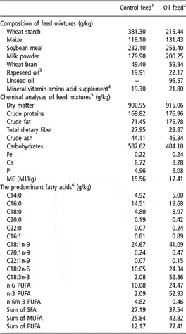
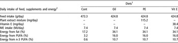
Thirty-two weaned castrated male crossbred pigs from a commercial herd (BW 10.9 ± 0.38 kg; n = 32) were penned individually in balance cages that allowed the separate collection of urine and feces under standard housing conditions. The experiment was divided into adaptation and experimental periods that lasted for 5 and 14 days, respectively. Animals were individually, restrictively, fed 2.7 times the maintenance requirements for energy (NRC, 1998). Water was provided ad libitum. Before starting the experimental period, pigs were randomly assigned to four dietary treatment groups. All animals were fed with isoenergetic daily rations. The negative control group (Cont) was fed with a low-antioxidant diet with 17.2% of its energy from fat (Control feed). The other three experimental groups were fed ‘Oil feed’ with the addition of linseed oil to induce the oxidative stress by elevating the amount of energy from fat to 34.1% (Tables 1 and 2). One of these groups served as a positive control group (Oil), one group was additionally supplemented with 271.2 mg/kg feed of a commercial preparation of PEs and one group with 90.4 mg/kg α-tocopheryl acetate (Vit E) (Table 2). The linseed oil was frozen until just before use and added to the feed directly before feeding to prevent the lipid peroxidation during storage. The live weight and feed intake of animals were monitored throughout the experiment.
Composition and analyses of feed mixtures

ME = metabolizable energy.
1‘Control feed’ was fed to the Control group.
2‘Oil feed’ was fed to other three experimental groups. Vitamin E and plant extract mixture were added to the Oil feed at a concentration of 90.4 mg/kg and 271.2 mg/kg for the Vit E and PE groups, respectively.
3Rapeseed oil was ‘food grade’ oil-low in C22:1n-9.
4Calculated to meet vitamin and mineral requirements according to NRC (1998). The mineral-vitamin-amino acid supplement provided marginal amounts of vitamin A (2000 IU/kg feed), vitamin E (13 IU/kg feed), Zn (90 mg/kg feed), and Se (0.20 mg/kg feed). It also contained 19.2% Ruekana, 43.5% limestone, 3.8% sodium chloride, 0.4% lysine and 7.3% methionine per kg of supplement.
5Proximate analysis and mineral analyses.
6Only predominant fatty acids are listed, but the sum of saturated fatty acids (SFAs), monounsaturated fatty acids (MUFAs) and polyunsaturated fatty acids (PUFAs) are computed from all fatty acids analysed.
Composition of daily ration and daily intake of energy and nutrients estimated for an 11 kg pig

ME = metabolizable energy; PUFA = polyunsaturated fatty acid.
1Diets: Cont = ‘Control feed’; Oil = ‘Oil feed’; PE = ‘Oil feed’; additionally supplemented with 271.2 mg/kg of plant extract mixture; Vit E = ‘Oil feed’; additionally supplemented with 90.4 mg/kg of α-tocopheryl acetate.
2The energy value of feedstuffs and diets was estimated according to GEH (Gesellschaft für Ernährungsphysiologie, Germany).
PE and vitamin E supplementation
The PE mixture, with the commercial name Xtract 6930 (Pancosma, Geneva, Swiss), contained 5% (wt/wt) carvacrol (naturally identical; Origanum spp.), 3% cinnamaldehyde (naturally identical; Cinnamonum spp.) and 2% capsicum oleoresin (natural; Capsicum annum) in the hydrogenated rapeseed oil matrix. The extract preparation was added to the feed at a concentration of 271.2 mg/kg. The amount of additional vitamin E supplementation in the Vit E group was calculated according to the expected increase of vitamin E requirements because of higher PUFA intake (Muggli, Reference Muggli1994). Vitamin E was added as α-tocopheryl acetate at a concentration of 90.4 mg/kg feed.
Sample collection
At the end of the 14-day experimental period, blood samples were taken from the jugular vein after overnight fasting and urine excreted in 48 h was collected.
Malondialdehyde (MDA) determination
Blood was centrifuged (1000 × g for 15 min) and plasma supernatants were transferred to microcentrifuge tubes and stored at −70°C. Urine was filtered into test tubes through filter paper (Schleicher & Schuell 520 A, Dassel, Germany), transferred to microcentrifuge tubes and stored at −70°C. The methodology of Wong et al. (Reference Wong, Knight, Hopfer, Zaharia, Leach and Sunderman1987) modified by Chirico (Reference Chirico1994) and Fukunaga et al. (Reference Fukunaga, Takama and Suzuki1995) was used to measure the concentrations of MDA in blood plasma and urine by HPLC using a reversed-phase chromatography column (HyperClone 5 μm ODS (C18) 120A, 4.6 × 150 mm; Phenomenex Inc., Torrance, CA, USA) and a C18 ODS guard column (4 mm × 3 mm; Phenomenex Inc.). Briefly, 100 μl of plasma or urine was hydrolyzed by 100 μl of 0.44 M orthophosphoric acid in the presence of 10 μl of 0.2% butylated hydroxytoluene (BHT). After the addition of 300 μl of pure ethanol, the samples were centrifuged for 15 min. The supernatant was mixed with thiobarbituric acid (TBA) and heated at 90°C for 60 min. The MDA-TBA2 complex was measured with a Waters Alliance 2690 apparatus (Milford, MA, USA) equipped with a Waters 474 scanning fluorescence detector. The results of the analysis were evaluated with the Millenium®32 Chromatography Manager program (Waters).
Urine isoprostanes (iPF2α-VI) determination
Filtered urine was stored at −70°C in the presence of 0.005% BHT. The samples were purified through solvent extraction. Briefly, samples were mixed with acetone and centrifuged at 3000 × g for 10 min. The supernatant was transferred to a clean test tube and evaporated under nitrogen steam. Samples were then incubated in an extraction buffer (1 M sodium citrate, pH 4, with 10% NaCl) for 90 min. Dichloromethane was added for the extraction of iPF2α-VI and the lipophilic fraction was transferred into a clean tube. This step was repeated once more and both dichloromethane fractions were pooled and evaporated under nitrogen steam. The residue was dissolved in a buffer and incubated at room temperature for 90 min. The amount of iPF2α-VI was determined by competitive enzyme-linked immunosorbent assay (ELISA) assay (kit iPF2α-VI EIA, Cayman Chemical, Ann Arbor, MI, USA). In each well, which had been pre-coated with mouse monoclonal anti-rabbit IgG, 50 μl of sample or standard was mixed with 50 μl of prepared iPF2α-VI-acetylcholinesterase (AChE) conjugate and 50 μl of antiserum-iPF2α. The plate was incubated for 18 h at 4°C. After washing, the plate was developed with 200 μl of Ellmans reagent per well for 45 min. The reaction between the AChE and Ellmans reagent was measured at 412 nm with an ELISA reader (EL 808, BIO-TEK, Winooski, VT, USA). The intensity of the color is proportional to the amount of iPF2α-VI-AChE conjugate, which is inversely proportional to the amount of iPF2α-VI in the sample. The results are presented in micrograms of iPF2α-VI excreted in 24 h.
Glutathione peroxidase (Gpx), total antioxidant status (TAS) of plasma
The whole blood for Gpx analysis and the plasma for TAS analyses were transferred to microcentrifuge tubes and stored at −70°C. The methodology of Paglia and Valentine (Reference Paglia and Valentine1967) was used for the measurement of Gpx and the methodology of Miller and Rice Evans (Reference Miller and Rice Evans1996) for the measurement of TAS. Samples were assayed with commercially available Gpx and TAS kits (Randox, Crumlin, UK) following the instructions of the kits. Briefly, for the Gpx assay, 50 μl of heparinised whole blood was diluted with 2 ml of diluting agent (Randox). The diluted sample (5 μl) was mixed with 200 μl of Ransel reagent 1 and 50 μl of cumene hydroperoxide in an RX Daytona biochemical analyser (Randox). Absorbance was measured at 340 nm. For TAS assay, 4 μl of plasma was mixed with 200 μl of chromogen (metmyoglobin and 2,2′-Azino-di-[3-ethylbenzthiazoline sulphonate]) and 40 μl of substrate (H2O2). Absorbance was measured at 600 nm. The intensity of color is inversely proportional to the antioxidant capacity of the sample.
Lymphocyte nuclear DNA damage
Lymphocytes were isolated from the blood samples according to a modified procedure described by Singh (Reference Singh1997). A partially modified procedure by Singh et al. (Reference Singh, McCoy, Tice and Schneider1988) was implemented for the Comet Assay, as described previously by Salobir et al. (Reference Salobir, Rezar, Pajk and Levart2005). An Olympus CH 50 epifluorescent microscope (Tokyo, Japan) at ×200 magnifications was used for the examination of lymphocyte nuclei in the microgels (100 W Hg lamp, excitation filter of 480 to 550 nm and barrier filter of 590 nm). The images were captured with a Hamamatsu Orca 1 CCD camera (Hamamatsu, Japan), analyzed and the nuclear DNA damage was estimated with the Comet 5 dedicated computer program (single cell gel electrophoresis, Kinetic Imaging Ltd, 2000, Bromborough, UK). The results are presented as the percentage of DNA in the tail of the comet and Olive tail moment (OTM), a parameter which is defined as the product of the amount of DNA in the tail and the mean distance of migration in the tail (higher values represent higher rates of DNA damage) (Olive et al., Reference Olive, Wlodek, Durand and Banath1992).
Urine 8-hydroxy-2′-deoxyguanosine (8-OHdG) excretion
Filtered urine was further centrifuged at 2000 × g for 10 min and stored at −70°C. Just before analysis it was diluted to 20 fold with Milli-Q ultrapure water (Millipore, Billerica, MA, USA). Urine 8-OHdG was determined by sensitive competitive ELISA (kit StressXpress, EKS 350, Stressgen, Victoria, Canada). The pre-coated wells were filled with 50 μl of standard or sample and 50 μl of anti-8-OHdG and were left to incubate for 1 h at room temperature. After washing, 100 μl of anti-mouse IgG-horseradish peroxidase (HRP) conjugate was added to the wells. One-hour incubation was followed by washing and the addition of 100 μl tetramethylbenzidine (TMB). After 15 min, 100 μl of acid stop solution was added to each well and the reaction between HRP and TMB was measured at 450 nm with an ELISA reader (EL 808, BIO-TEK). The results are presented in micrograms of 8-OHdG excreted in 24 h.
Statistical analysis
The totally randomized and balanced design enabled us to test the effect of diet on all measured parameters with the ANOVA using the General Linear Models procedure of SAS/STAT module (SAS 8e, 2000; SAS Inc., Cary, NC, USA). The blood samples were collected on two subsequent days, which is used as a covariable in the statistical analysis. The differences among groups were determined by using Tukey’s multiple comparison test. Significance was considered established at P < 0.05. The results in the tables are presented as least squares means ± SEM.
Results
Production parameters
All the animals remained healthy throughout the experiment. Because of the restricted feeding of isoenergetic daily rations, which were completely consumed by all the animals, there were no differences in BW among the different treatment groups. Mean live weight at the end of the experiment was 15.2 ± 0.67 kg (n = 32).
Lipid peroxidation and antioxidative status
The consumption of n-3 PUFA rich linseed oil increased plasma and urinary MDA concentrations for 1.4 and 2.3 fold, respectively (Table 3). Supplementation with PE and vitamin E did not significantly reduce the MDA levels in plasma and urine of pigs fed with linseed oil. The amount of iPF2α-VI excreted in the urine was not elevated by the addition of PUFA to the diet. The PE reduced the urinary iPF2α-VI for 40% in comparison to the Cont. The TAS of plasma and Gpx were not affected by any dietary treatment (Table 3).
Markers of lipid peroxidationFootnote 1 and antioxidant status of pigs fed a high PUFA diet with or without plant extract mixture or vitamin E supplementation

a,b Within a row, LS-means (least square-means) without a common superscript letter differ (P < 0.05).
1 MDA = malondialdehyde; iPF2α-VI = F2-isoprostanes; TAS = total antioxidant status of plasma; Gpx = erythrocyte glutathione peroxidase.
2 Diets: Cont = ‘Control feed’; Oil = ‘Oil feed’; PE = ‘Oil feed’; additionally supplemented with 271.2 mg/kg of plant extract mixture; Vit E = ‘Oil feed’; additionally supplemented with 90.4 mg/kg of α-tocopheryl acetate.
n = 8 animals per group.
Oxidative DNA damage
Linseed oil intake induced DNA damage in the pig’s peripheral blood lymphocytes. The PE and vitamin E reduced (P < 0.001) the percentage of DNA in the tail of the comet and OTM as measured by the Comet Assay (Table 4). The amount of 8-OHdG excreted in the urine in the Oil group was 1.8 fold higher in comparison to Cont group (P = 0.054). When PE mixture was added the 8-OHdG concentrations decreased back to the level of the Cont group (Table 4).
Lymphocyte DNA damage and urinary 8-OHdGFootnote 1 excretions of pigs fed a high PUFA diet with or without plant extract mixture or vitamin E supplementation

a,b Within a column, LS-means (least square-means) without a common superscript letter differ significantly (P < 0.05) or show a trend (P ⩾ 0.05 < 0.10).
1 8-hydroxy-2′-deoxyguanosine.
2 Diets: Cont = ‘Control feed’; Oil = ‘Oil feed’; PE = ‘Oil feed’; additionally supplemented with 271.2 mg/kg of plant extract mixture; Vit E = ‘Oil feed’; additionally supplemented with 90.4 mg/kg of α-tocopheryl acetate.
3 Olive tail moment – defined as the product of the amount of DNA in the tail and the mean distance of migration in the tail (higher values represent higher rates of DNA damage; no units).
n = 8 animals per group.
Discussion
‘Dietary oxidative stress’ can be provoked by the ingestion of highly oxidizable or prooxidative substances (PUFA, metals (Fe, Cu) and toxins) and by the low intake of antioxidants. The mixture of carvacrol, cinnamaldehyde and capsicum oleoresin, which is already present in the market of feed additives as appetite and digestion stimulant, was proven to be a modulator of the intestinal microbial ecosystem (Manzanilla et al., Reference Manzanilla, Perez, Martin, Kamel, Baucells and Gasa2004). It has not, however, been tested yet for its antioxidative potential in vivo, which could contribute to its health impact.
No studies had been documented that measured any kind of cumulative antioxidant effect of the mixture of cinnamaldehyde, capsicum oleoresin and carvacrol, however, there are quite some data on the in vitro antioxidant effect of each active component (Kogure et al., Reference Kogure, Goto, Nishimura, Yasumoto, Abe, Ohiwa, Sassa, Kusumi and Terada2002; Ahuja et al., Reference Ahuja, Kunde, Ball and Geraghty2006; Manjunatha and Srinivasan, Reference Manjunatha and Srinivasan2006; Singh et al., Reference Singh, Maurya, deLampasona and Catalan2007). In relevance to animal nutrition, most in vivo studies have been made on the antioxidant activity of oregano active components, showing improvement of the antioxidative stability of animal tissues during storage (Botsoglou et al., Reference Botsoglou, Fletouris, Florou-Paneri, Christaki and Spais2003; Papageorgiou et al., Reference Papageorgiou, Botsoglou, Govaris, Giannenas, Iliadis and Botsoglou2003; Janz et al., Reference Janz, Morel, Wilkinson and Purchas2007). In our study, a significantly higher concentration of MDA, a marker commonly used to determine lipid peroxidation in biological samples, was found in the plasma and urine of pigs fed with the linseed oil. The PE mixture and vitamin E supplementation did not decrease the MDA concentration in the plasma and its excretion in urine. Probably, the n-3 PUFA load from the feed was so immense that it required higher antioxidant protection than that provided by the amount of PE and vitamin E given.
Furthermore, linseed oil rich in n-3 PUFA did not increase the amount of 24 h iPF2α-VI excreted in the urine. Although it had been stated in the past that the formation of isoprostanes was not affected by the lipid content of the diet (Richelle et al., Reference Richelle, Turini, Guidoux, Tavazzi, Metairon and Fay1999; Willcox et al., Reference Willcox, Catignani and Roberts2003), it was recently shown that the abundant amount of n-3 PUFA in the organism leads to the formation of the family of F3-isoprostanes and decreases F2-isoprostanes (Gao et al., Reference Gao, Yin, Milne, Porter and Morrow2006). However, attempts to measure F3-isoprostanes have been so far met with only limited success, especially with noninvasively collected samples such as urine (Adiyaman et al., Reference Adiyaman, Lawson, Khanapure, FitzGerald and Rokach1998). Because in our study the pigs supplemented with linseed oil also received four times as much linoleic (n-6) acid as the pigs in the Cont, we assumed we could use iPF2α-VI as a marker of arachidonic acid peroxidation (a commonly used marker of oxidative stress). This investigation shows that the results of iPF2α-VI measurements should be interpreted with caution in studies where oxidative stress is induced by n-3 PUFA intake. In such circumstances it cannot be concluded that no oxidative stress was induced if F2-isoprostane levels were not elevated, as the reduction of F2-isoprostane levels after supplementation of n-3 PUFA might occur through several unidentified pathways (Nalsen et al., Reference Nalsen, Vessby, Berglund, Uusitupa, Hermansen, Riccardi, Rivellese, Storlien, Erkkila, Yla-Herttuala, Tapsell and Basu2006).
In contrast, when considering the effect of PE, we measured a significant reduction of iPF2α-VI in the PE group as compared with the Cont group (low fat). Up to this date there is no data available in the literature on the effect of carvacrol, cinnamaldehyde or capsicum oleoresin on iPF2α-VI formation to compare with our results. Vitamin E in our study was somehow less effective in the reduction of iPF2α-VI than PE. In the future it would be interesting to see if antioxidant substances also suppress the formation of F3-isoprostanes. As TAS did not differ among treatment groups, it could be presumed that the antioxidant defense system of the plasma that is measured by TAS (aqueous phase) was sufficient, although high n-3 PUFA intake increased the susceptibility of plasma to lipid peroxidation, which could lead to higher consumption of plasma antioxidants.
No increase in TAS was observed in the PE group when compared with both control groups (Cont and Oil). It is likely that the active components of the PE are rapidly metabolized and cleared from the blood (Michiels et al., Reference Michiels, Missotten, Dierick, Fremaut, Maene and De Smet2008) so they represent a smaller part of the molecules responsible for the antioxidant capacity of plasma measured by TAS (Gladine et al., Reference Gladine, Morand, Rock, Bauchart and Durand2007). Perhaps higher doses (if possible) and prolonged exposure would result in a positive effect of PE mixture and vitamin E on TAS values. No changes in Gpx activity in our study are not in accordance with the results of Manjunatha and Srinivasan (Reference Manjunatha and Srinivasan2006) who observed an increase in serum and hepatic Gpx activity in high-fat fed rats supplemented with capsaicin. Similar results were reported by Srihari et al. (Reference Srihari, Sengottuvelan and Nalini2008), where aqueous extract of oregano reversed the decreased Gpx in rats exposed to 1,2-dimethylhydrazine. It is worth mentioning that it is hard to directly compare the cited studies with our results because of the differences in extract preparation (different solvents, application), the amount of extract used, and the experimental design (method of oxidative stress induction, basic supply of nutrients involved in antioxidant defence (Se, Zn, vitamins), duration of the experiments and animal species used).
The n-3 PUFA-induced oxidative stress accelerates the formation of free radicals, which can also damage the DNA, resulting in single/double strand breaks or oxidized bases (Salobir et al., Reference Salobir, Rezar, Pajk and Levart2005). Genetic damage in the lymphocytes can induce alteration in the cell function which may lead to impaired immune response (Barnett and Barnett, Reference Barnett and Barnett1998). In our study, linseed oil increased DNA damage in pig peripheral blood lymphocytes. Percentage of DNA in the tail of the comet and OTM, parameters used to present DNA damage measured by Comet Assay, showed that PE and vitamin E have a protective effect on DNA. The PE also showed tendency towards a decrease (P = 0.054) in 8-OHdG excreted in the urine. In the in vitro study of Kapiszewska et al. (Reference Kapiszewska, Soltys, Visioli, Cierniak and Zajac2005) ethanolic extract of Origanum heracleoticum showed no protective effect against H2O2-induced DNA damage in human lymphocytes. King et al. (Reference King, Shaughnessy, Mure, Leszczynska, Ward, Umbach, Xu, Ducharme, Taylor, DeMarini and Klein2007) observed significant levels of DNA damage in colon cancer cells treated with cinnamaldehyde after 4 h of exposure. Although DNA fragmentation seen in Comet Assay can also be a consequence of excision and repair, rather than necessarily being irreversible final damage, authors suggest that cinnamaldehyde acts as an antimutagen through the enhancement of recombinational repair. It is hard to compare in vitro and in vivo studies in this respect because many of the physiological processes affecting the action of active substances (in this case antioxidants) are missing in the in vitro experiments. Of course there are also differences in the time of exposure, dosage and oxidative stress inducers. Additionally, the activity of PEs depends largely on the variety of active components in different plants and on different extraction procedures. The PE used in our study is a mixture of three purified active principles from which one is of natural origin and two are naturally identical molecules so results can hardly be directly compared with the results from the literature.
Based on the presented data, it could be concluded that the PE mixture composed of carvacrol, capsicum oleoresin and cinnamaldehyde showed some obvious antioxidative properties under dietary-induced oxidative stress conditions. The mixture effectively protected the pig’s blood lymphocytes against oxidative DNA damage at concentration of 271.2 mg/kg. Its effect was comparable to that of 90.4 mg/kg of vitamin E. The concentration of PE and vitamin E supplemented to pigs in this study was not sufficient to fully prevent the lipid peroxidation, which was the result of the high intake of lightly oxidizable PUFA. The results also underline the need for good antioxidant protection in practical conditions that may be underestimated or neglected, especially in the case of feeding diets containing high amounts of vegetable oils to create n-3 rich products. Further studies are necessary to evaluate also the efficiency of antioxidants in the case of feeding rancid fats.
Acknowledgements
This study was financially supported by the Slovenian Research Agency and Tanin Sevnica d.d, Slovenia.