Introduction
The middle cranial fossa dural plate forms the upper border of the mastoid cavity in tympanomastoidectomy operations. It is an important landmark in mastoidectomies, particularly in the presence of a Körner's septum (also termed the petrosquamous lamina) at the level of the antrum. During antrotomy, the risk of damaging the lateral semicircular canal or the facial nerve is reduced by direct visualisation of the middle cranial fossa dural plate.Reference Tos1 The middle cranial fossa dural plate is an important surgical landmark; however, the risk of dura injuries is increased in cases with a lower (i.e. more caudal) middle cranial fossa dural plate.
The aim of this study was to compare the position of the middle cranial fossa dural plate in patients with and without chronic otitis media, and to determine whether this structure is located lower in patients with this condition.
Materials and methods
We retrospectively assessed multislice computed tomography (CT) temporal bone images, obtained in 1 mm sections in the coronal plane, for 267 ears of 206 patients operated upon in our clinic with a diagnosis of chronic otitis media, between February 2011 and September 2012; these patients constituted the study group. We identified the middle cranial fossa dural plate in the affected ear scan, and then measured the depth of the dural plate beyond the point of greatest protrusion of the superior semicircular canal into the middle cranial fossa. If the dural plate extended to a point level with the semicircular canal, the depth was taken to be 0.0 mm; if it extended beyond this level, the length of the difference was recorded in millimetres.
We also retrospectively assessed the same parameter in similarly formatted, coronal temporal bone CT images of 222 ears of 111 patients who had presented to our ENT clinic over the same period with complaints other than chronic otitis media, such as vertigo and tinnitus. These patients constituted the control group.
Radiological measurements in the study and control groups were performed by the same investigator. Possible differences between the groups regarding the depth of the middle cranial fossa dural plate were investigated.
The level of statistical significance was set at 0.05, and all tests were two-tailed. Differences between categorical variables and between continuous variables without a normal distribution were calculated using the Pearson chi-square test and the Wilcoxon rank-sum test, respectively. All analyses were performed using the Stata version 12.0 statistical software program (StataCorp, College Station, Texas, USA).
Results and analysis
The study group included 267 ears of 206 patients. All these patients had undergone an operative intervention for chronic otitis media. Seventy-nine were male (38.35 per cent) and 127 were female (61.65 per cent). The age distribution was 7–64 years for male patients (mean age, 33.82 years) and 8–60 years for female patients (mean age, 34.94 years).
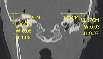
In the 79 male study group patients, the depth of the middle cranial fossa dural plate ranged from 0.0 to 12.4 mm (mean, 4.49 mm) in 49 right ears, and from 0.0 to 11.1 mm (mean, 3.65 mm) in 52 left ears. In the 127 female study group patients, the depth ranged from 0.0 to 14.0 mm (mean, 5.12 mm) in 83 right ears and from 0.0 to 15.2 mm (mean, 4.42 mm) in 83 left ears. Disregarding sidedness, the mean middle cranial fossa dural plate depth was 4.30 mm in male patients and 4.77 mm in female patients; this difference was not statistically significant (p = 0.0867). Disregarding gender, the mean middle cranial fossa dural plate depth was 5.08 mm on the right side and 4.12 mm on the left side; this difference was statistically significant (p = 0.002). The mean middle cranial fossa dural plate depth for all the chronic otitis media patient ears, disregarding sidedness and gender, was 4.59 mm (median, 4.7 mm; interquartile range, 2.9–6.3 mm). Figure 1 shows the depths of the middle cranial fossa dural plates in the affected and healthy ears of a patient with unilateral chronic otitis media.
Coronal computed tomography scan showing the location of the middle cranial fossa dural plate in the affected and healthy ears of a patient with unilateral chronic otitis media. Black arrows indicate the superior semicircular canals; yellow lines indicate the level and depth of the dural plates.

The control group comprised 222 ears of 111 patients without chronic otitis media. Thirty-five patients (31.53 per cent) were male and 76 (68.47 per cent) were female. The age distribution was 9–75 years for male patients (mean age, 50.11 years) and 10–75 years for female patients (mean age, 46.02 years).
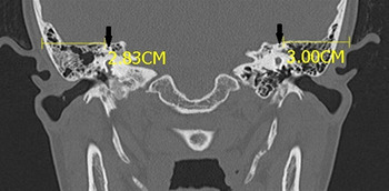
In the male control group patients, the depth of the middle cranial fossa dural plate ranged from 0.0 to 7.4 mm (mean, 3.12 mm) in the right ears and from 0.0 to 6.9 mm (mean, 2.63 mm) in the left ears. In the female control group patients, the depth ranged from 0.0 to 8.9 mm (mean, 3.18 mm) in the right ears and from 0.0 to 8.9 mm (mean, 2.09 mm) in the left ears. Disregarding sidedness, the mean depth was 2.88 mm in male patients and 2.64 mm in female patients; this difference was not statistically significant (p = 0.314). Disregarding gender, the mean depth in control group patients was 3.17 mm on the right side and 2.26 mm on the left side; this difference was statistically significant (p = 0.0026). The mean middle cranial fossa dural plate depth for all control group ears, disregarding sidedness and gender, was 2.71 mm (median, 2.6 mm; interquartile range, 0–0.43 mm). Figure 2 shows the location of the middle cranial fossa dural plates in a control group patient.
Coronal computed tomography scan showing the location of the middle cranial fossa dural plate in a control group patient. Black arrows indicate semicircular canals; yellow lines indicate the level of the dural plates.

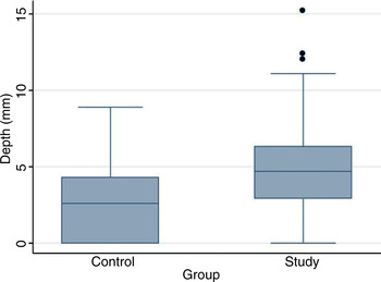
The difference between the overall mean middle cranial fossa dural plate depths of the study and control groups was found to be statistically significant (p < 0.001). Summary statistics for the middle cranial fossa dural plate depths observed in the study and control groups are presented in Figure 3.
Box plot for the middle cranial fossa dural plate depths in the study and control groups. Central bar = median; upper and lower borders = 1st and 3rd quartiles; whiskers = 95% confidence intervals; dots = outliers

Discussion
The middle cranial fossa dural plate is an important surgical landmark, particularly during tympanomastoidectomy in the presence of a Körner's septum at antrum level. However, there is a risk of iatrogenic dural plate trauma or other dural injuries, especially in patients with a low dural plate. This in turn may cause serious complications such as cerebrospinal fluid leakage, meningitis, epidural abscess, brain abscess, encephalocele or meningocele.Reference Emir, Kaptan, Göçmen and Samim2–Reference Wu, Jin, Yang, Liu and Duan6 Middle cranial fossa dural plate injuries may occur in the presence of chronic otitis media with or without cholesteatoma, and also due to surgical intervention, trauma, congenital cranial base tumours or radiotherapy.Reference Emir, Kaptan, Göçmen and Samim2–Reference Wu, Jin, Yang, Liu and Duan6 However, surgical trauma is the most frequent cause. Brain herniation into the tympanic cavity has been reported to occur following between 45.9 and 77 per cent of mastoidal surgery cases.Reference Emir, Kaptan, Göçmen and Samim2, Reference Mosnier, Fiky, Shahidi and Sterkers3, Reference Kizilay, Aladağ, Cokkeser and Ozturan7–Reference Jackson, Pappas, Manolidis, Glasscock, Von Doersten and Hampf10 Thus, it is important that surgeons are aware of the anatomy of the middle cranial fossa dural plate prior to surgery.
Computed tomography is the standard screening method for the temporal bone.Reference Alzoubi, Odat, Al-Balas and Saeed11 However, in patients with chronic otitis media it has a low specificity for the diagnosis of cholesteatoma due to chronic mucosal disease.Reference Alzoubi, Odat, Al-Balas and Saeed11, Reference Stasolla, Magliulo, Cortese, Roncacci and Marini12 Nevertheless, it is highly accurate in delineating middle cranial fossa dural plate anatomy, ossicular chain defects and internal ear fistulae.Reference Alzoubi, Odat, Al-Balas and Saeed11
The middle cranial fossa dural plate is generally separated from the external auditory canal by the tegmental air cells. However, when there is loss of air cell aeration, as in chronic otitis media or a more caudally located dural plate, the middle cranial fossa dural plate may be directly adjacent to the periosteum of the external auditory canal, obstructing the surgeon's access to the antrum and the epitympanum. Iatrogenic injury of the dura and/or temporal lobe may result.Reference Stasolla, Magliulo, Cortese, Roncacci and Marini12
The middle cranial fossa dural plate generally crosses the arcuate eminence just below the most protruding point of the superior semicircular canal into the middle cranial fossa (in the horizontal plane), in the mastoid and attic region. The lower edge of the middle cranial fossa dural plate forms a notch on the lateral aspect of the attic and the labyrinth. Inferior localisation of the middle cranial fossa dural plate is not common. However, when it occurs, the inferiorly located dural plate may cover the roof of the external auditory canal. If the surgeon is unaware of this situation, the cranial cavity may be penetrated during surgery.Reference Mafee, Valvassori and Becker13
In our study, the mean middle cranial fossa dural plate depth was 4.30 mm in male patients and 4.77 mm in female patients (disregarding sidedness), although this difference was not statistically significant. In our control group, the mean middle cranial fossa dural plate depth was 2.88 mm in males and 2.64 mm in females, a statistically insignificant difference. The overall mean middle cranial fossa dural plate depth was 4.59 mm in the study group and 2.71 mm in the control group; this difference was statistically significant. The mean dural plate depth was greater on the right side in both the study and control groups, compared with the left side, a statistically significant difference.
• This study assessed the depth of the middle cranial fossa dural plate on temporal bone computed tomography scans
• This dural plate was lower in patients with chronic otitis media
• It was also lower on the right side
• Computed tomography assessment of this dural plate is essential prior to temporal bone surgery
To our best knowledge, there has been no previous report describing a lower position of the middle cranial fossa dural plate in patients with chronic otitis media, as observed on temporal bone CT. Thus, we believe our findings are unique.
Conclusion
This study demonstrated that, in patients with chronic otitis media, the middle cranial fossa dural plate was located in a lower position, compared with its position in individuals without chronic otitis media. In addition, the middle cranial fossa dural plate was found to be located in a lower position on the right side compared with the left.
Therefore, surgeons should obtain information on the level of the middle cranial fossa dural plate from temporal CT scans taken prior to surgery, and should be aware that the level of this dural plate may be lower in patients with chronic otitis media, and on the right side, requiring extra caution.
Acknowledgement
The statistical analyses were performed by Dr Tayyar Alp Ozkan.