Many studies have demonstrated that environmental contamination with healthcare-associated pathogens is common in rooms of hospitalized patients and residents of long-term care facilities. Reference Donskey1–Reference Alhmidi, Cadnum and Koganti4 In contrast, relatively little information is available on contamination of surfaces outside patient rooms. Reference Hefzy, Wegdan and Abdel Wahed5–Reference Alhmidi, Cadnum, Koganti, Jencson, Wilson and Donskey8 Care areas and equipment outside patient rooms in healthcare facilities and in outpatient settings may represent an underappreciated reservoir of pathogen transmission. In contrast to patient rooms, these areas are typically shared by numerous patients each day, including some patients managed with contact precautions while in their hospital room. Moreover, cleaning in these areas may be suboptimal with limited monitoring of cleaning performance.
To develop effective control measures, there is a need for data on the burden of contamination with healthcare-associated pathogens in a variety of settings outside patient rooms in hospitals and in outpatient settings. To address this need, we examined the frequency of environmental contamination in multiple hospitals and outpatient facilities in northeastern Ohio. We also evaluated the thoroughness of cleaning by assessing removal of fluorescent markers applied to high-touch surfaces.
Methods
Study setting
The study was conducted in 4 Cleveland area hospitals and 5 outpatient healthcare facilities. The hospitals included a tertiary-care county hospital, a Veterans’ Affairs hospital, a community hospital, and a pediatric hospital. The outpatient facilities included 4 outpatient clinics and a surgery center. The institutional review boards affiliated with each facility approved the study protocol.
Culture collection and assessment of cleaning
We conducted 3 point-prevalence culture surveys in each facility with at least 1 month between each culture collection. In the hospitals, cultures were collected from multiple areas including radiology, physical therapy, endoscopy, emergency departments, waiting rooms, portable equipment, and subspecialty clinics. In the pediatric hospital, play areas with shared toys were also included. In outpatient clinics and the surgery center, cultures were collected from exam rooms with separate samples from patient and provider areas, patient bathrooms, waiting rooms, and portable equipment. Samples were collected on weekdays between 8 a.m. and 5 p.m.
In hospitals, ˜100 cultures were collected from high-touch surfaces during each point-prevalence survey. In the outpatient facilities, ˜20–70 cultures were collected during each point-prevalence survey with the number of cultures varying depending on the size of the facilities. For all facilities, 1 sample was also collected from the floor in each of the areas being sampled and another was collected from a sink in the patient care area.
The high-touch surface and floor cultures were collected from standardized 20 × 20-cm surface areas using cellulose sponges (Sponge Stick with neutralizing buffer, 3M, New Ulm, MN). The sink drains were sampled using BBL culture swabs (Becton Dickinson, Sparks, MD) inserted 2.5 cm below the strainer. Reference Jencson, Cadnum, Piedrahita and Donskey9
To assess thoroughness of cleaning, fluorescent solution (Tide laundry detergent) was applied using a cotton-tipped swab to high-touch surfaces as previously described. Reference Deshpande and Donskey10 The solution was allowed to air dry and research personnel returned 1 day later and used a black light to assess whether the fluorescent marker has been removed.
Microbiology and molecular typing
Sponges were processed as previously described using sterile phosphate-buffered saline with 0.02% Tween 80. Reference Tanner, Leecaster and Zhang2 Then 200-µL aliquots of eluate were plated on CHROMagar Staph aureus with 6 μg/mL cefoxitin for methicillin-resistant Staphylococcus aureus (MRSA), Enterococcosel agar with 20 μg/mL vancomycin for vancomycin-resistant enterococci (VRE), MacConkey agar for gram-negative bacilli, and Sabouraud dextrose agar for Candida spp. Reference Tanner, Leecaster and Zhang2,Reference Kumar, Eilertson and Cadnum11 Bacterial and Candida spp isolates were identified using matrix-assisted laser desorption/ionization time-of-flight (MALDI-TOF; Bruker Biotyper CA System, Bellerica, MA). Isolates of Enterobacteriaciae were screened for ceftazidime by plating on MacConkey agar containing 10 µg/mL ceftazidime. Isolates of Enterobacteriaciae, Pseudomonas aeruginosa, Acinetobacter baumanii, and Stenotrophomonas maltophilia were screened for carbapenem resistance by plating on MacConkey agar containing 1 μg/mL meropenem. Isolates recovered from selective media plates were subjected to susceptibility testing in accordance with Clinical Laboratory Standards Institute guidelines. 12
For recovery of toxin-producing Clostridioides difficile strains, broth enrichment cultures were performed by inoculating 200 µL eluate into C. difficile brucella broth with thioglycolic acid and L-cystine (CDBB-TC) broth as previously described. Reference Cadnum, Hurless, Deshpande, Nerandzic, Kundrapu and Donskey13 The cultures were incubated at 37°C for 72 hours. All specimens that underwent a color change from red to yellow were plated onto pre-reduced C. difficile brucella agar incubated anaerobically for 72 hours. Yellow colonies with the typical appearance were streaked for isolation onto blood plates and were confirmed to be C. difficile based on the typical odor and appearance of the colonies and by a positive reaction using a C. difficile latex agglutination assay (Microgen Bioproducts, Camberley, UK). Isolates identified as C. difficile were grown for 72 hours in CDBB-TC, and the culture medium was tested for production of toxins A and B using the Alere C. difficile Tox A/B II ELISA (TechLab, Blacksburg, VA, USA). Only toxin-producing strains of C. difficile were included in the analysis. Fluorescent polymerase chain reaction (PCR) ribotyping was performed as previously described. Reference Redmond, Silva, Wilson, Cadnum and Donskey14 For MRSA isolates, spa typing was performed using previously described methods. Reference Redmond, Pearlmutter and Ng-Wong15
Data analysis
The percentages of positive cultures were calculated by facility and area sampled for each pathogen. The mean colony-forming units (CFU) of the pathogens recovered was calculated for each area sampled. For high-touch surfaces, the percentage of sites positive for 1 or more of the bacterial pathogens including C. difficile, MRSA, VRE, and GNB was calculated. The thoroughness of cleaning was calculated as the percentage of sites with removal of the fluorescent marker 1 day after culture collection and marker placement.
Results
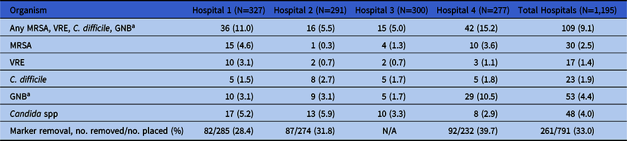
Table 1 shows the overall percentage of positive cultures of high-touch surfaces for the different pathogens in each study hospital and the percentage of fluorescent marker removal. The overall percentage of contamination with the composite of C. difficile, MRSA, VRE, and GNB was 9.1%, with a range of 5.0% to 15.5% for the 4 hospitals. Candida spp were recovered from 4.0% of surfaces, with a range of 2.9% to 5.9% for the 4 hospitals. Based on fluorescent marker removal, <40% of sites assessed had been cleaned.
Environmental Contamination in 4 Hospitals in Areas Outside Patient Rooms

Note. GNB, gram-negative bacilli; MRSA, methicillin-resistant Staphylococcus aureus; C. difficile, Clostridioides difficile; VRE, vancomycin-resistant enterococci.
a GNB included Enterobacteriaceae, Pseudomonas aeruginosa, Acinetobacter baumannii, and Stenotrophomonas maltophilia.
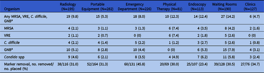
Table 2 shows the percentage of environmental contamination in the 4 hospitals stratified by the areas sampled. For the composite of bacterial pathogens, the waiting rooms had the highest frequency of contamination, and the clinics were the least contaminated (16.4% and 3.2%, respectively). The endoscopy units had the highest frequency of contamination with Candida spp (6.2%). The percentage of fluorescent marker removal ranged from 23.4% to 45.8%.
Environmental Contamination in 4 Hospitals by Areas Sampled

Note. GNB, gram-negative bacilli; MRSA, methicillin-resistant Staphylococcus aureus; C. difficile, Clostridioides difficile; VRE, vancomycin-resistant enterococci.
a GNB included Enterobacteriaceae, Pseudomonas aeruginosa, Acinetobacter baumanii, and Stenotrophomonas maltophilia.
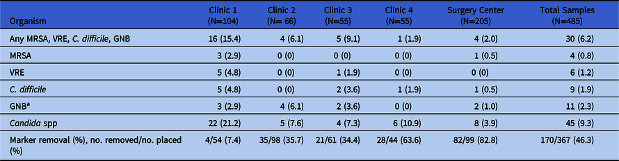
Table 3 shows the overall percentage of positive cultures of high-touch surfaces in the outpatient clinics and surgery center and the percentage of fluorescent marker removal. The percentage of contaminated surfaces for the composite of bacterial pathogens was lowest for clinic 4 and the surgery center (1.9% to 2%); the percentage of contamination with Candida spp was lowest for the surgery center (3.9%). The percentage of fluorescent marker removal was substantially higher in the surgery center than in the clinics (82.8% vs 7.4% to 63.6%, respectively).
Environmental Contamination in Outpatient Clinics

Note. GNB, gram-negative bacilli; MRSA, methicillin-resistant Staphylococcus aureus; C. difficile, Clostridioides difficile; VRE, vancomycin-resistant enterococci.
a GNB included Enterobacteriaceae, Pseudomonas aeruginosa, Acinetobacter baumanii, and Stenotrophomonas maltophilia.
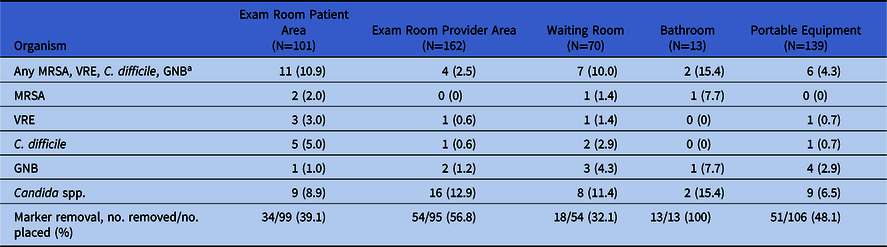
Table 4 shows the percentage of environmental contamination in the clinics stratified by the areas sampled. For the composite of bacterial pathogens, the bathrooms had the highest percentage of contamination; the percentage of fluorescent marker removal in bathrooms was 100%. In examination rooms, the patient area had more frequent contamination with the bacterial pathogens than the provider area. Fluorescent marker removal ranged from 32% to 100%.
Environmental Contamination in Outpatient Clinics by Areas Sampled

Note. GNB, gram-negative bacilli; MRSA, methicillin-resistant Staphylococcus aureus; C. difficile, Clostridioides difficile; VRE, vancomycin-resistant enterococci.
a GNB included Enterobacteriaceae, Pseudomonas aeruginosa, Acinetobacter baumanii, and Stenotrophomonas maltophilia.
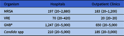
Table 5 shows the mean CFU of the pathogens recovered per positive high-touch surface. The concentration of GNB on surfaces was higher than the concentration of the other pathogens. The concentrations of the pathogens were similar in the hospital and outpatient clinic sites. For the high-touch surfaces, the 64 gram-negative bacterial species recovered included S. maltophilia (N = 24), Klebsiella pneumoniae or K. oxytoca (N = 15), P. aeruginosa (N = 12), A. baumanii (N = 7), Enterobacter cloacae (N = 4), Serratia marcescens (N = 1), and Proteus mirabilis (N = 1). Also, 18 (28%) of the isolates were carbapenem resistant including 7 S. maltophilia, 4, P. aeruginosa, 4 A. baumanii, and 3 Klebsiella spp. Two extended-spectrum β-lactamase (ESBL)–producing organisms were recovered, including 1 E. cloacae and 1 S. maltophilia. The most common Candida spp recovered were C. parapsilosis (39%), C. metapsilosis (11.9%), C. lusitaniae (9%), C. guilliermondii (7.5%), C. famata (7.5%), and C. albicans (6%). Spa typing was completed for 25 MRSA isolates recovered from high-touch surfaces. The most common spa types were t535 (N = 7), t002 (healthcare-associated MRSA clone) (N = 5), t064 (N = 3), and t024 (N = 2). Spa types with 1 isolate per type included t008 (community-associated MRSA clone), t334, t975, t4210, t127, and t216.
Mean (Range) Colony-Forming Units of Healthcare-Associated Pathogens Recovered From Culture Sites

Note. GNB, gram-negative bacilli; MRSA, methicillin-resistant Staphylococcus aureus; VRE, vancomycin-resistant enterococci.
a GNB included Enterobacteriaceae, Pseudomonas aeruginosa, Acinetobacter baumanii, and Stenotrophomonas maltophilia.
The overall percentages of positive cultures of floors for the composite of bacterial pathogens in the hospitals and outpatient clinics were 35.7% (30 of 84) and 31.8% (14 of 44), respectively. For hospitals, the percentages of contamination were as follows: MRSA (9.5%), VRE (14.3%), C. difficile (9.5%), and GNB (15.5%). For outpatient facilities, the percentages of contamination were as follows: MRSA (9.1%), VRE (4.5%), C. difficile (13.6%), and GNB (20.5%). The overall percentages of positive cultures of floors for Candida spp in hospitals and outpatient clinics were 19.0% and 11.4%, respectively. In total, 90 sinks were cultured, including 60 in hospitals and 30 in the outpatient clinics. Pseudomonas aeruginosa and other Pseudomonas spp were recovered from 62 (66%) of the sinks. No carbapenem-resistant Pseudomonas aeruginosa or Enterobacteriaceae were recovered.
Discussion
We examined the frequency of environmental contamination with healthcare-associated pathogens in 4 hospitals focusing on areas outside patient rooms and in 5 outpatient healthcare facilities in northeastern Ohio. In the hospitals, 9.1% of high-touch surfaces sampled were positive for 1 or more bacterial pathogens and 4.0% were positive for Candida spp. In outpatient facilities, 6.2% of sites were positive for 1 or more bacterial pathogens and 9.3% were positive for Candida spp. The thoroughness of cleaning was suboptimal with fluorescent markers removed from only 33% and 46.3% of high-touch surfaces in hospitals and outpatient facilities, respectively. These findings demonstrate that environmental contamination is common in hospitals outside patient rooms and in outpatient facilities and suggest a need for improved cleaning protocols.
Our findings are consistent with previous studies that have demonstrated substantial contamination with healthcare-associated pathogens in areas outside patient rooms. In a point-prevalence culture survey, Jury et al Reference Jury, Sitzlar and Kundrapu7 found that 14% of outpatient clinic and emergency department rooms were contaminated with C. difficile. Prior to a cleaning intervention, Hefzy et al Reference Hefzy, Wegdan and Abdel Wahed5 recovered MRSA and VRE from ˜20% of surfaces cultures in outpatient clinics affiliated with a hospital in Egypt. It has also been demonstrated that patients colonized or infected with VRE, C. difficile, and MRSA may shed these organisms during outpatient clinic visits and physical therapy appointments. Reference Smith, Iwen, Olson and Rupp6,Reference Jury, Sitzlar and Kundrapu7,Reference Li, Pinto-Herrera, Alhmidi, Cadnum, Silva and Donskey16,Reference Gontjes, Gibson, Lansing, Cassone and Mody17
Cleaning and disinfection are challenging in outpatient clinics and in areas outside patient rooms. Multiple patients pass through areas each day with limited time for cleaning between patients. Based on fluorescent marker removal from high-touch surfaces, cleaning procedures were suboptimal in most of the areas studied. Notably, contamination was uncommon in the surgery center which had a high frequency of fluorescent marker removal in comparison to the other areas studied (82.8% marker removal). However, in outpatient clinics, patient bathrooms had a high level of contamination despite 100% marker removal. Sites such as patient bathrooms may require more frequent or intensive cleaning than other areas.
Although contamination with GNB was relatively common, only 30% of isolates were resistant to carbapenems and only 3% were ESBL-producing organisms. Previous studies have suggested that recovery of multidrug-resistant GNB after procedures and patient care activities may be relatively infrequent in comparison to pathogens such as MRSA, possibly because many GNB survive less well on dry surfaces than gram-positive bacteria. Reference Alhmidi, Cadnum and Koganti3,Reference Alhmidi, Cadnum and Koganti4,Reference Alhmidi, Cadnum, Koganti, Jencson, Wilson and Donskey8 In addition, none of the study facilities reported outbreaks or high endemic rates of infections with multidrug-resistant GNB. Although sink contamination has been linked to transmission of multidrug-resistant GNB, Reference Jencson, Cadnum, Piedrahita and Donskey9 no carbapenem-resistant P. aeruginosa or Enterobacteriaceae were recovered from sinks in the current study.
Contaminated environmental surfaces have not traditionally been considered an important source for transmission of Candida spp. However, the emerging fungal pathogen Candida auris has frequently been recovered from environmental surfaces in rooms of colonized or infected patients, Reference Jencson, Cadnum, Piedrahita and Donskey9 and a recent report of an outbreak of C. auris in an intensive care unit linked transmission to shared temperature probes. Reference Eyre, Sheppard and Madder18 Some studies have also implicated environmental sources, including contaminated portable equipment, as a source for acquisition of other Candida spp. Reference Pfaller19–Reference Kumar, Eilertson and Cadnum21 For example, Sanchez et al Reference Sanchez, Vazquez, Barth-Jones, Dembry, Sobel and Zervos20 reported that a Candida parapsilosis strain causing infections was recovered from inanimate surfaces in a new intensive care unit before patients were admitted. The ability of C. auris, C. parapsilosis, and C. glabrata strains to survive for prolonged periods on dry and moist surfaces may increase the likelihood of transmission from the environment. Reference Jencson, Cadnum, Piedrahita and Donskey9
The finding that floors were more frequently contaminated that high-touch surfaces is consistent with many previous culture surveys. Reference Kumar, Eilertson and Cadnum21 The significance of floor contamination remains uncertain, but several recent studies have implicated contaminated floors as a potential source for dissemination of pathogens. Reference Redmond, Pearlmutter and Ng-Wong15,Reference Kumar, Eilertson and Cadnum21–Reference Koganti, Alhmidi, Tomas, Cadnum, Jencson and Donskey23 For example, contamination of hospital rooms with healthcare-associated pathogens typically progressed from floors to sock bottoms, bedding, and high-touch surfaces. Reference Redmond, Pearlmutter and Ng-Wong15
Our study has several limitations. Although the fluorescent marker results suggest that cleaning was suboptimal, it is possible that some high-touch items (eg, portable equipment) were not cleaned because they were not used. Because 3 separate point-prevalence surveys were collected, we cannot exclude the possibility that culture collection might have had an impact on cleaning processes during the study. However, there were no major differences in results for cultures collected at different time periods.
In summary, we found that environmental contamination was common in hospitals outside patient rooms and in outpatient healthcare facilities. Improvements in cleaning and disinfection practices are needed to reduce contamination.
Acknowledgments
We thank the nurses, environmental management services personnel, and patients at the Cleveland VA Medical Center who participated in the study.
Financial support
The authors acknowledge funding from the Centers for Disease Control and Prevention’s investments to combat antibiotic resistance (award no. 2019-06723 to C.J.D.).
Conflicts of interest
C.J.D. has received research grants from Clorox, Pfizer, and PDI. All other authors report no conflicts of interest relevant to this article.