Introduction
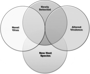
The term “emerging viruses” includes viruses that fit into several different categories (Fig. 1). There are completely novel viruses that suddenly appear in a host species without any historical precedence. However, emerging viruses also includes viruses that do not fit this description. Some viruses are labeled emerging because they have been recently detected in a different species. Others may be labeled as emerging because of increased transmission or severity of outbreaks due to a change in virulence. Still other ‘emerging’ viruses would be more accurately labeled as newly recognized rather than emerging. That is, these viruses have been in circulation for decades but increased surveillance or new diagnostic methods have recently allowed their detection in domestic or wildlife populations. Examples of all of these ‘emerging virus’ categories can be found within the pestivirus genus (Table 1).
Emerging viruses include novel viruses newly infecting populations, known viruses that have jumped to new species, known viruses with an altered virulence and viruses that have been in circulation for a period of time but have only recently been detected.

Categories of emerging pestiviruses

Until the early 1990s there were only three recognized species in the pestivirus genus, bovine viral diarrhea virus (BVDV), border disease virus (BDV), and classical swine fever virus (CSFV) (hereafter referred to as classic pestiviruses). Subsequently BVDV were divided into two different species, BVDV1 and BVDV2 and four additional putative pestivirus species have been identified. This genus has expanded for the following reasons. It was recognized that clinical presentations, not historically associated with pestivirus infections, such as porcine myocarditis syndrome in pigs (Finlaison et al., Reference Finlaison, King, Frost and Kirkland2009) and hemorrhagic syndrome in cattle (Corapi et al., Reference Corapi, French and Dubovi1989, Reference Corapi, Elliott, French, Arthur, Bezek and Dubovi1990) were caused by pestiviruses that were genetically and antigenically distinct from classic pestiviruses. In addition, it has become evident that clinical presentations associated with classic pestiviruses can also be caused by pestiviruses genetically and antigenetically distinct from classic pestiviruses (Pellerin et al., Reference Pellerin, van den Hurk, Lecomte and Tussen1994; Ridpath et al., Reference Ridpath, Bolin and Dubovi1994; Ridpath et al., Reference Ridpath, Neill, Frey and Landgraf2000; Decaro et al., Reference Decaro, Lucente, Cirone, Cordioli, Camero, Sciarretta, Losurdo, Lorusso and Buonavoglia2011, Reference Decaro, Lucente, Mari, Sciarretta, Pinto, Buonavoglia, Martella and Buonavoglia2012, Reference Decaro, Losurdo, Lucente, Sciarretta, Mari, Larocca, Elia, Cavaliere, Martella, Fasanella and Buonavoglia2013a; Ridpath et al., Reference Ridpath, Falkenberg, Bauermann, Vanderley, Do, Flores, Rodman and Neill2013). Increased surveillance of wildlife species and phylogenetic analysis of pestiviruses isolated from those species has led to the recognition that unique pestiviruses are circulating in wildlife (Depner et al., Reference Depner, Hubschle and Liess1991; Vilcek et al., Reference Vilcek, Ridpath, Van Campen, Cavender and Warg2005). Furthermore, it has been shown that classic pestiviruses infect multiple host species resulting in clinical presentations that vary based on the host species (Cabezon et al.; Marco et al., Reference Marco, Lopez-Olvera, Rosell, Vidal, Hurtado, Juste, Pumarola and Lavin2007; Marco et al., Reference Marco, Cabezon, Rosell, Fernandez-Sirera, Allepuz and Lavin2011) and that within classic pestiviruses species there are variations in virulence (Moennig et al., Reference Moennig, Floegel-Niesmann and Greiser-Wilke2003; Ridpath, Reference Ridpath2003).
Novel pestivirus
Bungowannah virus: a novel pestivirus with a novel clinical presentation
The ‘classical’ or historic disease presentations associated with the four recognized species of pestiviruses include pyrexia (BVDV, CSFV, and BDV), diarrhea (BVDV), acute hemorrhagic syndrome (CSFV), reproductive failure (CSFV, BVDV, and BDV) and congenital defects of the nervous system, skeletal system, immune system and hair coat (BVDV and BDV). In 2003, an outbreak of sudden deaths was observed 3–4-week-old piglets in New South Wales, Australia. Pathological changes consisted of multifocal non-suppurative myocarditis (Kirkland et al., Reference Kirkland, Frost, Finlaison, King, Ridpath and Gu2007). Pestiviruses were not on the initial list of possible pathogens. Firstly, myocarditis was not a lesion commonly associated with pestivirus infection at that point in time. Secondly, while virus isolation was considered the gold standard for identifying pestiviruses, initial attempts to culture this pathogen were unsuccessful. Sequence-independent single-primer amplification (SISPA) was used to generate viral sequences from tissues from affected animals. The causative agent was identified as a pestivirus that was subsequently named Bungowannah virus. Bungowannah virus is the most divergent of the pestiviruses identified to date. While there is antigenic cross reactivity between this virus and other pestiviruses it is relatively limited compared to antigenic cross reactivity among the other members of the genus.
Pestiviruses with altered virulence
Clinical presentation was initially used to differentiate the first three recognized species of pestivirus [hog cholera virus (later renamed classical swine fever virus), BVDV and border disease virus]. In the intervening years, it has become evident that clinical presentation following pestivirus infection is highly variable depending on the age, reproductive and immune status of the host and the virulence of the infecting pestivirus. Sometimes variation in pestivirus virulence can impact control programs as illustrated by CSFV strains with reduced virulence and BVDV strains with enhanced virulence.
Reduced virulence CSFV
Classical swine fever, historically known as hog cholera, was initially known as a highly contagious, highly virulent viral disease of pigs. The ‘classic’ symptoms of classical swine fever were high fevers (as high as 41.7 C), conjunctivitis, enlarged and discolored lymph nodes and hemorrhages of the skin usually observed on the ear, tail, abdomen and the inner side of the limbs (Moennig et al., Reference Moennig, Floegel-Niesmann and Greiser-Wilke2003). Neurological signs are also frequently seen with death occurring within 5–14 days following the onset of illness. As early as the 1970s it was recognized that CSFV isolates varied in virulence (Carbrey et al., Reference Carbrey, Steward, Kresse, Snyder and Liess1977) and that infection with some strains resulted in very mild clinical symptoms. The existence of low-virulence CSFV strains has caused problems with CSF control programs. This is because clinically mild disease on farms may not prompt producers to test for CSFV. Failure to recognize that clinically mild disease may be caused by CSFV leads to reduced and delayed testing. Reduced and/or delayed testing results in longer lasting and more widespread outbreaks (Sandvik et al., Reference Sandvik, Drew and Paton2000; Terpstra and de Smit, Reference Terpstra and de Smit2000).
BVDV with enhanced virulence
When first described by Cornell University researchers in 1946 (Olafson et al., Reference Olafson, McCallum and Fox1946) bovine viral diarrhea (BVD) was characterized as an acute transmissible viral infection marked by severe leucopenia, high fever, depression, diarrhea, gastrointestinal erosions, and hemorrhages. However, in the ensuing years acute BVD infections came to be regarded as clinically unimportant and the prevailing wisdom was that transmission of the virus between healthy immunocompetent cattle was probably insignificant (Radostits et al., Reference Radostits, Blood and Gay1994). However, this began to change in the late 1980s when severe acute BVD once more came to the forefront. In 1987 (Perdrizet et al., Reference Perdrizet, Rebhun, Dubovi and Donis1987), reported thrombocytopenia with hemorrhage associated with BVD virus infection occurring in dairy herds in the Northeastern USA. Case records of cattle admitted to the Cornell College of Veterinary Medicine for the years 1977–1987 revealed that 10% of clinically acute BVD infections in adult cattle were associated with thrombocytopenia (Corapi et al., Reference Corapi, French and Dubovi1989). Severe acute BVD cases were reported with increasing frequency in North America in the early 1990s. These outbreaks were particularly devastating in the Canadian provinces of Quebec and Ontario (Carman et al., Reference Carman, van Dreumel, Ridpath, Hazlett, Alves, Dubovi, Tremblay, Bolin, Godkin and Anderson1998). In the province of Quebec a BVD epidemic starting in early 1993 resulted in the death of 32,000 out of 143,000 (22.4%) animals in the 1993 veal calf crop (Pellerin et al., Reference Pellerin, van den Hurk, Lecomte and Tussen1994). Clinical signs included high temperatures, bloody diarrhea, hemorrhages, and prolonged bleeding from venipuncture sites. This disease came to be regarded as a distinct form of severe acute BVD termed hemorrhagic syndrome. While the ‘BVDV infections are mostly subclinical’ trope is still frequently repeated it should be noted that there is a continuum of virulence among BVDV field strains. To date the most virulent strains belong to the BVDV2 species, however enhanced virulence viruses are recognized among BVDV1 strains (Ridpath et al., Reference Ridpath, Neill and Peterhans2007) and outbreaks of severe disease have been associated with BVDV1 strains in the field (Blanchard et al., Reference Blanchard, Ridpath, Walker and Hietala2010).
Phylogenetic analysis revealed that several outbreaks of severe acute BVD observed between 1993 and 1995 in North America that could be traced to a single strain of BVDV that spread explosively following acute infection (Ridpath et al., Reference Ridpath, Neill, Vilcek, Dubovi and Carman2006). This observation called into question the conventional wisdom that BVD outbreaks are subclinical in healthy animals, can nearly always be traced to contact with a persistently infected (PI) animal and that transmission of the virus following acute infection is insignificant to the spread of virus. These findings suggest that management of BVDV outbreaks need to take virulence into account.
Classic pestiviruses in new species
The focus of pestivirus control programs has predominately been on controlling infections in cattle and pigs and to a lesser extent in sheep and goats. However, there are other commodity groups such as farmed cervids and camelids that are impacted by pestivirus infections. In addition, it has become apparent that management of wildlife populations must consider the impact of pestiviruses acquired via contact with domestic animals. The emerging problem of pestivirus transmission from the major domestic species to other species is illustrated by the adaptation of BDV infections in free ranging chamois and the emergence of BVDV infections in domestic alpaca.
BDV in Chamois: Change in virulence of ‘classic’ pestivirus species occurring in a new host resulting in a novel presentation
BDV is known primarily as a pathogen of sheep and, to a lesser extent, goats. Infection of non-pregnant adults is typically subclinical. However, if a pregnant animal contracts the virus, BDV is able to cross the placenta and cause abortions or congenital defects in the lamb or kid. The resulting weak offspring, born with hair rather than wool and suffering from CNS lesions accompanied by ataxia, are known as ‘hairy shakers’. The impact on herds is mainly a decrease in the number of healthy offspring born during the birthing season (Nettleton et al., Reference Nettleton, Gilray, Russo and Dlissi1998).
An aberration in this ‘typical’ clinical presentation was observed when BDV strains, apparently transmitted from domestic sheep, underwent a change in virulence following replication in wild chamois populations. It appears that prior to 2000, BDV circulated among wild chamois populations in the Pyrenees without notable negative effects. However, an outbreak of severe clinical disease in southern chamois (Rupicapra pyrenaica), associated with BDV infections, was noted starting in 2001 (Marco et al., Reference Marco, Lopez-Olvera, Rosell, Vidal, Hurtado, Juste, Pumarola and Lavin2007, Reference Marco, Rosell, Cabezon, Mentaberre, Casas, Velarde and Lavin2009). Clinical presentation included depression, weakness, movement difficulties and varying degrees of alopecia with skin hyperpigmentation, resulting in a greater than 40% reduction in chamois populations across all age groups. The causative agent was identified as a BDV. Other ruminant species in the contact area were serology positive for BDV exposure, but did not develop the clinical presentation observed in chamois (Fernandez-Sirera et al., Reference Fernandez-Sirera, Cabezon, Allepuz, Rosell, Riquelme, Serrano, Lavin and Marco2012a, Reference Fernandez-Sirera, Riba, Cabezon, Rosell, Serrano, Lavin and Marcob). Since the first outbreaks, BDV has become endemic in some herds, which has a negative impact on population dynamics, while it has been cleared from other herds, which is accompanied by a quick recovery of population numbers (Fernandez-Sirera et al., Reference Fernandez-Sirera, Cabezon, Allepuz, Rosell, Riquelme, Serrano, Lavin and Marco2012a). The outbreak of severe disease with significant impact on population numbers in a wildlife species associated with virus that has minimal effect on domestic species highlights some of the problems that might be faced as expansion of range or agriculture or the reintroduction of wildlife species results in increased contact between domestic and wild ruminants.
BVDV in alpaca
New world camelids, such as alpacas and llamas, are rarely seropositive to BVDV, unless they have exposure to cattle (Belknap et al., Reference Belknap, Collins, Larsen and Conrad2000; Wentz et al., Reference Wentz, Belknap, Brock, Collins and Pugh2003). Prior to 2002 it was thought that transmission was of BVDV to llamas was inefficient and that llama fetuses were resistant to BVDV infection (Mattson, Reference Mattson1994). However, in that year Goyal et al. (Reference Goyal, Bouljihad, Haugerud and Ridpath2002) reported the isolation of BVDV from an aborted alpaca fetus and in 2005 Carman et al., (Reference Carman, Carr, DeLay, Baxi, Deregt and Hazlett2005) confirmed persistent BVDV infection in an alpaca cria. The birth of the PI cria was associated with an outbreak in Eastern Ontario in which clinical signs of BVDV (anorexia, lethargy, and reproductive loss) were observed in an alpaca herd starting 2.5 months after the addition of a chronically ill cria and its dam to the farm (Carman et al., Reference Carman, Carr, DeLay, Baxi, Deregt and Hazlett2005). The chronically ill cria, which died at 8 months of age, suffered from intermittent bouts of diarrhea, pneumonia and nasal discharge. No samples were collected from this cria. The cria's dam had been on four different farms during her pregnancy. Two of these farms reported outbreaks of reproductive disease, including abortions, stillborn cria, and the birth of weak cria. Subsequently testing of alpacas from these farms found that they had antibodies against BVDV but had no record of contact with cattle or vaccination. The alpaca industry recognized that PI cria represented a significant hazard in that dams with cria at foot are commonly sent to different farms for breeding. The recognition that BVDV can establish persistent infection in alpacas led to significant changes across the alpaca industry, including changes in show rules and testing for persistent infection before entry onto new farms.
Newly recognized pestiviruses
As previously stated, some pestiviruses that are labeled as emerging have apparently been in circulation for a significant period of time, but were just recently recognized as distinct from classic pestiviruses. Frequently this new recognition is the result of phylogenetic analysis. Failure to recognize these viruses earlier may be due to similarity in clinical presentation to classic pestiviruses, as is the case with HoBi-like viruses, or to limited disease surveillance in wildlife populations, as is the case with Pronghorn virus.
HoBi-like viruses
Recently, a new species of pestivirus, which infects cattle, has emerged. Known variously as HoBi-like virus, atypical pestivirus and BVDV3, this new group of viruses is associated with reproductive and/or respiratory disease outbreaks in South America, Southeast Asia, Asia, and Europe (Stahl et al., Reference Stahl, Kampa, Alenius, Persson Wadman, Baule, Aiumlamai and Belak2007; Decaro et al., Reference Decaro, Lucente, Cirone, Cordioli, Camero, Sciarretta, Losurdo, Lorusso and Buonavoglia2011, Reference Decaro, Lucente, Mari, Sciarretta, Pinto, Buonavoglia, Martella and Buonavoglia2012; Bauermann et al., Reference Bauermann, Ridpath, Weiblen and Flores2013b; Mishra et al., Reference Mishra, Rajukumar, Pateriya, Kumar, Dubey, Behera, Verma, Bhardwaj, Kulkarni, Vijaykrishna and Reddy2014). Under controlled conditions, clinical presentations following infection with HoBi-like viruses are indistinguishable from those associated with field strains of BVDV1 or BVDV2 of typical virulence including immunosuppression and persistent infection (Ridpath et al., Reference Ridpath, Falkenberg, Bauermann, Vanderley, Do, Flores, Rodman and Neill2013; Bauermann et al., Reference Bauermann, Falkenberg, Vander Ley, Decaro, Brodersen, Harmon, Hessman, Flores and Ridpath2014a). Diagnostic tests designed for BVDV screening have limited usefulness for detecting HoBi-like viruses (Bauermann et al., Reference Bauermann, Flores and Ridpath2012) and current BVDV vaccines only offer limited cross-protection against HoBi-like viruses (Bauermann et al., Reference Bauermann, Harmon, Flores, Falkenberg, Reecy and Ridpath2013a). A survey of fetal bovine serum collected from animals in the USA did not detect the presence of HoBi-like viruses or antibodies against HoBi-like viruses suggesting that these viruses have not yet invaded the USA (Bauermann et al., Reference Bauermann, Flores, Falkenberg, Weiblen and Ridpath2014b). In contrast, it was demonstrated that HoBi-like viruses had been in circulation in Europe earlier than previously reported (Decaro et al., Reference Decaro, Mari, Lucente, Sciarretta, Elia, Ridpath and Buonavoglia2013b). These studies suggest that the introduction of HoBi-like viruses poses a threat for US dairy and beef producers and that control of this threat will require the development of new reagents and vaccines.
Pestiviruses in wildlife: Pronghorn virus (PHV)
PHV is a pestivirus that was first isolated from an immature, blind pronghorn antelope found in the wild in Wyoming (Vilcek et al., Reference Vilcek, Ridpath, Van Campen, Cavender and Warg2005). Phylogenetic analysis shows that PHV and Bungowannah virus are the most divergent of the pestiviruses identified to date (Neill et al., Reference Neill, Ridpath, Fischer, Grundhoff, Postel and Becher2014). For over a decade no other detection of PHV was reported. However, in a survey of samples collected in the state of Nevada from wildlife between 2011 and 2014, PHV was detected in mule deer, big horn sheep, and mountain goat samples (manuscript in preparation). It is unknown at this time whether PHV will infect domestic animals that share range with these wildlife species or what effect PHV infections have on wildlife populations.
Acknowledgments
This manuscript was prepared as part of the duties of the author as an employee for the United States Department of Agriculture. The author has no protected, financial, occupational or other personal interests in a product, service and/or a company which could influence the content or opinions presented in the manuscript.