INTRODUCTION
Embryos of fish are free-living and thus develop in an environment that may be characterized by variable temperature, salinity and oxygen availability. This means that they usually are exposed to the same or a similar environment as the adults (Pelster, Reference Pelster1999). Physiological changes and adaptations to reduced oxygen availability in adult fish have been analysed in a large number of studies. They show that hypoxic conditions provoke adaptations in the central circulatory system, such as hypoxic bradycardia (a decrease in heart rate), combined with an increase in stroke volume and peripheral resistance (Randall, Reference Randall1982; Fritsche, Reference Fritsche1990; Burggren, Reference Burggren and Pinder1991). In respect to embryos and larvae, several studies demonstrate that, as a probably general trait in development, the nervous system which controls or modifies cardiac activity becomes functional only late during larval development (Kimmel, Reference Kimmel1992; Protas & Leontieva, Reference Protas and Leontieva1992; Fritsche, Reference Fritsche1997). A coordinated response to environmental hypoxia is established only in late larval stages, although the sensors for oxygen are present and functional much earlier (Pelster, Reference Pelster1999). Fish which form burrows in intertidal sediments experience more extreme conditions of hypoxia at low tide when irrigation of the burrow is impossible (Atkinson & Taylor, Reference Atkinson and Taylor1991). However, there have been few studies concerning the tolerance to hypoxia of burrowing fish embryos.
The Chinese black sleeper (Bostrichthys sinensis Lacépède), a commercially important species in China, belongs to the family Eleotridae. It is a carnivorous fish and is distributed in the coastal waters and estuaries of south-eastern China (Hong & Zhang, Reference Hong and Zhang2003). On the surface of mudflats, fish build ‘Y’-shaped muddy burrows of 40–65 cm depth, with one entrance and one exit (Hong et al., Reference Hong, Chen, Zhang and Zheng2006). It is a seasonally breeding fish, living in the intertidal zone. During the non-spawning season, females and males live in individual burrows, but during the spawning season, they mate and spawn as pairs inside the same burrow (Hong, Reference Hong2005). Eggs adhere to the substrate at the bottom of the burrow with attaching-filaments. After fertilization, the eggs swell and form a perivitelline space, and embryonic development proceeds inside the burrow (Chen et al., Reference Chen, Hong, Zhou and Zhang2006a). The larvae hatch 144 hours and 30 minutes after fertilization at 22.5–25.0°C. The dissolved oxygen (DO) concentrations inside the burrows of the Chinese black sleeper are as low as 0.20–1.57 mg/l at ebb tide (Hong, Reference Hong2005).
To elucidate how Chinese black sleeper embryos cope with extremely low oxygen concentrations, tolerance of hypoxia at heartbeat stage has been examined at different oxygen concentrations (Chen et al., Reference Chen, Hong, Zhou and Zhang2006a). The data show that the exposure of the embryos to 0.2 mg/l DO at 25°C causes a periodic heartbeat (including a period of heartbeat and a period of silence). This raises two questions, whether the periodic heartbeat is a special phenomenon that only occurs at the heartbeat stage of Chinese black sleeper, and what is the nature of the physiological basis for periodic heartbeat. The assumption of this study was that the physiological changes caused to Chinese black sleeper embryos by an insufficient oxygen supply were reflected in the development of bradycardia and the occurrence of a periodic heartbeat.
It is well known that in fish, in the final stage of incubation, oxygen consumption of developmentally advanced and active embryos reaches its maximum (Braum, Reference Braum1973; Kioerboe & Moehlenberg, Reference Kioerboe and Moehlenberg1987; Rombough, Reference Rombough1988). At that time, the egg envelope becomes a barrier limiting the oxygen supply to the embryo. However, little is known about the impact of the egg envelope and perivitelline liquid on embryonic development under hypoxic conditions.
The aim of this study was to investigate the cardiac performance of the Chinese black sleeper at early stages of development (the heartbeat, blood circulation and newly hatched larval stages) under hypoxic conditions. The tolerance of the embryos to hypoxia at different stages of development was compared. Whole egg embryos (the egg membranes were intact) and excised embryos (the egg membranes were peeled off) were exposed to hypoxic conditions in order to study the function of the perivitelline liquid during the embryonic development of the burrowing fish. The response of the embryos and larvae to hypoxia was expressed in terms of the occurrence of periodic heartbeat and the intensity of bradycardia.
MATERIALS AND METHODS
The experimental fish were caught in the intertidal zone at Longhai, Fujian, China. The average standard length and weight were 16.94±1.10 cm and 117.25±28.52 g. HCG and LHRH-A (Ningbo Sansheng Pharmaceutical Co., Ltd., Zhejiang, People's Republic of China) were used to induce maturation and ovulation. The hormones were dissolved in saline. Both female and male were injected with HCG at a dose of 100IU and LHRH-A at a dose of 1ug/ind. A semi-dry method was used for artificial fertilization. Fertilized eggs were washed three times and then incubated in membrane filtered (0.22 mm) seawater at 15‰ salinity and 25°C. One hundred eggs were incubated in a container filled with 200 ml seawater.
The methods described by Czerkies et al. (Reference Czerkies, Kordalski, Golas, Krysinski and Luczynski2002) and Chen et al. (Reference Chen, Hong, Zhou and Zhang2006a) were applied in the present study. Nitrogen gas was bubbled through the water column to bring the water to the desired oxygen concentrations (Bardega et al., Reference Bardega, Czerkies, Golas and Luczynski1992). Experimental water flow to the inlet and from the outlet manifold travelled through glass pipes submerged in a thermoregulated water bath. Each test chamber was a Petri dish 3.5 cm in diameter and 20 ml capacity filled with water of the desired oxygen concentration. The shape of the test chamber prevented embryos from escaping with the out-flowing water. Water flow was 10 ml/minute and was controlled using a peristaltic pump. Water temperature and oxygen concentration were measured at the outflow of the chambers and bottles (Czerkies et al., Reference Czerkies, Kordalski, Golas, Krysinski and Luczynski2002; Chen et al., Reference Chen, Hong, Zhang, Wu and Wang2006b). DO concentrations were measured using an YSI 55 oxygen electrode, with an accuracy of 0.01 mg/l.
Before the test, embryos at the heartbeat stage (19.26 hours after fertilization) and blood circulation stage (27.14 hours after fertilization) were checked with a low-power stereomicroscope and those without visible malformations were chosen for the experiments. Some chosen embryos were excised from their envelope using forceps. For each test and in each DO level, one embryo or one larva was used. Successive observations of the embryos were carried out at 0.2 mg/l DO, and of the newly hatched larvae at 0.2, 0.1, and 0.02 mg/l DO, at 25°C. In the control treatment, embryos and larvae were exposed to normoxic water at 6.0 mg/l DO. After exposure for 60 minutes to water of the desired oxygen concentration, embryos and larvae were removed to the normoxic water. Each embryo was used once only.
The test chamber was placed under an inverted microscope (Leica-DMIL) connected to a camera (Canon, Power Shot S45) and the time from exposure to periodic heartbeat occurrence was recorded. The heart rates of the newly hatched larvae were recorded every 5 minutes after exposure. The time taken for 50 heartbeats was recorded and the frequency of heartbeats (F) was calculated. The intensity of bradycardia (B) was defined as the relative decrease in heart rates:

where Fn is the heart rate in normoxia; and Fh is the heart rate in hypoxia.
The term breakdance performance (a kind of arrhythmic heartbeat, i.e. the atrium contracts twice while the ventricle beats only once) described by Chen et al. (Reference Chen, Haffter, Odenthal, Vogelsang, Brand, van Eeden, Furutani-Seiki, Granato, Hammerschmidt, Heisenberg, Jiang, Kane, Kelsh, Mullins and Nüsslein-Volhard1996) was used in the present study to characterize the heartbeat of the newly hatched larvae exposed to hypoxia.
The values are presented as mean±standard deviation (SD). A t-test was used to analyse the differences in the values between different experimental groups at the significance level of 0.05. All statistics were performed with the statistical software SPSS for Windows (SPSS, Chicago, IL, USA).
RESULTS
All tested embryos survived and hatched normally without recognizable malfunctions after the experiment.
Heart rates of the embryos exposed to normoxic water remained unchanged during the test. Embryos exposed to hypoxic conditions displayed bradycardia. Exposure of embryos at both the heartbeat and blood circulation stages to a DO concentration of 0.2 mg/l caused a periodic heartbeat. The occurrence of periodic heartbeat in whole egg embryos at the heartbeat stage (44.0±7.7 minutes) was significantly (t-test, P < 0.05) later after exposure than that at the blood circulation stage (25.4±5.0 minutes). Furthermore, at the heartbeat stage, whole egg embryos displayed periodic heartbeat after 44±7.7 minutes of exposure, which was significantly (t-test, P < 0.05) later than excised embryos (after 31.4±3.3 minutes of exposure). Similarly, at the blood circulation stage, the occurrence of periodic heartbeat in whole egg embryos (25.4±5 minutes) was significantly (t-test, P < 0.05) later than that in excised embryos (20.2±4.9 minutes). Periodic heartbeat in newly hatched larvae was not observed at a DO concentration of 0.2 mg/l. However, exposure of the newly hatched larvae to DO concentrations of 0.1 mg/l and 0.02 mg/l caused periodic heartbeat. Periodic heartbeat was displayed significantly (t-test, P < 0.05) earlier (after 13.2±3.4 minutes of exposure) in 0.02 mg/l DO than that (after 23.3±6.2 minutes of exposure) in 0.1 mg/l DO in newly hatched larvae.
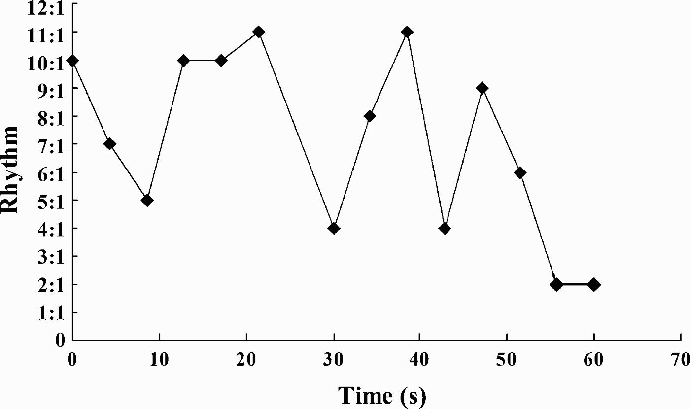
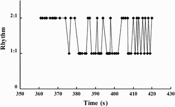
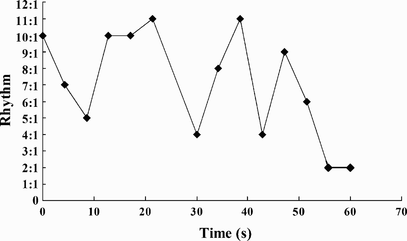
Exposed to a DO concentration of 0.02 mg/l at 25°C, a breakdance performance was observed in 18.75% of the total number of the newly hatched larvae, and those that showed breakdance did not display periodic heartbeat. After about 15 minutes of exposure the larvae began to display breakdance and it lasted for 10 minutes. Two typical periods of breakdance are shown in Figures 1 & 2. The first period occurred during the first 60 seconds of the 10 minute breakdance. During this period, the larva started with a 10:1 rhythm (where the atrium contracted 10 times while the ventricle beat only once), and then, the heart rates switched without any regular pattern but ranging from an 11:1 rhythm to a 2:1 rhythm (Figure 1). The second period occurred from 360 seconds to 420 seconds within the 10 minute breakdance (Figure 2). Within this 60 seconds, the heart rate varied without any regular pattern between a 2:1 and a 1:1 rhythm. It switched 28 times from the 2:1 rhythm to the 1:1 rhythm and then back to the 2:1. After 10 minutes of breakdance, the heartbeat rates became normal (1:1 rhythm) once again.
The first 60 seconds of the first period of the 10 minute breakdance performance in a Bostrichthys sinensis newly hatched larva exposed to a DO concentration of 0.02 mg/l at 25°C. Data are presented as one sample.
The second period of the 10 minute breakdance performance (from 360 seconds to 420 seconds) in a Bostrichthys sinensis newly hatched larva exposed to a DO concentration of 0.02 mg/l at 25°C. Data are presented as one sample.

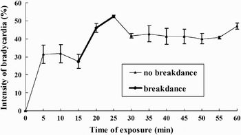
Figure 3 shows the development of bradycardia in Bostrichthys sinensis newly hatched larvae that displayed breakdance at a DO concentration of 0.02 mg/l at 25°C. Bradycardia developed within the first 5 minutes of exposure to hypoxia. This was followed by a plateau, which lasted for 5 minutes. Then the B values decreased to their lowest point, but increased rapidly after 15 minutes of exposure and reached their peak after 25 minutes of exposure. Then the B values decreased until a plateau was attained after 30 minutes of exposure, which lasted until termination of the hypoxia. The breakdance performance was observed during the exposure period (15–25 minutes) when the B values increased rapidly.
Development of bradycardia of Bostrichthys sinensis newly hatched larvae, exposed to DO of 0.02 mg/l at 25°C. Data are presented as mean±SD of four to six replicate measurements.

DISCUSSION
Our previous study demonstrates that the rates of oxygen consumption increase with the development of embryos incubated at 25.0±0.1 in Bostriththys sinensis. The lowest rate of oxygen consumption (1.65±0.66 nmol O2/(ind.·h)) is recorded at gastrulation. Then the rate of oxygen consumption increases significantly and reaches 6.68±0.3 nmol O2/(ind.·h) (P < 0.01) from the initiation of blood circulation to the presence of oxygen-carrying erythrocytes throughout the circulatory system. Afterwards the oxygen consumption rate reaches its maximum immediately before hatching (8.26±1.7 nmol O2/(ind.·h)) (P < 0.05) (Chen et al., Reference Chen, Hong, Zhang, Wu and Wang2006b). As a result, it is understood that the periodic heartbeat of Bostriththys sinensis embryos at the blood circulation stage occurred earlier than that at the heartbeat stage, since the embryos at the blood circulation stage consumed more oxygen than at the heartbeat stage. It can be inferred that embryos at the blood circulation stage became unadaptable earlier than those at the heartbeat stage when they were exposed to a hypoxic DO concentration of 0.2 mg/l.
The sites of gas exchange vary during the early development of fish (Pelster, Reference Pelster1999). At the embryonic stage, Bostrichthys sinensis embryos may use the vascular net around the yolk sac and skin to carry out gas exchange. It is hypothesized that, with the appearance of the gill slits, the newly hatched larvae obtained more oxygen through the gill slits and then their tolerance to hypoxia improved. Hence, the newly hatched larvae did not display periodic heartbeat (as embryos did) at a DO concentration of 0.2 mg/l. However, when exposed to lower DO concentrations (0.1 mg/l and 0.02 mg/l), they displayed periodic heartbeat. The threshold of DO concentration, which caused periodic heartbeat of the newly hatched larvae, needs to be investigated further.
The roles of the egg envelopes and perivitelline fluid in oxygen supply for fish embryos under hypoxic conditions vary with different species. Vendace (Coregonus albula) and whitefish (C. lavaretus) embryos respond to hypoxic conditions with bradycardia. The intensity of bradycardia increases at decreasing partial pressures of oxygen and it is always higher in whole-egg embryos than in the excised ones (Czerkies et al., Reference Czerkies, Kordalski, Golas, Krysinski and Luczynski2002). This suggests that the egg envelope and perivitelline fluid have a disadvantageous impact on the gas exchange processes of embryos under hypoxic conditions in these two species. However, in the present study, the periodic heartbeat in whole eggs occurred later than that in excised ones, at both the heartbeat and blood circulation stages, at a DO concentration of 0.2 mg/l, indicating that the tolerance to hypoxia of embryos remaining within their egg envelopes was higher than that of excised ones and that egg envelope and perivitelline fluid had an advantageous impact on the gas exchange processes of the embryos in Bostrichthys sinensis. It can be inferred from our results that in Bostrichthys sinensis the perivitelline fluid served as a temporary buffer to maintain the pO2 for a period of time until the DO concentration in the perivitelline fluid was the same as that in the hypoxic water.
Several QT-prolonging drugs added to the bathing medium elicited bradycardia and arrhythmia in zebrafish (Danio rerio) embryos. The 2:1 heart block chemotype is intriguingly similar to the phenotype of the breakdance mutant of zebrafish (Langheinrich et al., Reference Langheinrich, Vacun and Wagner2003). The breakdance mutant of zebrafish was isolated in the Tübingen screen in 1996 (Chen et al., Reference Chen, Haffter, Odenthal, Vogelsang, Brand, van Eeden, Furutani-Seiki, Granato, Hammerschmidt, Heisenberg, Jiang, Kane, Kelsh, Mullins and Nüsslein-Volhard1996). The only available physiological information about this mutant is that the atrium and ventricle contract with a 2:1 rhythm. In the present study, we found that the breakdance performance occurred in newly hatched larvae under acute hypoxic conditions (0.02 mg/l) in Bostrichthys sinensis. The rhythm of the breakdance switched without any regular pattern from an 11:1 rhythm to a 1:1 rhythm. Compared with zebrafish, the rhythms of breakdance are more complicated in Bostrichthys sinensis. Breakdance has been described in human beings and linked to the human ether-a-go-go related gene (HERG) which has been shown to encode the rapidly activating delayed rectifier K+ current IKr (Curran et al., Reference Curran, Splawski, Timothy, Vincent, Green and Keating1995; Sanguinetti et al., Reference Sanguinetti, Jiang, Curran and Keating1995; Piippo et al., Reference Piippo, Laitinen, Swan, Tiovonen, Viitasalo, Pasternack, Paavonen, Chapman, Wann, Hirvela, Sajantila and Kontula2000; Vandenberg et al., Reference Vandenberg, Walker and Campbell2002). It has been documented that a number of K+ channels can respond to alteration in oxygen tension and many hypoxia-sensitive channels have been identified in cardiac myocytes (Ju et al., Reference Ju, Saint and Gage1994, Reference Ju, Saint and Gage1996; Fearon et al., Reference Fearon, Palmer, Balmforth, Ball, Mikala, Schwartz and Peers1997, Reference Fearon, Palmer, Balmforth, Ball, Varadi and Peers1999, Reference Fearon, Varadi, Koch, Isaacsohn, Ball and Peers2000; Hool, Reference Hool2000, Reference Hool2001; Hool & Arthur, Reference Hool and Arthur2002). Also, longer-term hypoxia alters ion channel expression and cellular excitability as a result of the induction of hypoxia-responsive genes (Peers, Reference Peers2002). Based on these findings, it is hypothesized that the acute hypoxic condition restrained the IKr's activity and this caused the breakdance.
ACKNOWLEDGEMENTS
This work was supported by the National Natural Science Foundation of China (No. 40476056). Professor John Hodgkiss is thanked for his help with the English.