Introduction
Schizophrenia has frequently been linked to a higher prevalence of non–right-handedness as well as reduced left-hemispheric language dominance (Collinson, Mackay, James, & Crow, Reference Collinson, Mackay, James and Crow2009; Dragovic & Hammond, Reference Dragovic and Hammond2005; Sommer, Ramsey, Kahn, Aleman, & Bouma, Reference Sommer, Ramsey, Kahn, Aleman and Bouma2001) and several authors have suggested that atypical language lateralization constitutes a biological risk factor for schizophrenia (Angrilli et al., Reference Angrilli, Spironelli, Elbert, Crow, Marano and Stegagno2009; Crow, Reference Crow2000; Oertel et al., Reference Oertel, Knöchel, Rotarska-Jagiela, Schönmeyer, Lindner, van de Ven and Linden2010). However, while reduced language lateralization in schizophrenia patients compared to healthy controls has been reported in both neuroimaging studies comparing speech-related brain activations in the left and right hemispheres (Bleich-Cohen, Hendler, Kotler, & Strous, Reference Bleich-Cohen, Hendler, Kotler and Strous2009; Bleich-Cohen et al., Reference Bleich-Cohen, Sharon, Weizman, Poyurovsky, Faragian and Hendler2012; van Veelen et al., Reference van Veelen, Vink, Ramsey, Sommer, van Buuren, Hoogendam and Kahn2011) and behavioral studies (Hugdahl et al., Reference Hugdahl, Løberg, Specht, Steen, van Wageningen and Jørgensen2007), there are also several studies that have found no effect of reduced lateralization on neuroimaging activation (Razafimandimby, Tzourio-Mazoyer, Mazoyer, Maïza, & Dollfus, Reference Razafimandimby, Tzourio-Mazoyer, Mazoyer, Maïza and Dollfus2011) or in behavioral studies (Løberg, Jørgensen, & Hugdahl, Reference Løberg, Jørgensen and Hugdahl2002). Although it has been shown that some of this heterogeneity can be attributed to methodological differences between studies (Sommer et al., Reference Sommer, Ramsey, Kahn, Aleman and Bouma2001) it has recently been suggested that the presence of positive symptoms (Collinson et al., Reference Collinson, Mackay, James and Crow2009) and particularly the experience of auditory hallucinations are factors that could determine whether a schizophrenia patient shows reduced language lateralization or not (Hugdahl et al., Reference Hugdahl, Løberg, Specht, Steen, van Wageningen and Jørgensen2007, Reference Hugdahl, Løberg, Jørgensen, Lundervold, Lund, Green and Rund2008). This assumption is supported by several studies that have applied the dichotic listening task, a widely used paradigm to assess language lateralization (Kimura, Reference Kimura2011). In this task, participants wear headphones and two different spoken stimuli (e.g., syllables or words) are presented at the same time, one to the left and one to the right ear. Participants are instructed to indicate the stimulus they heard best on each trials and most individuals show a right ear advantage, which reflects a left-hemispheric dominance for the processing of verbal information (Bryden, Reference Bryden1988). Studies using this task include for example the finding that young and stabilized schizophrenia patients with few positive symptoms do not show reduced language lateralization (Løberg et al., Reference Løberg, Jørgensen and Hugdahl2002). Moreover, patients with ongoing auditory hallucinations have been shown to exhibit a stronger reduction of the left-hemispheric language dominance than patients with previous experience of auditory hallucinations, but with no current hallucinations (Løberg, Jørgensen, & Hugdahl, Reference Løberg, Jørgensen and Hugdahl2004). Furthermore, it has been shown that increasing frequency of auditory hallucinations is related to a gradual decrease in left hemisphere language dominance in schizophrenia patients (Hugdahl et al., Reference Hugdahl, Løberg, Jørgensen, Lundervold, Lund, Green and Rund2008; Plaze et al., Reference Plaze, Bartrés-Faz, Martinot, Januel, Bellivier, De Beaurepaire and Paillère-Martinot2006).
While these studies have yielded important insights into the relation of schizophrenia, auditory hallucinations and language lateralization, their sample sizes are typically small. Thus, to evaluate generalizability of the effects, the current study seeks to systematically analyze and statistically integrate studies investigating language lateralization using the dichotic listening task in schizophrenia patients. To this end, two different meta-analyses were performed. In a first step, studies that used the dichotic listening task in schizophrenia patients and healthy controls were analyzed to investigate, whether schizophrenia patients in general show a reduced left-hemispheric language dominance compared to healthy individuals. Moreover, a second meta-analysis was performed which compared schizophrenia patients experiencing auditory hallucinations with non-hallucinating controls to evaluate the specific impact of auditory hallucinations on language lateralization in schizophrenia patients.
Methods
Study Selection
Initial search for relevant studies was performed using the Pubmed (ncbi.nlm.nih.gov/pubmed), ISIWeb of Knowledge (apps.isiknowledge.com), and PsycInfo (apa.org/pubs/databases/psycinfo) databases using the search terms ‘dichotic listening’ and ‘schizophrenia’. In addition, relevant papers listed or reviewed by review articles about language lateralization and schizophrenia were included in the initial study selection (Bruder, Reference Bruder1983; Collinson et al., Reference Collinson, Mackay, James and Crow2009; Sakuma, Hoff, & DeLisi, Reference Sakuma, Hoff and DeLisi1996; Sommer et al., Reference Sommer, Ramsey, Kahn, Aleman and Bouma2001). Additional studies were located from the reference lists of published articles. This initial search revealed 61 possibly relevant articles that used dichotic listening to investigate language lateralization in schizophrenia patients. Those studies were then screened and included in the first meta-analysis, if they met the following inclusion criteria:
1. Both a schizophrenia group and a healthy control group were included in the study.
2. A standard version of the dichotic listening task (i.e., consonant-vowel or fused-words paradigm) was used.
3. Verbal stimuli (i.e., syllables or words) were used.
4. The stimuli were spoken by a natural voice and had no emotional connotations.
5. Calculation of an effect size measure for a group comparison between schizophrenia patients and control participants was possible.
6. The paradigm was described in enough detail to be able to extract all information necessary and to perform the analysis (e.g., sample sizes for the different groups were given).
7. The results had not been published in another included report (e.g., if two studies analyzed the same sample, only one was included).
8. The publication was written in English.
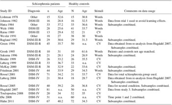
Application of these criteria reduced the number of studies to 21, published between 1978 and 2011. A list of the included studies can be found in Appendix A and an overview of study characteristics in Table 1. Of the 21 studies included, the DSM IV was used in nine studies to diagnose schizophrenia, the DSM-III-R was used in six studies and the DSM-III was used in three studies. In three studies, other criteria (e.g., psychiatry expertise) were applied for diagnosis.
Characteristics of studies included in the first meta-analysis

For the second meta-analysis the same inclusion criteria were used as for the first with the exception that here criterion one was:
1. Both a schizophrenia group experiencing auditory hallucinations and a non-hallucinating control group (e.g., healthy controls or non-hallucinating patients with schizophrenia) were included in the study.
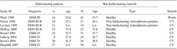
Auditory hallucinations were operationalized as a trait variable for the present study. For example, patients were included in the hallucinating group if they frequently experienced auditory hallucinations but they did not necessarily needed to do so during testing. Application of these criteria reduced the number of studies to eight, published between 1988 and 2007. A list of the included studies can be found in Appendix B and an overview about study characteristics in Table 2. Of the eight studies included, four used the DSM IV, three used the DSM-III-R and one used the DSM-III to diagnose schizophrenia.
Characteristics of studies included in the second meta-analysis

Dependent Variables and Calculation of Effect Sizes
The right ear advantage in dichotic listening studies is usually defined as the difference between the number of correctly identified stimuli presented to the right ear compared to the number of correctly identified stimuli presented to the left ear. The included studies reported the right ear advantage in three different ways:
1. The mean number of correctly identified stimuli for the left (NL) and right ear (NR) was reported.
2. A non-standardized mean difference value was reported, for example, NR − NL
3. A standardized difference value (corrected for overall performance) was reported: (NR − NL) / (NR + NL) * 100 (this measure is called laterality index in some studies).
As a result of the use of different dependent variables in different studies described above, Hedges’ g (Hedges & Olkin, Reference Hedges and Olkin1985), the standardized mean difference between groups corrected for the sample bias, was calculated as an unbiased estimate of the population effect. Depending on the available statistical information four different procedures were used to calculate Hedges’ g:
1. Both the mean and a variance measure (e.g., standard deviation) of some form of laterality index indicating the extent of the right ear advantage in a single value were given for both groups. In this case, Hedges’ g could directly be calculated.
2. The mean and a variance measure (e.g., standard deviation) for the number of correctly identified stimuli presented to the right and the left ear were given, but not the standardized laterality index. In this case, the mean of the laterality index was calculated for both groups using the formula described above. The standard deviation of the laterality index was then estimated by adding the standard deviation for the left ear items to the standard deviation for the right ear items. Hedges’ g was then calculated as described in 1.
3. In a few studies, individual raw data or group means and standard deviations were only available in diagrams. In this case, the means and standard deviations were read out using a ruler and Hedges’ g was subsequently calculated as described in 1 or 2 above.
4. Test statistics (F or t values) for the group comparison, but no descriptive statistics were given. In this case, Hedges’ g was converted from these test-statistics (Cohen, Reference Cohen1988).
Some of the studies included in the meta-analyses used the forced attention version of the dichotic listening task (Hugdahl & Andersson, Reference Hugdahl and Andersson1986). This variant of the dichotic listening task consists of three different attention instruction conditions. The non-forced attention condition resembles the standard dichotic listening paradigm, while in the other two conditions participants are asked to attend to and report the stimuli presented in the left or right ear. For the present meta-analyses, only data from the non-forced condition were analyzed. Moreover, in studies comparing emotional to non-emotional dichotic listening stimuli (e.g., syllables spoken sadly or neutrally), only the emotionally neutral condition was analyzed. When studies reported separate results for subsamples of their schizophrenia sample (e.g., for smoking and non-smoking or male and female patients) a weighted mean of these results was calculated so that every study was represented by a single effect size in the analyses.
Meta-analyses
For both meta-analyses the individual study effect sizes obtained for the group comparisons regarding the laterality index in dichotic listening were used as dependent variables. Negative effect sizes indicate a smaller laterality index in patients/hallucinating patients compared to healthy/non-hallucinating controls. Assuming that all included studies provide estimates of the same latent population effect, the meta-analyses were performed using a fixed-effects model to determine the weighted mean of Hedges’ g (M(g)) across all included studies. Subsequently, it was tested whether M(g) significantly deviates from zero, with the threshold of statistical significance being set α = 0.05. For all significant effects, the fail safe N was calculated according to the procedure described by Rosenberg (Reference Rosenberg2005). This measure gives an estimate for the number of unpublished non-significant studies that would be needed to nullify the reported significant effect which is based on the published material. Furthermore, a homogeneity test with a significance threshold of α = 0.05 was performed to test whether homogeneity can be assumed for the studies included in the analysis. As a measure of homogeneity, the I2 index was calculated, which indicates the variability between effect sizes due to inhomogeneity between the studies in percent.
Results
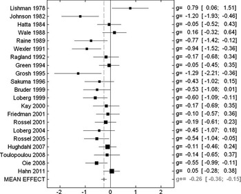
In the first meta-analysis, k = 21 different studies were included (see Figure 1 for effect sizes and corresponding confidence intervals for all included studies). The overall sample consisted of 700 schizophrenia patients and 707 healthy controls. The analysis revealed a mean weighted effect size of Hedges’ g = −0.26 (95% confidence interval −0.36 to −0.15), significantly different from zero (Z = −4.69; p < .00001), indicating that schizophrenia patients had a lower mean laterality index than healthy controls. The fail safe N for this comparison was 150. The homogeneity test reached significance (Q(20) = 42.81; p < .01; I2 = 53.3%), possibly indicating that different sub-samples, for example hallucinating versus non-hallucinating patients, exist in the schizophrenia group.
Forest plot of the study effect sizes (squares) and 95%-confidence intervals (horizontal line) of the difference between schizophrenia patients and healthy controls regarding language lateralization. Negative values indicate that patients had a lower laterality index than controls. The mean effect over all studies is indicated by the gray diamond in the bottom row and the size of the squares representing the individual studies indicates the weight of the study in the calculation of the mean effect. The numbers at the right margin represent the individual study effect size with the numbers in parentheses indicating the lower and upper boundaries of the 95%-confidence interval.

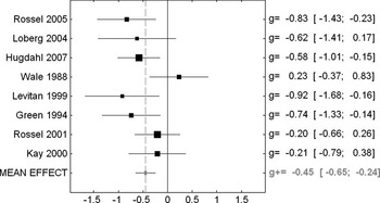
In the second meta-analysis, k = 8 different studies were included (see Figure 2 for effect sizes and corresponding confidence intervals for all included studies). The overall sample consisted of 179 schizophrenia patients with auditory hallucinations and 228 non-hallucinating control subjects. The analysis revealed a mean weighted effect size of Hedges’ g = −0.45 (95% confidence interval −0.65 to −0.25), significantly different from zero (Z = −4.45; p < .00001) indicating that schizophrenia patients had a lower mean laterality index than healthy controls. The fail safe N for this comparison was 49. In contrast to the first meta-analysis, the homogeneity test did not reach significance for this sample (Q(7) = 11.17; p = .13; I2 = 37.3%), so that homogeneity of the included study effects can be assumed. The assumed homogeneity of the included studies in the second meta-analysis thus support the assumption that the failure of the homogeneity test in the first meta-analysis was due to sub-samples in the schizophrenia group.
Forest plot of the study effect sizes (squares) and 95%-confidence intervals (horizontal line) of the difference between schizophrenia patients that experience auditory hallucinations and non-hallucinating controls regarding language lateralization. Negative values indicate that hallucinators had a lower laterality index than non-hallucinators. The mean effect over all studies is indicated by the gray diamond in the bottom row and the size of the squares representing the individual studies indicates the weight of the study in the calculation of the mean effect. The numbers at the right margin represent the individual study effect size with the numbers in parentheses indicating the lower and upper boundaries of the 95%-confidence interval.

Discussion
The first meta-analysis indicated that schizophrenia patients show significantly reduced laterality indices in the dichotic listening task when compared to healthy controls. Thus, this analysis supports the assumption that schizophrenia is associated with reduced left-hemispheric language dominance. This result is then in line with the results reported by Sommer et al. (2001) who also found significantly decreased language lateralization in schizophrenia patients in a combined analysis of the results of six dichotic listening studies that used the consonant-vowel or fused rhymed words version of the dichotic listening task. However, while the group comparison effect in the present study was significant, the effect size of g = −0.26 can be characterized as small and a significant homogeneity test indicated substantial inter-study variability. This finding supports the idea that schizophrenia patients cannot be treated as a homogenous group regarding language lateralization. This assumption fits well with the findings that there are sub-groups within the overall schizophrenia population that show the same lateralization patterns as healthy controls, e.g., young and stabilized schizophrenia patients with few positive symptoms (Løberg et al., Reference Løberg, Jørgensen and Hugdahl2002).
In addition to the general effect, the study also confirmed an effect of the experience of auditory hallucinations on language lateralization as a possible mediating factor in explaining language lateralization in schizophrenia. The second meta-analysis indicated that schizophrenia patients which experience auditory hallucinations show a significantly reduced right ear advantage in the dichotic listening task when compared to non-hallucinating controls. Interestingly, the effect size of g = −0.45 in this meta-analysis was substantially larger than the one observed for the comparison of all schizophrenia patients with healthy controls. Moreover, the homogeneity test for this comparison was not significant, although it has to be taken into account that the sample sized in the second comparison also was smaller, leading to reduced power compared to the homogeneity test for the first meta-analysis. However, since I2 also was substantially smaller than for the first meta-analysis, greater homogeneity of the hallucinating group than for schizophrenia patients in general can be assumed. Thus, the present study indicates that schizophrenia patients that experience auditory hallucinations show a substantially larger reduction of left-hemispheric language lateralization in comparison to non-hallucinating controls than schizophrenia patients in general compared to healthy controls. This finding yields further support for the assumption that the experience of auditory hallucinations is a factor that is mediating whether a schizophrenia patient shows reduced language lateralization or not. It is, however, important to note that this does not necessarily suggest that reduced language lateralization is linked to the experience of auditory hallucinations itself without the context of psychosis. A recent functional magnetic resonance imaging study comparing language lateralization in psychotic patients (mostly diagnosed with schizophrenia), non-psychotic subjects with auditory hallucinations and non-psychotic subjects without auditory hallucinations found that language lateralization was only reduced in the psychotic group, but not in the two other groups (Diederen et al., Reference Diederen, De Weijer, Daalman, Blom, Neggers, Kahn and Sommer2010). These findings are, however, not necessarily in contradiction with the results of the present study. While auditory hallucinations in psychotic and non-psychotic individuals have been found to activate roughly the same cortical networks (Diederen et al., Reference Diederen, Daalman, de Weijer, Neggers, van Gastel, Blom and Sommer2011), the much younger age at onset of auditory hallucinations in non-psychotic compared to psychotic individuals has been suggested to indicate a different underlying pathophysiology for the two phenomena (Daalman et al., Reference Daalman, Boks, Diederen, de Weijer, Blom, Kahn and Sommer2011). Taking these findings into account, the results of the present study thus indicate that reduced language lateralization is a trait marker factor for the experience of auditory hallucinations in schizophrenia patients, but possibly not in healthy subjects.
Although it has been suggested that reduced language lateralization may be a potential cause of schizophrenia (Angrilli et al., Reference Angrilli, Spironelli, Elbert, Crow, Marano and Stegagno2009), so that individuals showing atypical language lateralization are at increased risk of becoming schizophrenia or developed typical symptoms like auditory hallucinations, it is not possible based on the present data to conclude whether reduced language lateralization is cause or effect of schizophrenia or the experience of auditory hallucinations.
It could be argued that the experience of auditory hallucinations during testing may act as a distractor and therefore influence test performance in the dichotic listening task. However, voxel-based morphometry studies provide evidence for grey matter volume reductions specifically in left-hemispheric language areas in hallucinating patients relative to healthy controls. For example, Neckelmann et al. (Reference Neckelmann, Specht, Lund, Ersland, Smievoll, Neckelmann and Hugdahl2006) reported that the rate and frequency of auditory verbal hallucination correlated negatively with grey matter volume reductions in the in the left superior temporal gyrus, left thalamus, and left and right cerebellum. Moreover, a recent meta-analysis on the neuroanatomy of auditory verbal hallucinations in schizophrenia by Modinos et al. (Reference Modinos, Costafreda, van Tol, McGuire, Aleman and Allen2012) found that the severity of auditory verbal hallucinations was significantly associated with grey matter volume reductions in the left superior temporal gyrus. This pronounced grey matter volume loss in left-hemispheric language areas argues for a structural basis for the here observed relation between language lateralization and auditory verbal hallucinations.
According to a model suggested by Hugdahl et al. (Reference Hugdahl, Løberg, Jørgensen, Lundervold, Lund, Green and Rund2008), auditory hallucinations are internally generated speech misrepresentations involving mainly speech perception areas of the left temporal lobe (Kompus, Westerhausen, & Hugdahl, Reference Kompus, Westerhausen and Hugdahl2011). Thus, it should be particularly problematic for patients experiencing hallucinations to identify external speech sounds presented to the right ear which are mainly processed by left-sided areas. Therefore, the experience of auditory hallucinations should then lead to decreased lateralization. This model has been supported by the finding that an increasing frequency of hallucinations in schizophrenia patients is linked to a gradual decrease in the ability to process and report the right ear stimulus in the dichotic listening task (Hugdahl et al., Reference Hugdahl, Løberg, Jørgensen, Lundervold, Lund, Green and Rund2008). Furthermore, it is also supported by the results of a meta-analysis of five studies that investigated brain activations in schizophrenia patients during the experience of auditory verbal hallucinations and found the left-hemispheric language-related areas were significantly more activated during hallucinations than the homotope right-sided regions (Sommer, Aleman, & Kahn, Reference Sommer, Aleman and Kahn2003; but also see Bentaleb, Beauregard, Liddle, & Stip, Reference Bentaleb, Beauregard, Liddle and Stip2002). Similar results were also reported by a more recent meta-analysis that analyzed twelve different neuroimaging studies of schizophrenia patients experiencing auditory verbal hallucinations and resting in the absence of auditory stimulation (Kompus et al., Reference Kompus, Westerhausen and Hugdahl2011). The analysis showed increased activation in the left primary auditory cortex, but also in the right rostral prefrontal cortex during the experience of hallucinations without external auditory stimulation.
One methodological aspect of the present study that needs discussion is the greater diversity of the non-hallucinating control group in the second meta-analysis compared to the healthy controls in the first meta-analysis. In this group both healthy controls and schizophrenia patients without auditory hallucinations were included, so that in theory one could argue that the greater effect size of this meta-analysis compared to the first might possibly be a control group effect. However, this is unlikely since for six out of the eight studies included in this analysis, the same healthy control groups as included for the same study in the first meta-analysis were used. In these cases, only the schizophrenia group was changed, from the overall patient group of each study in the first analysis to the hallucinating subgroup in the second. Moreover, in the two studies that used non-hallucinating schizophrenia patients as control group, these control groups showed a normal right ear advantage, comparable to the healthy controls in other studies (study ID's: Green Reference Green, Hugdahl and Mitchell1994 and Levitan Reference Levitan, Ward and Catts1999, see Appendix B for details). Thus, it appears that the increased effect size could be attributed to an effect of hallucinations rather than to a control group effect.
Another methodological issue of the present study is the fact that for most studies included in the first meta-analysis it was not indicated whether and to what extent the patients included in the respective samples showed auditory hallucinations or not. Since auditory hallucinations are a common positive symptom of schizophrenia and affect more than 70% of the patients (Wing, Cooper, & Sartorius, Reference Wing, Cooper and Sartorius1974), it is likely, that a fair amount of patients experiencing auditory hallucinations were included in several of the studies in the first meta-analysis. Therefore, it is not certain whether the significant group difference observed in the first meta-analysis is indeed indicative of a general illness effect on language lateralization or whether it is solely caused by those patients that experience auditory hallucinations. This question could be answered by conducting a meta-analysis on dichotic listening performance in non-hallucinating schizophrenia patients, but unfortunately only very few studies (e.g., Løberg et al., Reference Løberg, Jørgensen and Hugdahl2002) specifically investigated this group, so that there is insufficient material for a meta-analytic integration. Thus, based on the present findings it would be a particularly interesting endeavor for future research to compare language lateralization in schizophrenia patients with and without auditory hallucinations to healthy, non-hallucinating controls as well as non-clinical hallucinators (Badcock & Hugdahl, Reference Badcock and Hugdahl2012; Diederen, van Lutterveld, & Sommer, Reference Diederen, van Lutterveld and Sommer2012). This experimental design would allow to further disentangle general illness effects from those specific for auditory hallucinations.
Conclusion
lt has been proposed that reduced left-hemispheric language lateralization constitutes a trait marker for schizophrenia (Angrilli et al., Reference Angrilli, Spironelli, Elbert, Crow, Marano and Stegagno2009; Crow, Reference Crow2000; Oertel et al., Reference Oertel, Knöchel, Rotarska-Jagiela, Schönmeyer, Lindner, van de Ven and Linden2010) and the present study yield further support for this assumption by showing a significant relation between schizophrenia and reduced language lateralization on a meta-analytical level. However, while the effect size suggest that reduced language lateralization is a weak trait marker for schizophrenia as such, a second meta-analysis revealed that it represents a much stronger trait marker for patients experiencing auditory hallucinations within the schizophrenia population.
Acknowledgments
All authors declare no conflicts of interest. The manuscript (or parts thereof) has not been previously published elsewhere and also is not under consideration for publication elsewhere. This work was supported by a fellowship within the Postdoc-Programme of the German Academic Exchange Service (DAAD) (S.O); and a grant from The European Research Council (ERC) (K.H., grant number # 249516).