INTRODUCTION
Bovine babesiosis is a well-recognized, tick-borne protozoan disease of cattle in most tropical and subtropical as well as some temperate regions of the world, often resulting in great economic losses (Kuttler, Reference Kuttler and Ristic1988; Bock et al. Reference Bock, Jackson, de Vos and Jorgensen2004). Babesia bovis, one of the major causative agents of bovine babesiosis, induces severe clinical symptoms, such as fever, anaemia, haemoglobinuria, and hypotensive shock syndrome, with the occasional death (Bock et al. Reference Bock, Jackson, de Vos and Jorgensen2004; Vial and Gorenflot, Reference Vial and Gorenflot2006). Babesia sporozoites directly invade host erythrocytes after the animal has been bitten by an infected tick. The intraerythrocytic parasites multiply by forming 2–4 merozoites to establish a seemingly perpetual cycle of asexual reproduction (Vial and Gorenflot, Reference Vial and Gorenflot2006). The pathogenicity of B. bovis in the host animals is related to the erythrocytic stage of the parasite (Wright and Goodger, Reference Wright, Goodger and Ristic1988; Bock et al. Reference Bock, Jackson, de Vos and Jorgensen2004). A better understanding of the biological processes involved in the growth cycle at the erythrocytic stage is required in order to develop effective therapeutic tools against bovine babesiosis.
Cell cycle progression is essential for the survival of any organism, and cyclin-dependent kinases (CDKs), in particular, are responsible for the control of cell cycle progression in cooperation with other regulatory molecules, called cyclins (Morgan, Reference Morgan1995). The CDK/cyclin activity is frequently up-regulated in human cancer cells, which has stimulated the interest in the chemical inhibition of these enzymes (Senderowicz and Sausville, Reference Senderowicz and Sausville2000). After purine-derivative compounds were discovered to be inhibitors of CDK1, a large variety of chemical compounds have been extensively investigated as CDK inhibitors (Chang et al. Reference Chang, Gray, Rosania, Sutherlin, Kwon, Norman, Sarohia, Leost, Meijer and Schultz1999; Knockaert et al. Reference Knockaert, Greengard and Meijer2002). Several CDK inhibitors suppress tumor growth although the specificity for CDK activities of these inhibitors in treated cells is still controversial (Knockaert et al. Reference Knockaert, Greengard and Meijer2002; Becker et al. Reference Becker, Murthi, Smith, Come, Costa-Roldán, Kaufmann, Hanke, Degenhart, Baumann, Wallner, Huber, Dedier, Dill, Kinsman, Hediger, Bockovich, Meier-Ewert, Kluge and Kley2004).
The role of CDKs in protozoan parasites remains unclear. In Plasmodium falciparum, a protozoan parasite that belongs to the same phylum Apicomplexa as Babesia spp., the asexual growth was significantly suppressed by the addition of chemical inhibitors for the CDK/cyclin activity into the in vitro culture (Harmse et al. Reference Harmse, van Zyl, Gray, Schultz, Leclerc, Meijer, Doerig and Havlik2001). This report suggested that several CDK inhibitors have chemotherapeutic potential for malaria. The homologous genes coding for CDKs in P. falciparum have been identified. Experiments using recombinant enzyme systems showed that CDK inhibitors significantly inhibited the enzymatic activities of these CDK homologues (Geyer et al. Reference Geyer, Prigge and Waters2005). These facts suggest that CDKs play an essential role in the asexual growth of P. falciparum. However, the effective doses of the CDK inhibitors against the parasite's growth were different from those obtained in an in vitro assay using purified higher eukaryotic CDK/cyclin complex (Harmse et al. Reference Harmse, van Zyl, Gray, Schultz, Leclerc, Meijer, Doerig and Havlik2001). Further, it has been reported that purvalanol B, one of the representative CDK inhibitors, interacts with casein kinase rather than CDKs as an intracellular target in protozoan parasites such as Plasmodium, Toxoplasma, Trypanosoma and Leishmania (Knockaert et al. Reference Knockaert, Gray, Damiens, Chang, Grellier, Grant, Fergusson, Mottram, Soete, Dubremetz, Le Roch, Doerig, Schultz and Meijer2000). These reports suggest that target molecules and biological mechanisms of CDK inhibitors in protozoan parasites might differ from those observed in higher eukaryotic cells. These differences are encouraging with regard to the identification of a parasite-specific inhibitor.
In the case of B. bovis, the effect of CDK inhibitors on the parasite's growth is unknown. Recently, an EST sequence database of B. bovis has been published (de Vries et al. Reference De Vries, Corton, Harris, Cornelissen and Berriman2006). In this database, the 3 kinds of CDK gene homologues (annotated as cdc2 (CDK1)-like kinase or cyclin-dependent serine/threonine protein kinase) and a cyclin gene homologue (annotated as cyclin-putative) can be observed. Therefore, the CDKs potentially act as key molecular switches for the cell cycle progression of B. bovis. In this study, we evaluated the inhibitory effects of selected chemical compounds, referred to as CDK inhibitors, on the asexual growth of B. bovis.
MATERIALS AND METHODS
Chemical compounds
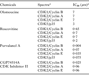
Five purine derivatives, olomoucine, roscovitine, purvalanol A, CGP74514A, and CDK2 Inhibitor II, were purchased from Calbiochem-Novabiochem Co. (Darmstadt, Germany). They have high permeability to cells and selective inhibitory activities against purified eukaryotic CDKs through the competitive binding to the ATP-binding pocket of CDKs (Vesely et al. Reference Vesely, Havlicek, Strnad, Blow, Donella-Deana, Pinna, Letham, Kato, Detivaud, LeClerc and Meijer1994; Meijer et al. Reference Meijer, Borgne, Mulner, Chong, Blow, Inagaki, Inagaki, Delcros and Moulinoux1997; Gray et al. Reference Gray, Wodicka, Thunnissen, Norman, Kwon, Espinoza, Morgan, Barnes, LeClerc, Meijer, Kim, Lockhart and Schultz1998; Davis et al. Reference Davis, Benson, Bramson, Chapman, Dickerson, Dold, Eberwein, Edelstein, Frye, Gampe, Griffin, Harris, Hassell, Holmes, Hunter, Knick, Lackey, Lovejoy, Luzzio, Murray, Parker, Rocque, Shewchuk, Veal, Walker and Kuyper2001; Dai et al. Reference Dai, Dent and Grant2002). Spectra and 50% inhibitory concentration (IC50) against the purified CDKs of each compound, as evaluated in previous research, are summarized in Table 1.
Compounds used in this study

a These data are available in a CALBIOCHEM® online catalogue (www.calbiochem.com).
In vitro growth inhibition assay
The Texas strain of B. bovis was maintained in purified bovine erythrocytes (RBC) with a serum-free GIT medium (Wako Pure Chemical Industrial, Ltd, Osaka, Japan) as described previously (Bork et al. Reference Bork, Okamura, Matsuo, Kumar, Yokoyama and Igarashi2005). The in vitro growth inhibition assay followed the method previously described by Bork et al. (Bork et al. Reference Bork, Yokoyama, Matsuo, Clavelia, Fujisaki and Igarashi2003, Reference Bork, Yokoyama, Ikehara, Kumar, Sugimoto and Igarashi2004) with some modifications. Parasitized RBC were mixed with uninfected RBC to obtain RBC packs with 1% parasitaemia, and GIT medium containing an appropriate concentration of each compound was added into each of 3 wells. Dimethyl sulfoxide (DMSO), which was used as a solvent for each drug, was added at the same final concentration into control wells. All cultures were incubated at 37°C in a humidified multi-gas incubator. Every 24 h for a period of 3 days, the medium was replaced with a fresh medium containing the appropriate compound. Thin blood smears were periodically prepared, stained with a Giemsa's solution, and then the number of infected RBC was counted out of a total of 1000 RBC for the calculation of parasitaemia. Counting RBC was performed without any information about the samples to eliminate any bias. The IC50 values of each compound were determined by interpolation after curve-fitting using a Prism® program (GraphPad Software, Inc., San Diego, CA, USA) based on the parasitaemia observed at 72 h after the treatment.
RBC invasion assay
The RBC invasion assay was performed according to the procedure described previously (Franssen et al. Reference Franssen, Gaffar, Yatsuda and de Vries2003; Okubo et al. Reference Okubo, Yokoyama, Takabatake, Okamura and Igarashi2006). PBS (pH 8·0) containing 1 mm CaCl2 (PBS/Ca2+) was selected as a culture maintenance buffer instead of GIT medium to reduce the influence of medium components on the drug activity. After B. bovis-infected RBC were suspended in an equal volume of a PBS/Ca2+, the mixture of 400 μl was subjected to 5 intermittent (10 sec, 0°C) high-voltage pulses (1·5 kV, 400 Ω, 25 μF) in a Gene Pulser II (Bio-Rad Laboratories, Hercules, CA, USA) with a 0·2 cm pulser cuvette (Bio-Rad Laboratories) to purify the intraerythrocytic parasites from infected RBC artificially. After washing with the PBS/Ca2+, the parasites were distributed into a 10% RBC suspension in PBS/Ca2+ with an appropriate concentration of each compound. After incubation at 37°C for 1 h, the number of infected RBC was counted out of a total of 1000 RBC for calculation of parasitaemia. Percentage inhibition of RBC invasion by treatment with each of the compounds was determined by comparison of parasitaemia observed in the treated culture with that in the control culture. The ratio of extraerythrocytic merozoites to total number of merozoites was also calculated to determine invasion efficiency more precisely.
Parasite-replication assay
An inhibitory effect on the intraerythrocytic development of B. bovis was examined by a parasite-replication assay. Briefly, purified parasites were prepared from the infected RBC by high-voltage pulsing, as described above, and incubated with uninfected RBC. After incubation for 1 h to allow the parasites to invade the RBC, each compound was added into the culture. After an additional 5-h incubation, Giemsa-stained smears were made. The ratio of RBC containing degenerated parasites to the total number of infected RBC was calculated to evaluate intracellular killing effect by the compounds. The ratio of divided parasites to the total number of live parasites was also calculated to ascertain the effect of each drug on intraerythrocytic replication.
RESULTS
Inhibitory effect of each compound on the in vitro growth of B. bovis
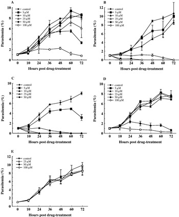
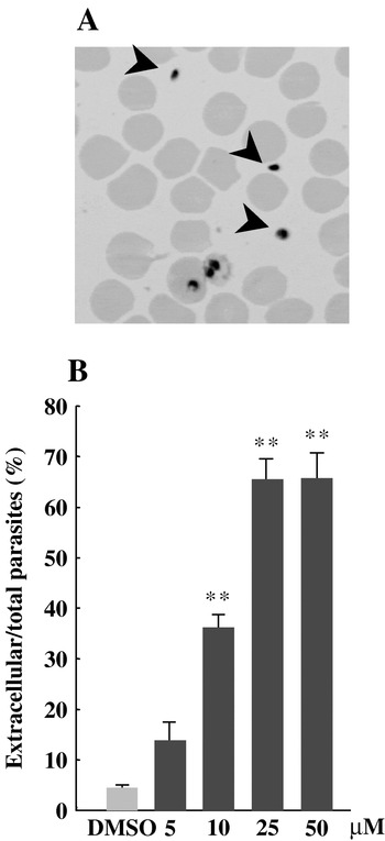
Treatment with roscovitine, purvalanol A, CGP74514A, and CDK2 Inhibitor II showed a reduction of the in vitro growth of B. bovis (Fig. 1A–D) with IC50 values (at 72 h) of 45·1, 11·0, 4·9 and 42·7 μm, respectively, whereas olomoucine did not show a clear reduction even at a 100 μm concentration (Fig. 1E). In the CGP74514A-treated culture only, an abnormal accumulation of extraerythrocytic merozoites was prominently observed at 10 h after addition of the compound (Fig. 2A). The percentage of extraerythrocytic merozoites in the total number of observed parasites reached about 70% when cultures were treated with a high concentration (25 or 50 μm) of the compound (Fig. 2B). The accumulation of extraerythrocytic merozoites suggested that this compound was affecting RBC invasion.
In vitro growth curve of Babesia bovis in the presence of different concentrations of roscovitine (A), purvalanol A (B), CGP74514A (C), CDK2 Inhibitor II (D) and olomoucine (E) based on the dynamics of parasitaemia. Each value represents the mean±s.d. in triplicate. These curves represent the results of 1 representative experiment out of similar experiments repeated twice.

Abnormal accumulation of extraerythrocytic merozoites (arrowheads) observed at 10 h post-treatment with 10–50 μm CGP74514A (A). The percentages of extraerythrocytic to the total number of merozoites in the cultures treated with CGP74514A (B). Each value represents the mean±s.d. in triplicate.

To confirm that the effects of the compounds were not due to their action on RBC, we performed culture experiments using RBC that had been pre-treated with the compounds. Briefly, RBC were incubated with a high concentration of each compound (100 μm for roscovitine and CDK2 Inhibitor II, 50 μm for purvalanol A and CGP74514A) at 37°C for 3 h, washed with a normal GIT medium 3 times, and then used for standard parasite cultivation. It was found that there was normal growth of B. bovis in all cultures with pre-treated RBC, similar to the control (data not shown), indicating that these compounds directly act on the parasites, but not on the host RBC.
Inhibition of RBC invasion or intraerythrocytic development of B. bovis by each compound
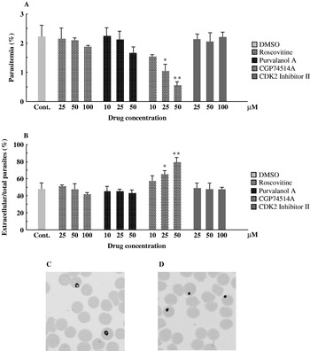
To determine which step during the asexual growth cycle of B. bovis was influenced by the treatment with these compounds, we first performed an RBC invasion assay. In the control culture, 52·0±6·8% (mean±standard deviation, s.d.) of merozoites invaded RBC, reaching 1·8–2·8% absolute parasitaemia after incubation for 1 h (Fig. 3A). Treatment with 25 μm CGP74514A showed 52·9±9·3% inhibition of RBC invasion compared to that of control (P<0·05). A higher inhibitory effect (75·4±5·4% compared to control, P<0·01) was observed in the 50 μm CGP74514A-treated culture (Fig. 3A). Both concentrations of CGP74514A also induced a significant increase in the ratio of extraerythrocytic merozoites to total merozoites (Fig. 3B). Treatment with 10 μm CGP74514A showed 30·9±2·7% inhibition which was not significant (Fig. 3A). A 15·5±1·5% or 25·6±8·6% (not significant) decrease of parasitized RBC was observed in the cultures treated with 100 μm roscovitine or 50 μm purvalanol A, respectively (Fig. 3A). However, a decreased number of parasitized RBC in these cultures was possibly due to a direct killing effect by these compounds because the number of extraerythrocytic merozoites was not increased (Fig. 3B). In contrast, invasion efficiency observed in cultures treated with CDK2 Inhibitor II was similar to that of control (Fig. 3A and B). These results could support the hypothesis that CGP74514A, especially in high concentration, affected RBC invasion by merozoites.
Effects of the inhibitory compounds on the invasion of RBC by Babesia bovis (A). The percentages of extraerythrocytic to the total number of merozoites in the cultures treated with each of the compounds (B). Asterisks indicate significant differences (Student's t-test; * P<0·05, ** P<0·01) between the drug-treated and control cultures. Each value represents the mean±s.d. in triplicate. Ring-shaped parasites observed in a control culture immediately after the RBC invasion (C). Dot-shaped parasites observed in a 25 μm Purvalanol A-treated culture (D).

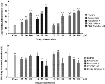
In general, B. bovis forms a ring-shape after the parasite's invasion into host RBC (Fig. 3C), which is then followed by an intraerythrocytic replication as it divides. However, the parasites treated with roscovitine, purvalanol A, and CDK2 Inhibitor II hardly ever formed the typical ring-shape within the infected RBC, but they did show the parasite's degeneration into a ‘dot-shape’ parasite (Fig. 3D). These results suggested that these compounds might affect the subsequent step of RBC invasion of B. bovis. Therefore, an inhibitory effect on the intraerythrocytic development of B. bovis was examined by a parasite-replication assay. The ratio of extraerythrocytic merozoites to total merozoites at 5 h after drug-treatment was the same as that immediately before drug-treatment. It indicated that mature merozoites had completed RBC invasion within 1 h pre-incubation of the culture, whereas immature merozoites liberated by a high-voltage pulse had remained outside the RBC. Therefore, an influence on RBC invasion by the treatment with each of the compounds could be ruled out in this assay. In the control culture, degenerated parasites were seen at the ratio of 13·5±0·9% to total parasites, and 23·5±1·3% of total live parasites developed into dividing-forms (Fig. 4A and B). An increased number of degenerated parasites was observed in a dose-dependent manner by treatment with roscovitine, purvalanol A and CDK2 Inhibitor II (Fig. 4A). The appearance of dividing-form parasites was significantly (P<0·05) decreased in 25 and 50 μm roscovitine-, 25 and 50 μm purvalanol A-, and 100 μm CDK2 Inhibitor II-treated cultures (Fig. 4B). These results suggested that the early stages of intraerythrocytic development of B. bovis were affected by treatment with these compounds. A concentration less than 25 μm of CGP74514A did not affect the viability of the intraerythrocytic parasites or the replication capacity (Fig. 4A and B). However, the addition of 50 μm CGP74514A to the culture caused intracellular killing of the parasites (Fig. 4A). A suppression of in vitro growth observed in 50 μm CGP74514A-treated culture could be promoted by this killing effect.
An increase of degenerated parasites (A), and a decrease of the dividing form parasites (B) by treatment with each of the compounds. Asterisks indicate significant differences (Student's t-test; * P<0·05, ** P<0·01) between the drug-treated and control cultures. Each value represents the mean±s.d. in triplicate.

DISCUSSION
In this study, 4 different CDK inhibitors, roscovitine, purvalanol A, CGP74514A, and CDK2 Inhibitor II, significantly suppressed the in vitro growth of B. bovis, suggesting the chemotherapeutic potential of CDK inhibitors for bovine babesiosis. Olomoucine did not show any inhibitory effect on in vitro growth of B. bovis in this study. Since this drug shows only moderate inhibitory effect in the in vitro kinase inhibition assay (Vesely et al. Reference Vesely, Havlicek, Strnad, Blow, Donella-Deana, Pinna, Letham, Kato, Detivaud, LeClerc and Meijer1994), the concentrations tested in this study were probably too low to have any activity in an in vitro culture system.
The IC50 values of the inhibitors obtained in this study were considerably higher than the values shown in previous studies. One possible reason for this discrepancy is the low efficiency of transporting the drugs into parasites. The differences in target molecules and/or mode of action of these inhibitors in B. bovis from those in higher eukaryotic cells also might explain the higher IC50 values. Although the differences between higher eukaryotic CDKs and those of B. bovis are not understood, the activity of the inhibitors might be affected by the diversity of CDKs. Further, it has been reported that purvalanol B, an analogue of purvalanol A, interacts with more casein kinase than CDKs as an intracellular target in related protozoa such as Plasmodium, Toxoplasma, and also Trypanosoma and Leishmania (Knockaert et al. Reference Knockaert, Gray, Damiens, Chang, Grellier, Grant, Fergusson, Mottram, Soete, Dubremetz, Le Roch, Doerig, Schultz and Meijer2000). The compounds used in this study possibly act against completely distinct targets from those reported in higher eukaryotic cells.
Treatment with roscovitine, purvalanol A, and CDK2 Inhibitor II blocked the early stages of intraerythrocytic development of B. bovis. These compounds potentially target the molecule(s) involved in intraerythrocytic development of B. bovis. Since CDK homologues of B. bovis possibly participate in the intraerythrocytic development, they are suspected to be the candidates of intracellular target of these compounds.
Interestingly, CGP74514A was shown to inhibit the RBC invasion of B. bovis. Because higher eukaryotic cells need not invade other cells for their replication, the inhibition of the RBC invasion by B. bovis is considered to be a very characteristic effect of CGP74514A. Although the reason why CGP74514A showed a different effect than other inhibitors is unclear, CGP74514A could have different targets in B. bovis. Inhibition of RBC invasion by treatment with another kind of kinase inhibitor, Staurosporine, has been reported in B. bovis as well as Plasmodium spp. (Bork et al. Reference Bork, Das, Okubo, Yokoyama and Igarashi2006; Ward et al. Reference Ward, Fujioka, Aikawa and Miller1994), indicating that protein phosphorylation necessarily participates in the RBC invasion step of the parasites. CGP74514A may target the kinase family that participates in phosphorylation of the proteins involved in RBC invasion.
In conclusion, our data revealed that CDK inhibitors had growth inhibitory effects on the erythrocytic stage of B. bovis by targeting the molecules that participated in the process of successful RBC invasion and intraerythrocytic development. Further studies for elucidation of the target molecules and mode of action of CDK inhibitors in B. bovis, as well as verification of the chemotherapeutic effect of the CDK inhibitors in an in vivo rodent model of Babesia infection are being pursued.
This study was supported by Grants-in-Aid for Scientific Research from the Japan Society for the Promotion of Science, Grants from the Promotion of Basic Research Activities for Innovative Biosciences (PROBRAIN), and The 21st Century COE Program (A-1), Ministry of Education, Culture, Sports, Science and Technology, Japan.