Alzheimer disease (AD) is the leading cause of senile dementia in the US, where it affects 15% of people over 65 and almost 50% of those over 85 (Ref. Reference Smith1). AD is characterised by severe neurodegeneration and cognitive impairment, but the precise pathogenesis has not been fully elucidated. The hallmark features of AD, neurofibrillary tangles (NFTs) and amyloid-β (Aβ) plaques, although not the sole players in neurodegeneration, are crucial to disease development and progression.
Aβ is the major component of senile plaques characteristic of AD and is derived from the amyloid-β protein precursor (APP) encoded on chromosome 21 (Ref. Reference Korenberg2). Mutations in the APP gene are directly linked to the onset of familial AD (Ref. Reference Hellstrom-Lindahl, Viitanen and Marutle3). Interestingly, recent evidence indicates that Aβ can provide oxidative protection (Ref. Reference Moreira4). In vitro studies of Aβ in its soluble, non-aggregated form (Refs Reference Nunomura5, Reference Kontush6), demonstrate its antioxidant capacity, probably through its function as a high-valence metal chelator (Refs Reference Nunomura5, Reference Zou7). The eventual accumulation of Aβ in neuronal tissue, however, is likely to be detrimental to the cell. Specifically, Aβ-containing senile plaques elicit inflammatory responses from surrounding microglial and astrocytic cells (Refs Reference Morgan8, Reference Piazza and Lynch9) and ultimately increase oxidative damage and further selfaggregation (Ref. Reference Ong and Farooqui10). Of note here, APP is upregulated by mitogenic stimulation and APP metabolism is controlled by cell-cycle-dependent changes (Refs Reference Schubert11, Reference Milward12, Reference Copani13). Moreover, Aβ itself has been identified to be mitogenic in vitro (Refs Reference Schubert11, Reference Milward12). Similarly, the hyperphosphorylated form of the microtubule-associated protein tau (the primary component of NFTs) (Refs Reference Iqbal14, Reference Grundke-Iqbal15), produces neuronal dysfunction in AD through microtubule destabilisation. Although tau phosphorylation is known to be effected through the action of various kinases including cdk5 and GSK-3β, similarly hyperphosphorylated tau present in mitotically active cells is thought to be partly driven by the activity of cell cycle proteins (Refs Reference Brion16, Reference Brion, Octave and Couck17).
Oxidative stress has become increasingly significant in the pathogenesis of AD over the past few years, and has also been identified as concordant with markers of cell cycle re-entry (Ref. Reference Zhu18). Although the exact origins of oxidative stress remain uncertain, it has been demonstrated to be one of the primary role-players in the onset of AD and in its development. Hence, markers of oxidative stress, such as 8-hydroxyguanosine (8OHG), precede the general signs of AD in immunohistochemically stained neurons by decades (Refs Reference Zhu18, Reference Nunomura19), and it is not until gross accumulations of oxidative stress elicit overbearing amounts of Aβ that the Aβ-induced pathogenesis of AD occurs.
The pathological significance of cell cycle alteration in Alzheimer disease
The four phases of the cell cycle consist of elaborate feedback mechanisms and regulatory checkpoints that ensure its competency. The phases are: S-phase, where DNA replication takes place; M-phase, where mitosis, or cell division, occurs; and the gap phases G1 and G2, which separate the two. Quiescent cells, such as neurons in the adult hippocampus, exist in the nondividing, silent G0 phase. Such cells are terminally differentiated and generally thought to be incapable of re-entering the cell cycle (Ref. Reference McShea, Wahl and Smith20).
Importantly, the transition through these phases is controlled by an array of special cell cycle proteins, the cyclins and the cyclin-dependant kinases (CDKs), which fluctuate in their expression and activity as the cell cycle progresses. For instance, the expression and activation of the cyclin D1 (encoded by CCND1)–CDK4/6 complex, triggered by the presence of mitotic growth factors, controls the re-entry of resting (G0) cells into the G1 phase of cell cycle (Ref. Reference Sherr21). The G1-S transition is then controlled by the activation of the cyclin E1 (CCNE1)–CDK2 complex, among others (Ref. Reference Sherr21), such that the absence of cyclin E1 and/or the inhibition of the cyclin-E1–CDK2 complex by p21 (CDNK1A), p27 (CDKN1B) and p53 (TP53) will cause the cell cycle to be arrested at the G1 checkpoint. The subsequent fate of the G1-arrested cells depends on the presence or absence of cyclin A1 (CCNA1) (Ref. Reference Grana and Reddy22) such that, in the absence of cyclin A1, the cells return to G0 and redifferentiate. However, if allowed to proceed in the presence of cyclin A1, the cells are committed to division, lack the ability to redifferentiate and, if unable to complete the cell cycle, die via an apoptotic pathway (Ref. Reference Meikrantz and Schlegel23). Therefore, once beyond late G1, when cyclin A1 is expressed, cell cycle arrest will lead to cell death.
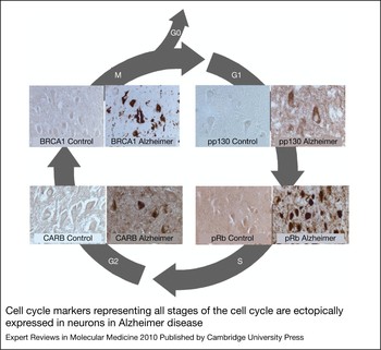
Notably, AD involves the erroneous departure of quiescent G0 neurons into G1 phase and beyond (Refs Reference McShea24, Reference Nagy, Esiri and Smith25, Reference Smith and Lippa26). Although the exact reasons for this re-entry are unknown, there is much pathological evidence to support it (Fig. 1). In addition to the proteins described in Figure 1, AD neurons exhibit significantly elevated levels of the cell cycle markers cyclin D1, CDK4 and Ki67 (MKI67), as well as those of the cyclin-E1–CDK4 complex, when compared with age-matched controls (Refs Reference Zhu18, Reference McShea24, Reference Nagy, Esiri and Smith25, Reference Vincent, Rosado and Davies27, Reference Vincent28, Reference Nagy29), indicating a departure from quiescence. Moreover, markers of DNA replication that demonstrate a transition through S-phase, such as the chromosome-maintenance protein MCM2, are also elevated in AD neurons (Refs Reference Bonda30, Reference Yang, Geldmacher and Herrup31, Reference Mosch32, Reference Zhu33, Reference Spremo-Potparevic34), and the mitotic signalling G-protein Ras has also been found to have altered levels in AD (Refs Reference Gartner, Holzer and Arendt35, Reference McShea36). This latter protein is especially involved in the cellular transition from G0 to G1 phase through its interactions with cyclin D1 (Ref. Reference Zhu, Özben and Chevion37), and its downstream mediators, MAPK, Raf and MEK1/2, have all been found to be activated in AD neurons (Refs Reference Zhu38, Reference Zhu39).
Cell cycle markers representing all stages of the cell cycle are ectopically expressed in neurons in Alzheimer disease. Phosphorylated p130 (pp130), a marker of G1 phase, is selectively increased in neurons in Alzheimer disease (AD) and therefore may not be able to prevent cell cycle re-entry (Ref. Reference Previll68). Phosphorylated retinoblastoma protein Rb (pRb), which is normally expressed during the G1-S transition, is present in nuclei, neurofibrillary tangles (NFTs) and other pathological structures (Ref. Reference Thakur69). CARB (CIP-1-associated regulator of cyclin B), a regulator of cyclin B in G2 phase, is also increased in NFTs (Ref. Reference Zhu70), as is BRCA1, an M-phase marker (Ref. Reference Evans71). Reproduced from Ref. Reference Kubiak, Smith, Di Napoli and Wojcik72, with permission from Nova Science Publishers, Inc. © 2010

Interestingly, a genetic link between AD and mitotic malfunction also exists: the genes associated with a predisposition to AD, specifically APP and presenilin-1 and presenilin-2 (PS1 and PS2) (Refs Reference Manzano40, Reference Prat41, Reference Varvel42), are also role players in the cell cycle and its control, and, as such, these proteins are both mitogenic in vitro (Ref. Reference Schubert11, Reference Milward12). Moreover, the adaptor protein involved in the cleavage of APP, APP-BP1 (NAE1), is a cell cycle protein that regulates mitotic transition from S- to M-phase (Ref. Reference Neve and McPhie43). Overexpression of APP-BP1 would therefore push neurons into the S-phase and cause DNA replication and expression of corresponding cell cycle markers CDC2 and cyclin B1 (CCNB1) (Ref. Reference Yang, Geldmacher and Herrup31, Reference Zhu, Özben and Chevion37, Reference Zhu, Raina and Smith44). Similarly, PS1 and PS2 are responsible for the proteolytic cleavage of APP (Ref. Reference Zhu, Özben and Chevion37) and are thus implicated in cell cycle control. Specifically, PS1 and PS2 have been associated with the centromere and centrosome of dividing cells (Ref. Reference Li45): overexpression of these genes in transfected HeLa cells elicited G1 arrest in the cell cycle (Refs Reference Zhu, Özben and Chevion37, Reference Janicki, Stabler and Monteiro46, Reference Janicki and Monteiro47), whereas their deficiency produces accelerated transgression from G1 through S-phase (Ref. Reference Soriano48).
Such evidence for mitotic alteration suggests a pivotal role of cell cycle re-entry in AD. As these markers preclude the appearance of gross indicators of the disease, such as NFTs and Aβ plaques (Ref. Reference Zhu18), that role may in fact be primary. Therefore, aberrant cell cycle regulation is probably a causal factor in disease progression rather than an epiphenomenon, actually eliciting neurodegeneration rather than being a result of it (Ref. Reference McShea, Wahl and Smith20).
The consequence of neuronal cell cycle re-entry in neurodegeneration: in vitro and in vivo studies
The early appearance of aberrant cell cycle markers in AD neurons suggests that cell cycle re-entry is a causal phenomenon in neurodegeneration. Notably, although various mitotic markers are upregulated in vulnerable neurons in AD, no evidence of actual mitosis has ever been found (Ref. Reference Zhu33). Many AD neurons do exhibit DNA replication, expression of chromosome maintenance protein (Ref. Reference Bonda30) and show binucleation events (Ref. Reference Zhu33), indicating a completion of, or at least entrance into S-phase (Refs Reference Bonda30, Reference Mosch32). In addition, some cells have been observed in G2-phase (Refs Reference Vincent28, Reference Nagy29); however, no studies demonstrate an entrance into or completion of M-phase (Ref. Reference Ogawa49). It seems as though these cells encounter a ‘mitotic catastrophe’ during which they are unable to complete the cell cycle as a result of inadequate control and halted protein expression (Ref. Reference Ogawa49). Importantly, once cyclin A1 is expressed (i.e. after G1 progression), the cells become committed to division and lack the ability to return to G0. They must therefore either complete the cycle or die. Given the lack of evidence for successful completion of the cell cycle, it is likely that the reactivation of cell cycle machinery in postmitotic neurons leads, albeit slowly, to their death (Refs Reference Zhu50, Reference Perry51).
Accordingly, when a powerful oncogene, the SV40 T antigen, is expressed specifically in maturing Purkinje cells in transgenic mice, the cells replicate their DNA (i.e. initiated cell cycle) but then degenerate and die (Ref. Reference Feddersen52). A recent study using transgenic mice that inducibly expressed SV40T in neurons confirmed the degenerative effect of cell cycle re-entry and noted the concurrent appearance of AD-type pathologies – NFT-like profiles and Aβ deposits (Ref. Reference Park53). Similarly, the expression of SV40T by the rhodopsin gene promoter causes photoreceptor degeneration, again associated with cell cycle reactivation and DNA synthesis (Ref. Reference al-Ubaidi54).
It is well known that the activation of cell cycle processes is part of the mechanism by which developmental failure of trophic support leads to neuronal cell death. Additional evidence supports a role for cell cycle regulators in neuronal death evoked by various stressors (Refs Reference Giovanni55, Reference Park56). However, the aforementioned studies in transgenic mouse and cell culture fail to accurately mimic age-related neurodegenerative diseases. In particular, the embryo-derived primary neuronal cultures used in cell culture studies do not accurately model adult neurons, and the expression of transgenes during embryogenesis (combined with neurodegeneration) at an early age in the transgenic mice might be more representative of developmental error than age-related degeneration. In addition, SV40T is derived from a virus and is not a pathophysiologically relevant entity and thus, its relevance to AD is unclear.
Recently, we circumvented these shortcomings with the generation of bitransgenic mice (CaMKII-MYC) that could be induced to overexpress human c-Myc (MYC) under the control of the CaMKII gene promoter that drives high transgene expression selectively in forebrain neurons (Ref. Reference Lee57). MYC is known for its oncogenic activity and its overexpression is frequently linked to tumorigenesis (Ref. Reference Dang, Le and Gao58). Importantly, levels of MYC protein are increased in the vulnerable neurons in patients with AD (Ref. Reference Ferrer59). In fact, MYC induction in CaMKII-MYC mice induced neuronal-specific cell cycle re-entry, neurodegeneration and cognitive decline. Therefore, these findings strongly support the idea that aberrant neuronal cell cycle re-entry is an important pathogenic mechanism responsible for the neurodegeneration in AD (Ref. Reference McShea24).
The candidate inducers of cell cycle re-entry in neurons
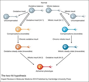
The molecular mechanisms behind the cell death associated with cell cycle re-entry seem to be closely involved with oxidative stress. As both phenomena are manifest early in the pathogenesis of AD, and as mature cases of AD exhibit markers of both (Refs Reference Zhu18, Reference Zhu60), it seems clear that there is a relationship between the two. The ‘two-hit’ hypothesis, in particular, suggests that either of the two cellular detriments (oxidative stress or mitotic dysfunction) initiates a cycle in which the first factor produces a compensatory steady state that enables its proliferation under the stressed conditions. Consequently, the weakened state of a cell makes it vulnerable to the effects of the additional insult (Ref. Reference Zhu60). In other words, one ‘hit’ instigates a cellular adaptation that can last for decades with coherent neuronal functioning, and adaptations of this new steady state produce vulnerabilities in affected neurons that lead to insults of the second ‘hit’. Consequently, the combination produces cellular dysfunction and death (Ref. Reference Zhu18). It is our hypothesis that these two hits are oxidative stress and cell cycle aberration, and the sequence of events can begin with either one, leading to the other. Figure 2 presents a pathway of the mitotic steady state as the primary factor that ultimately leads to neurodegeneration.
The two-hit hypothesis. An initial insult, whether oxidative or mitotic, that is chronic and above threshold limits leads to a new steady state (either oxidative steady state or mitotic steady state). In this new steady state neurons are vulnerable to the subsequent second hit, which causes the Alzheimer disease phenotype. Reproduced from Ref. Reference Zhu60, with permission from Elsevier © 2004.

Notably, in regard to mitotic abnormalities, it seems as though the APP mutant phenotype (of familial AD) essentially produces a ‘mitotic steady state’ that exposes affected cells to future insult through oxidative damage. Indeed, mutations in APP as well as in PS1 or PS2 elicit alterations in cell cycle control and functioning mechanisms. Interestingly, as studies with several transgenic mouse models indicate, neuronal cell cycle re-entry (as a result of mutations in APP, PS1 or PS2) precedes amyloid deposition, and thus full AD pathogenesis, by several months and occurs in an anatomical pattern that reproduces the neuronal vulnerability seen in AD (Refs Reference Zhu18, Reference Yang61). It is thus clear that, although these mice demonstrated significant cell cycle re-entry as early as 6 months of age, they maintained cognition and cellular functioning at a near-normal level for long periods of time (Ref. Reference Zhu18). In other words, those mice exhibited a ‘mitotic steady state’ that eventually made the cells vulnerable to further insult, as the neurons became increasingly susceptible to molecular attack, for example, by reactive oxidative species (ROS).
However, in the much more prevalent, sporadic form of AD (or late-onset AD), an ‘oxidative steady state’ has been demonstrated, which exposes affected neurons to other insults that are due to mitotic abnormalities (Ref. Reference Zhu18). Although neurons are generally capable of sequestering acute levels of oxidative stress in the brain, widespread accumulation of reactive oxidative species eventually requires cellular adaptations that describe the oxidative steady state (Refs Reference Zhu62, Reference Ogawa63). Specifically, affected neurons combat repetitive oxidative insults through permanent instalments of sequestration proteins and antioxidants, such as Aβ. Although these proteins do enable neurons to survive for decades under the repetitive oxidative stress (Ref. Reference Morsch, Simon and Coleman64), their eventual accumulation might subject surrounding neurons to certain vulnerabilities, including mitotic abnormalities. Consequently, once the cells begin altered mitotic functioning in their already weakened states, they inevitably succumb to more severe dysfunction and eventual cell death. Inflammation has also been postulated as a progenitor of cell cycle re-entry in AD. A recent study using APP transgenic mice (R1.40 Tg) demonstrated that alterations in brain microglia are coincident with the first evidence of cell cycle re-entry events at 4–6 months of age, approximately 6 months before Aβ deposition. The microglial activation was found to be dependent on Aβ generation, because microglial activation was limited in mice lacking β-secretase 1 (BACE1) (Ref. Reference Varvel65). Additionally, treatment of young R1.40 animals with nonsteroidal anti-inflammatory drugs (NSAIDs) effectively blocked the activation of microglia in the rat brains, and thus blocked neuronal re-entry into the cell cycle.
Therapeutic potential of targeting cell cycle re-entry in Alzheimer disease
The definitive role of the cell cycle in AD affords many access points for therapeutic intervention (Ref. Reference Woods, Snape and Smith66). Importantly, because the aberrant re-entry into the cell cycle appears so early in the disease progression, such treatment strategies might eventually enable the prevention or reversal of disease.
In conventional models of neurodegeneration, compounds sharing the ability to abrogate cell cycle progression from G0 to G1 have been shown to provide neuroprotection in rodent models of acute ischemic stroke and traumatic brain injury (Refs Reference Park53, Reference Lee57, Reference Varvel65, Reference Woods, Snape and Smith66). However, formal tests of the cell cycle treatment have not yet been undertaken in clinical studies. The results of retrospective and prospective analyses will have a bearing on interpretation of the usefulness of the hypothesis for development of novel therapeutics. The retrospective study reported by Breuer and Anderson (Ref. Reference Breuer and Anderson67) provides one such example. They found that the incidence of AD was one-third lower in elderly female nursing-home residents who were treated with Tamoxifen, a well-tolerated antiproliferative cancer medication that crosses the blood–brain barrier, than in untreated residents. Importantly, the cognitive skills for daily decision-making were also significantly better in residents taking Tamoxifen. An array of other compounds which provide neuroprotective properties through the action of cell cycle inhibition includes retinoic acid, flavopiridol, simvastatin, rosiglitazone, amongst others (Ref. Reference Woods, Snape and Smith66).
A number of prospective studies with possible use as incidental tests of the cell cycle hypothesis are currently ongoing or planned. A Phase II trial of epigallocatechin-gallate (ClinicalTrials.gov identifier: NCT00951834) is due to start in the near future. Epigallocatechin-gallate is a natural product that has been shown to abrogate the cell cycle at the G0–G1 phase in various tissues and is listed as a cell cycle inhibitor. This study is due to complete in the first half of 2011. Additionally, the VALID study (ClinicalTrials.gov identifier: NCT00071721) is one of number of studies investigating the effect of valproate in Alzheimer disease. Although valproate has a number of pharmacological actions, like epigallocatechin-gallate, it can abrogate the cell cycle. The VALID study is reported to be completing in the near future.
Conclusions
Although the precise origins of mitotic dysfunction in AD are not fully understood, genetic, inflammatory and oxidative defects can induce the pathways from which the altered mitotic signaling could arise, and, as is the case in AD, the complex interaction between oxidative stress and cell cycle re-entry and dysfunction seems pertinent, with mitotic abnormalities being of particular importance in familial AD. In any case, however, the cell cycle is a clear progenitor of neuronal dysfunction and as such, provides a new and possibly beneficial therapeutic avenue for disease control. Future investigation on the matter is thus of great importance.
Acknowledgments
Work in the authors' laboratories is partly supported by the National Institutes of Health (AG031364, AG028679 and AG030096) and by the Alzheimer's Association (NIRG-07-60164). M.S. is a paid consultant for or owns equity or stock options in Medivation, Neurotez, Neuropharm, Panacea Pharmaceuticals and Voyager Pharmaceuticals. X.Z. is a paid consultant for and received grant funding from Medivation. We are extremely grateful and thank the peer reviewers for their helpful comments, which greatly improved the impact and focus of our paper.