To address the anatomic and physiologic issues created in the patient in whom an atriopulmonary connection has failed, the strategy of conversion was first reported by Laks et al.8 Our group added the intraoperative ablation of potential circuits for arrhythmias to the therapeutic strategy.9 The primary principles of conversion are to excise the greater portion of the often massively dilated right atrium, and treat the underlying atrial arrhythmias. The cardiac anatomy is then restored, either with an extracardiac conduit or a lateral tunnel cavopulmonary connection.10, 11 The atrial arrhythmias are treated by a right-sided maze for patients with atrial flutter,12 and the third variant of the Cox maze for patients with atrial fibrillation.13 In our series of patients, essentially all have received an epicardial pacemaker for further management of their arrhythmias, be these tachycardic or bradycardic.
These surgical procedures are fairly daunting, with most patients requiring two, and sometimes three or four, median sternotomies. Dense adhesions from the multiple previous operations contribute to the degree of difficulty of the reoperation for conversion. In addition, these patients are typically in low cardiac output, and can have extremely low output when they develop their atrial arrhythmias. Because of the long-standing abnormal physiology of the Fontan circuit, they often have hepatic and renal dysfunction, with poor coagulation factors. The high central venous pressure leads to difficulties with bleeding at the time of sternal reentry. The electrophysiologic management of these patients is also often difficult, and requires an experienced and dedicated paediatric electrophysiology team.
Our purpose is to review the indications for conversion, and to present the results of these procedures at Children's Memorial Hospital. We then compare our results with other published series of conversion, and discuss the results of cardiac transplantation in this population of patients.
Methods
We reviewed the computerized database for cardiac surgical procedures held at Children's Memorial Hospital to identify all patients undergoing conversion of a previously existing Fontan circulation. We also included those patients undergoing orthotopic cardiac transplantation because of a failed Fontan circuit. Our study was approved by the institutional review board. Between 1994 and 2004, 78 patients underwent conversion of their Fontan circulation. Between 1990 and 2004, we performed orthotopic cardiac transplantation in an additional eight patients because of failure of the Fontan circuit, four having failed an attempt at conversion. Of the patients, 21 were classified in the second grade of the system of the New York Heart Association, 43 were in the third grade and 14 were in the worst grade. The majority, 49, had undergone but one previous sternotomy, but 20 had required two, seven needed three, and two patients had received four prior sternotomies. They were on a median of three anti-arrhythmic medications, with three suffering protein-losing enteropathy, and 13 identified with moderate or severe atrioventricular valvar insufficiency. Stenosis of the atriopulmonary connection was seen in nine, and 12 of the patients had initially been referred for cardiac transplantation. Elevated end-diastolic pressures of more than 12 millimetres of mercury were found in 14 patients. We discovered atrial reentry as the cause of supraventricular tachycardia in 43 patients, and atrial fibrillation in 32. The mean age at the onset of supraventricular tachycardia was 14.3 plus or minus 8.0 years. The mean age at the original operation creating the Fontan circuit was 7.1 plus or minus 5.7 years, and the mean age at conversion was 20.9 plus or minus 8.1 years. This resulted in a mean interval from the initial procedure to conversion of 12.6 plus or minus 4.5 years. Surgery to correct atrial reentry tachycardia has evolved over the period from cryoablation of the isthmus, performed in nine, to a right-sided maze procedure carried out in 39. For all 30 patients with atrial fibrillation, we performed the third variant of the Cox maze operation. Having initially installed atrial pacemakers in 43 patients, we have now inserted dual-chamber epicardial pacemakers in 32 patients.
Operative technique
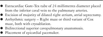
The main steps of the operative procedures are shown in Table 1. All patients were carefully evaluated preoperatively by electrophysiology with, in nearly all cases, preoperative cardiac catheterization with haemodynamic assessment, angiography for anatomic definition, and intracardiac electrophysiologic mapping. Many patients were maintained on intravenous milrinone for 36–72 hours prior to the operative procedure. The operation in all cases was performed with the use of transesophageal echocardiography. In 12 patients, four of these after entry to cavities or vessels, we used femoral cannulation prior to sternal reentry. In two further patients, the femoral vessels were exposed but a femoral approach was not used, or our attempt at femoral cannulation proved unsuccessful due to arterial stenosis. After successfully opening the sternum, we proceed to aortic and direct caval venous cannulation, using right-angle cannulas inserted high in the superior caval vein, and straight cannulas in the inferior caval vein (Fig. 1). In patients without a shunt at atrial level, the right atrial resection is performed with the heart beating. The next step is to transect the inferior caval vein, anastomosing to it a 24-millimetre Gore-Tex stretch tube graft. A vent is then placed in the right superior pulmonary vein. The aorta is cross-clamped and cold blood cardioplegia delivered.
Table 1. Operative steps of conversion.


Figure 1. Technique of cannulation technique for conversion. Note direct caval venous cannulation. Projected right atrial resection is shown (dotted lines).
With the heart arrested, we create an atrial septectomy and perform the right-sided maze (Fig. 2), excising the remnants of the right atrial appendage. Linear cryoablation lesions are placed with a Surgifrost® device (CryoCath Technologies, Inc., Kirkland, Quebec, Canada), using temperatures of −160 degree centigrade for 1 minute. The lesions extend from the base of the right atrial appendage to the cut edge of the atrial septal defect, across the terminal crest from the cut edge of the atrium to the atrial septal defect, and from the cut inferior caval vein to the coronary sinus. In addition, if a tricuspid valvar orifice is present, a lesion is created from the inferior caval vein to the annulus of the tricuspid valve. More recently a lesion is also placed from the base of the coronary sinus to the atrial septal defect (Fig. 3).

Figure 2. The modified right atrial maze procedure. Sites of cryoablative lesions are indicated (dotted lines). ASD: atrial septal defect; CS: coronary sinus; IVC: inferior caval vein; RAA: right atrial appendage; SVC: superior caval vein; TV: tricuspid valve.

Figure 3. Lesion from coronary sinus to edge of atrial septal defect with Surgifrost device (−160 degrees centigrade for 1 minute).
In patients with atrial fibrillation, we perform the third variant of the Cox maze procedure (Fig. 4). The left atrial appendage is excised. In most patients, a partial encircling atriotomy is performed, with scissors dividing the atrium adjacent to the right upper and lower pulmonary veins. This atrial incision generally encompasses one-half of the left atrial circumference. Cryoablation lesions are then placed to encircle the left upper and left lower pulmonary veins, connecting with the surgical incisions (Fig. 5). Another lesion is placed along the base of the left atrial appendage, from the cut portion of the excised left atrial appendage to the lesion placed around the left pulmonary veins. Another lesion is placed from the encircling atriotomy lesion to the annulus of the mitral valve, directing it at the infero-medial scallop. A lesion, created for 2 minutes, is placed on the epicardial surface of the coronary sinus directly opposite that involving the mitral valve. In some patients with dense adhesions in the region of the left atrial appendage, we did not excise the appendage, instead creating a circular lesion around its base.

Figure 4. The third variant of the Cox maze. Illustration of opened atrium. Note the superior and inferior caval veins have been transected. Right pulmonary veins have been “isolated” by an incision. Left pulmonary veins have been isolated by cryoablation (see Fig. 5). There are lesions from the base of the excised left atrium to the pulmonary vein lesion and from the mitral annulus to the pulmonary venous lesion. The epicardial lesion on the coronary sinus (shown) is created over 2 minutes.

Figure 5. Cryoablation lesion isolating the left pulmonary veins. This single long lesion completes the pulmonary venous isolation.
At the conclusion of the third variant of the Cox maze procedure, the right atrium is closed with running polypropylene, the heart de-aired, and the cross clamp removed. At this point, the bidirectional Glenn anastomosis is performed as the heart is being rewarmed, and the Gore-Tex tube is anastomosed to the undersurface of the pulmonary artery, frequently at the site of the prior atriopulmonary connection. The patient is weaned from cardiopulmonary bypass on low-dose dopamine and low-dose dobutamine, at 3 micrograms per kilogram per minute, and intravenous milrinone at 0.3–0.5 micrograms per kilogram per minute. Modified ultrafiltration is performed. We then place bipolar atrial and ventricular epicardial pacing leads (Fig. 6). These sites are sometimes difficult to obtain because of the scarring from previous operations. An excellent site for the atrial lead is at the dome of the left atrium beneath the ascending aorta. A frequently used site for the ventricular pacemaker lead is on the diaphragmatic surface of the heart, where there are often fewer adhesions than elsewhere. We currently attach these leads to the AT 500™ (Medtronic, Inc., Minneapolis, MN) pacemaker, which has dual-chamber pacing and atrial anti-tachycardia capabilities should the patient have recurrence of their atrial tachycardia.

Figure 6. Placement of pacemaker leads. Bipolar leads are necessary for the pacemaker used. Note bidirectional superior cavopulmonary anastomosis and 24-millimetre Gore-Tex graft from inferior caval vein to pulmonary artery.
The mean duration of the operation, from skin incision to closure, is 10 plus or minus 2 hours, with a median of 10 hours, and a range from 6.3 to 16.3 hours. All patients are taken to the intensive care unit ventilated, and typically are not extubated for at least 36 to 48 hours. Some patients require prolonged ventilation for up to 3 to 5 days because of the lengthy nature of the procedure. All patients receive intravenous amiodarone in the perioperative period, and oral amiodarone for 3 months postoperatively. The patients who have had a right maze undergo postoperative electrophysiologic evaluation. The patients undergoing the Cox procedure are not studied postoperatively.
Results
One patient died 2 months subsequent to conversion. This patient was 15-years old, and died as a result of hepatic and renal failure, and persistent low cardiac output. Mean stay in hospital for the overall group was 14.9 plus or minus 14.5 days, and chest tubes were required for 9.4 plus or minus 5.5 days. We undertook reoperation in one patient for bleeding. Another patient developed recurrent atrial fibrillation and low cardiac output, and had placement of a ventricular assist device followed by successful cardiac transplantation. Significant sternal infections developed in two patients, with one treated with vacuum-assisted closure, and the other with muscle flaps. Acute renal failure developed in nine patients, five requiring dialysis. The others, who had transient rises in enzymes, recovered without dialysis. Of the five patients who needed dialysis, one patient had a renal transplant. The other four patients who did not recover included the single patient who died in hospital, and three others who died at 5, 7, and 11 months subsequent to conversion. Another patient died from coronary arterial thrombosis 26 months postoperatively.
With regard to control of arrhythmias, in the initial group of nine patients who had only block at the isthmus block, four of eight had recurrence of their atrial reentry tachycardia, and one patient had recurrence of atrial fibrillation. Of the 39 patients who underwent a right-sided maze procedure for atrial reentry tachycardia only, three suffered recurrence of the arrhythmia. In that same group, 3 of 39 do not have long-term follow-up because they had early orthotopic cardiac transplantation. Of the patients who had a Cox procedure, one received orthotopic cardiac transplantation, and 6 of 30 had recurring atrial reentry tachycardia, but no atrial fibrillation, easily treated with a single anti-arrhythmic medication.
In our analysis of the outcomes,14 univariate regression analysis of risk factors for death, transplant, or dialysis, showed significant risk factors to be severe ventricular dysfunction, ischaemic time greater than 100 minutes, age greater than 25 years, atrioventricular valvar regurgitation more than mild, cardiopulmonary bypass time greater than 240 minutes, dominant ventricle of right or indeterminate morphology, and at least mild ventricular dysfunction. Multivariate logistic regression analysis showed significant risk factors for death, transplant, or dialysis to be severe ventricular dysfunction, age greater than 25 years, a dominant ventricle of right or indeterminate morphology, and ischaemic times greater than 100 minutes. Following conversion, the great majority of patients had a dramatic improvement in their classification within the system of the New York Heart Association (Fig. 7).

Figure 7. Improvement in the classification of the New York Heart Association from preoperative to postoperative assessment. (From: Mavroudis C, Deal BJ, Backer CL. The beneficial effects of total cavopulmonary conversion and arrhythmia surgery for the failed Fontan. Semin Thorac Cardiovasc Surg Pediatr Card Surg Annu 2002; 5: 12–24. Reproduced with permission.)
Cardiac transplantation
The mean age of the eight patients undergoing transplantation was 18.6 plus or minus 12.4 years. Of note, three of these patients had failed a prior conversion at 7 days, 11 months, and 33 months prior to transplantation. Of the eight, one patient was on a ventricular assist device, three were on ventilators, one was on inotropes, and three had protein-losing enteropathy. We lost one of the patients immediately after transplantation, with two further patients dying late, one from renal failure after 1 year, and one from non-compliance 5 years after the transplant. Of interest, only one of these patients had rejection greater than grade 1A, and the rate of survival at 5 years is 50%.
Discussion
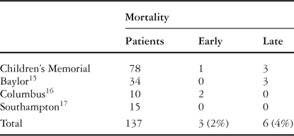
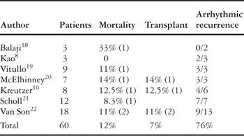
In our series of 78 patients with a failed atriopulmonary connection and arrhythmias, the conversion was accomplished with low operative and late mortality, and a distinct improvement in exercise tolerance and quality of life. We have used the term “conversion” rather than “revision” of the Fontan circuit as we believe the strategy to be more than a revision. The use of “conversion” includes not only the anatomic rerouting, but also the arrhythmic surgery and placement of pacemakers. The results of conversion have also been good at several other institutions (Table 2). Notably, Baylor University,15 Columbus Children's Hospital,16 and Southampton17 have reported excellent results. The benefit of adding arrhythmic surgery to the conversion of the Fontan circuit is now quite evident. Reported series without arrhythmic surgery, such as the partial experience reported from Southampton, show a high incidence of recurrence of arrhythmias, with its debilitating results.8, 10, 18–22 In a compilation of seven series, recurrence has been observed in three-quarters of 60 patients (Table 3).
Table 2. Results of conversion.

Table 3. Results of conversion without arrhythmic surgery.

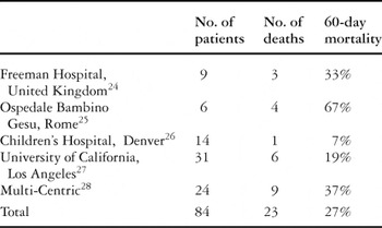
In comparison to the strategy of conversion, orthotopic transplantation for the patient with a failed Fontan circuit has some additional hazards that need to be taken into consideration. These include death while awaiting a donor heart, perioperative mortality, the complications of immunosuppression, and finally, compliance issues in the population of adolescents and young adults. In the Pediatric Heart Transplant Study Group, of patients who underwent transplantation for a failed Fontan circuit between 1993 and 2001, 14% of patients on the waiting list had died at 6 months, and 16% had died by 12 months following listing for cardiac transplantation.23 In a compilation of five separate studies, the 60-day mortality rate for transplantation for the failed Fontan circuit is 27% (Table 4). At the Freeman Hospital, in Newcastle, United Kingdom, the 24-hour mortality was 33%.24 At the Bambino Gesu Hospital, Rome, the operative mortality was 33% for patients having the Fontan circulation.25 In the Denver Children's Hospital series, three patients either died late or required re-transplantation, with two of these three patients having problems with non-compliance.26 In the series reported from Los Angeles, the 30-day mortality was 19%.27 Finally, a recently published multi-centric study revealed 37% mortality at 60 days.28 Of course, the known complications of immunosuppression, which would not be a prob-lem after conversion, include infection, rejection, transplant coronary arterial disease, side effects of steroids, and again, the issue with regard to non-compliance in this population of adolescents and young adults.29
Table 4. Results of cardiac transplantation for failing Fontan circuit.

Conclusions
Cardiac transplantation for a patient with a failed Fontan circuit has a 6-month waiting list mortality of 14%, an operative mortality of 27%, a life-time risk of immunosuppression, and the significant potential for non-compliance.
Conversion to an extracardiac cavopulmonary connection with concomitant arrhythmic surgery and placement of a pacemaker, in contrast, not only in our hands but also in the hands of others, has been demonstrated to be excellent therapy for selected patients in whom an atriopulmonary Fontan circuit has failed. Conversion must include surgical ablation of the arrhythmic circuit or circuits. This approach is associated with low operative mortality, improvement in exercise tolerance, improvement in classification in the system of the New York Heart Association, and has a low incidence of recurrent arrhythmias. Conversion does not exclude a patient from future orthotopic cardiac transplantation, and we believe it should be considered the primary therapy of choice in most patients with a failed atriopulmonary Fontan circuit.