Introduction
The use of biomaterials to design cellular mesh scaffolds with the ability to mimic the tissue properties have been investigated in the last years. In this matter, the scaffolds must be capable to restore and repair the damaged tissue, it being achieved when the scaffold establishes a regenerative microenvironment in the damaged area, creating the favorable conditions for adhesion, proliferation, and differentiation of cells.[Reference Stratton, Shelke, Hoshino, Rudraiah and Kumbar1–Reference Jaiswal and Jaiswal3] Therefore, it is of great relevance to find new methods capable of producing nanofibers (NFs) with the objective to promote successful culture and cell growth. Forcespinning is a technique which uses centrifugal forces to produce NFs from polymeric solutions and its configuration can produce large quantities of NFs. Electrospinning is the most used technique to produce NFs,[Reference Chang, Wang and Chen4, Reference Sarkar, Gómez, Zambrano, Ramírez, de Hoyos, Vásquez and Lozano5] but the Forcespinning technique allows the production of NFs from non-conductive polymeric solutions including biopolymer as Poly-Lactic Acid,[Reference Hong, Gao, Xie, Gong and Shen6] Polyvinyl Alcohol[Reference Rwei and Huang7] and Polycaprolactone (PCL)[Reference Yu, Jia, Yao and Lu8]; which are the ideal materials (accepted by the FDA) for cellular and muscle tissue regeneration.[Reference Caramella, Conti, Modena, Ferrari, Bonferoni, Genta, Rossi, Torre, Sandri, Sorrenti, Catenacci, Dorati and Tripodo9–Reference Zachary McEachin13]
Specifically, PCL is a polymer with a good biocompatibility, slow degradability and good thermal stability that are desirable attributes for tissue engineering applications.[Reference Siqueira, de Moura, de Barros Machado, Backes, Roberto Passador and de Sousa Trichês14, Reference Tan, Pingguan-Murphy, Ahmad and Akbar15] The use of TiO2 nanoparticles (NPs) as a dopant in a PCL polymeric matrix is of relevance due to the fact that TiO2 NPs possess an inert property and its behavior as an inhibitor of the microorganisms proliferation is ideal for biologic applications.[Reference Tan, Pingguan-Murphy, Ahmad and Akbar15, Reference Haider, Al-Anbari, Kadhim and Salame16] On the other hand, the use of a material capable of removing devitalized dead tissue (caused by burns, ulcers, scars or transplants) in a short period of time and capable of growth and cell culture is relevant for tissue engineering. The collagenase meets this requirement perfectly, it being a proteinase that hydrolyzes collagen, setting an optimal environment for cell growth.[Reference Fierro-Arias, Campos-Cornejo, Contreras-Ruiz, Espinosa-Maceda, López-Gehrke, Márquez-Cárdenas, Ramírez-Padilla, Veras-Castillo and Rodríguez-Alcocer17–Reference Melmed, Polonsky, Reed Lorsen and Kronenberg19]
The following types of collagenase can be found in mammals: MMP-1, MMP-8, and MMP-13. The MMP-1 collagenase type I degrades the collagen fibrils in two types (I and III), while the bacterial collagenase degrades the collagen in five types (I, II, III, IV, and V).[Reference Melmed, Polonsky, Reed Lorsen and Kronenberg19] Therefore, it is important to find the optimal conditions to balance the properties of a biocompatible PCL polymeric matrix, TiO2 and collagenase for the designing of NFs mesh scaffolds suitable to promote cell regeneration.
This research focuses on the study of the physical and chemical properties of PCL-TiO2-collagenase NFs mesh scaffolds produced by Forcespinning. The mixture was prepared by incorporating collagenase and TiO2 NPs in low concentrations to the PCL matrix solution. The morphology of the forcespun NFs was investigated by SEM and it was found that the fiber diameter size distributions ranged between 100 and 300 nm. Furthermore, collagenase agglomerations were observed due to its low dispersion in the polymeric solution. The degree of crystallinity and structural information of the mesh scaffolds were investigated by differential scanning calorimetry (DSC), showing differences in the crystalline domains induced by the dopants in the PCL polymeric matrix. The dependence on the crystal domains was studied by XRD, detecting a shift to lower angles associated to compressive stresses. Functional groups observed in the mesh scaffolds were studied by Fourier transform infrared (FTIR) in which the main contribution is given by PCL. Also, Raman spectroscopy was used to detect the presence of TiO2 NPs in the developed mesh scaffold. Contact angle (CA) and tensile tests were performed in order to determine the permeability and the mechanical strength of the developed scaffolds. Collected experimental data showed an improvement in both the hydrophobicity and mechanical strength of the PCL-TiO2-collagenase mesh scaffold. It is believed that the results obtained in this study could be of great relevance for tissue engineering applications.
Materials
The materials used in this study are Titanium (IV) butoxide (grade 97%), nitric acid (grade 95%), acetic acid ACS (grade 99.7%), chloroform (anhydrous 90%), DMF N, N-Dimethylformamide (anhydrous 99.8%), PCL (Mw 80,000), Titanium (IV) oxide Nanopowder (21 nm primary) P25 Degussa, all purchased from Sigma Aldrich, México; and the collagenase (type I lyophilized), Trypsin-EDTA that were purchased from Gibco, México. All these materials were used without any further purification.
Methods
TiO2 synthesis by Sol-gel method
The synthesis was carried out by mixing deionized water, nitric acid, and titanium (IV) butoxide in a 3-mouth flask at 25 °C under magnetic agitation. Subsequently, the solution was heated up to 70–90 °C for 8–12 h and assisted with a water cooler reflux. The gel obtained was dried at 80 °C for 10 h and thermally annealed at 400 °C for 4 h to remove organic material, solvents, and humidity. The synthesis procedure followed in this research paper is similar to the one described in Ref. Reference Del Ángel-Sánchez, Vázquez-Cuchillo, Salazar-Villanueva, Sánchez-Ramírez, Cruz-López and Aguilar-Elguezabal20.
NFs production
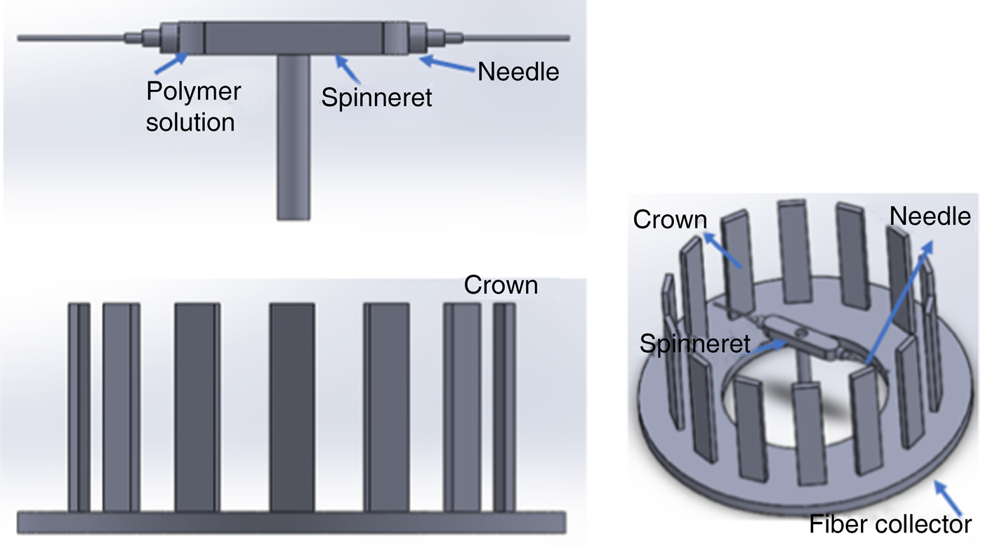
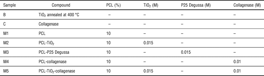
The solutions used for the NFs production consisted in PCL diluted with chloroform and DMF in a ratio of 80:20 (v/v) to obtain the 10% polymeric matrix solution. TiO2, P25 Degussa, and collagenase were added in different concentrations into the polymeric matrix solution to obtain the doped samples. The solutions used for the NFs production are listed in Table I. The NFs were produced by using a Forcespinning machine with a 30-gauge needle operating at the angular velocity of 6000 rpm. The NFs were collected using a 16-stick metal crown (located at 30 cm from the needle) and removed with a metallic frame (1 in × 1 in) to obtain the mesh scaffold structure. A schematic diagram of the Forcespinning setup is shown in Fig. 1.
Schematic diagram of the centrifugal force machine set-up.

Description of the solutions used to produce nanofibers by centrifugal force.

Differential scanning calorimetry
The degree of crystallinity and melting temperature of PCL were measured by using a DSC Perkin-Elmer Pyris 8000 equipment. The melting temperature (T m) was determined at the maximum of the melting endotherm. The degree of crystallinity (χ c) was estimated using the enthalpy of melting change according to the equation:  $\chi _{\rm c} = (\Delta H_{\rm m})/(1-w)\Delta H_{\rm m}^0 $ where H m and ΔH m0 are the melting and the fully crystalline melting (139.5 J/g) enthalpies of the PCL, respectively, and w is the dopants weight fraction. All of the samples (average weight 10 mg) were held in standard aluminum pans and covers. The specimens were scanned from 50 °C to 70 °C with a heating increase rate of 10 °C/min, in a nitrogen gas atmosphere. The DSC analysis curves for the material samples was carried out for the materials as-received (first run data).
x-ray diffraction (XRD)
The XRD measurements of developed NFs mesh scaffolds and bare materials were carried out using a PanAnalytical X'Pert Pro PW1800 diffractometer with a scanning rate of 2°/min and by using Cu Kα radiation. The system was operated at 45 mA and 40 kV and the XRD data was collected in the 2θ range of 10°–85°.
Fourier transform infrared
A FTIR (Perkin-Elmer Frontier) equipment with an UATR accessory was used to carry out the IR analysis. The procedure consisted in placing the developed mesh scaffolds on the ZnSe-diamond crystal of the UATR and pressed with a tip to assure a good contact between the sample and the incident IR beam. The IR spectra were measured in the interval range of 4000 to 380 cm−1 with a resolution of 4 cm−1, and by considering an average of 16 scans.
Raman analysis
The Raman spectra were carried out using a JY-Xplora plus microscope at room temperature using a Pumped Solid-State Diode (532 nm) as an excitation line. The measurements were performed with a microscope objective of 20x, setting the diameter of the laser beam around of 1 µm and the laser power was fixed to 1.4 mW in order to avoid damage in the developed mesh scaffolds. The spectra were measured in a range from 100 to 3100 cm−1 with an integration time of 30 s, 3 accumulations and 1800 lines/mm grid diffraction.
Contact angle
CA measurements were performed using a Data physics (OCA, 15EC) system. The CA was measured between the mesh scaffold surface and a water droplet using SCA20_U software. The procedure consisted in placing a water droplet, from a dispenser with a volume capacity of 10 µL and deposition ratio of 2 µL/s, onto the mesh scaffold surface. The CA value retrieved from the calculation corresponds to the average considering the value calculated from the right and left side of the droplet.
Mechanical test measurements
Tensile tests were made using a universal testing machine (INSTRON 3365) and in accordance with the ASTM standard F2150. The measurements were performed at room temperature and by using samples with an area of 1 in × 1 in.
Scanning electron microscopy (SEM) and transmission electron microscopy (TEM)
A SEM (ZEISS model EVO MA 25) was used with an accelerating voltage of 10.00 kV and a work distance of 7.0 mm to observe the morphology and state of aggregation of the samples in the form of powders and fibers of the synthesized compounds. A TEM (JEOL 2100) was used to observe the internal morphology to determine the diameter of the synthetized TiO2 NPs.
Size distribution
The size distribution of the TiO2 NPs was obtained by a Zetasizer (Nano Z, Malvern MPT-2 Multi-Purpose) equipment. The measurements were performed using a mixture of ethylene glycol and the TiO2 NPs to enhance their dispersion.
Cell culture
Stem cells were obtained from abdominal adipose tissue harvested from healthy donors ages 30–40 years old, following the procedure described in Ref. Reference Van der Bogt, Sheikh, Schrepfer, Hoyt, Cao, Ransohotf, Swijnenburg, Pearl, Lee, Fischbein, Contact, Robbins and Wu21. Mononuclear cells were seeded directly into 25 cm2 flasks containing DMEM-F12 supplemented with 10% FBS, 50 U/mL penicillin, and 50 µg/mL streptomycin. When the culture reached confluence, cells were detached with 0.25% Trypsin-EDTA and centrifuged at 37 °C for 10 min. The obtained cell pellet was suspended in a medium maintenance and then quantified on 24-well microplates containing samples M2 and M4. The cultures were followed with an inverted microscope.
Results and discussion
Morphology of forcespun PCL NFs
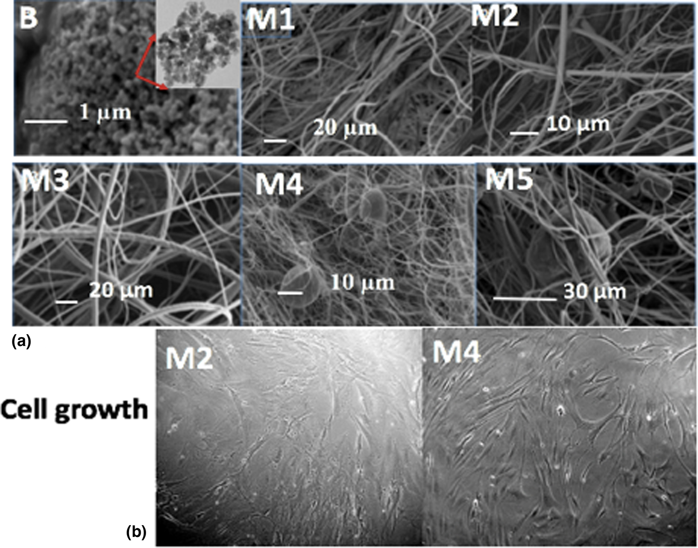
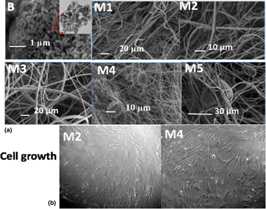
The morphology of the TiO2 NPs and the PCL composites mesh scaffolds are shown in Fig. 2 and their corresponding size distributions are shown in Fig. S1 (Supplementary Information). The image for the synthesized TiO2 NPs shows agglomerations with ranged sizes between 150 and 300 nm and in the inset, there is a TEM image of isolated NPs with average sizes of less than 10 nm. Here the SEM images for scaffolds M1, M2, and M3 show beadles NFs with average diameters between 200 and 500 nm.
(a) SEM images of sample B and developed mesh scaffolds M1–M5. A TEM image of B was also shown in the inset. (b) Optical micrographs of mesenchymal cells after 14 days, for scaffolds M2 and M4.

The collagenase was carefully diluted in the solutions used for mesh scaffolds in M4 and M5, however, it was difficult to obtain a good particle dispersion producing NFs with agglomerations of PCL-collagenase with diameters of around 30 µm, as shown in Fig. 2(a). Notice from Fig. 2(a) that in some regions of the developed mesh scaffold there are collagenase agglomerations which indicates that not all collagenase is embedded in the polymeric NFs. The use of collagenase in mesh scaffold has been previously reported by Phipps et al. in Ref. Reference Phipps, Clem, Grunda, Clines and Bellis22, in which a collagenase solution was used to increase the porosity of the scaffold structures to favor the cell culture. However, in this research paper, the collagenase was incorporated into the NFs solution in order to obtain a homogeneous mixture capable of promoting the desired conditions for the cell culture, avoiding a post-treatment.
Cell growth on NFs scaffolds
The images of the cells, taken by an inverted microscope, after 14 days of culture for mesh scaffolds M2 and M4 are shown in Fig. 2(b). It is possible to note that the cells uniformly cover surfaces of the mesh scaffolds with a tendency to grow in groups. Additionally, the Mesenchymal cells on the mesh scaffolds were well spread and elongated with their characteristic morphology. Such cell distribution is attributed to the collagenase and PCL-collagenase agglomerations which creates the necessary morphology to attach the cells and promote their proliferation.
DSC and XRD
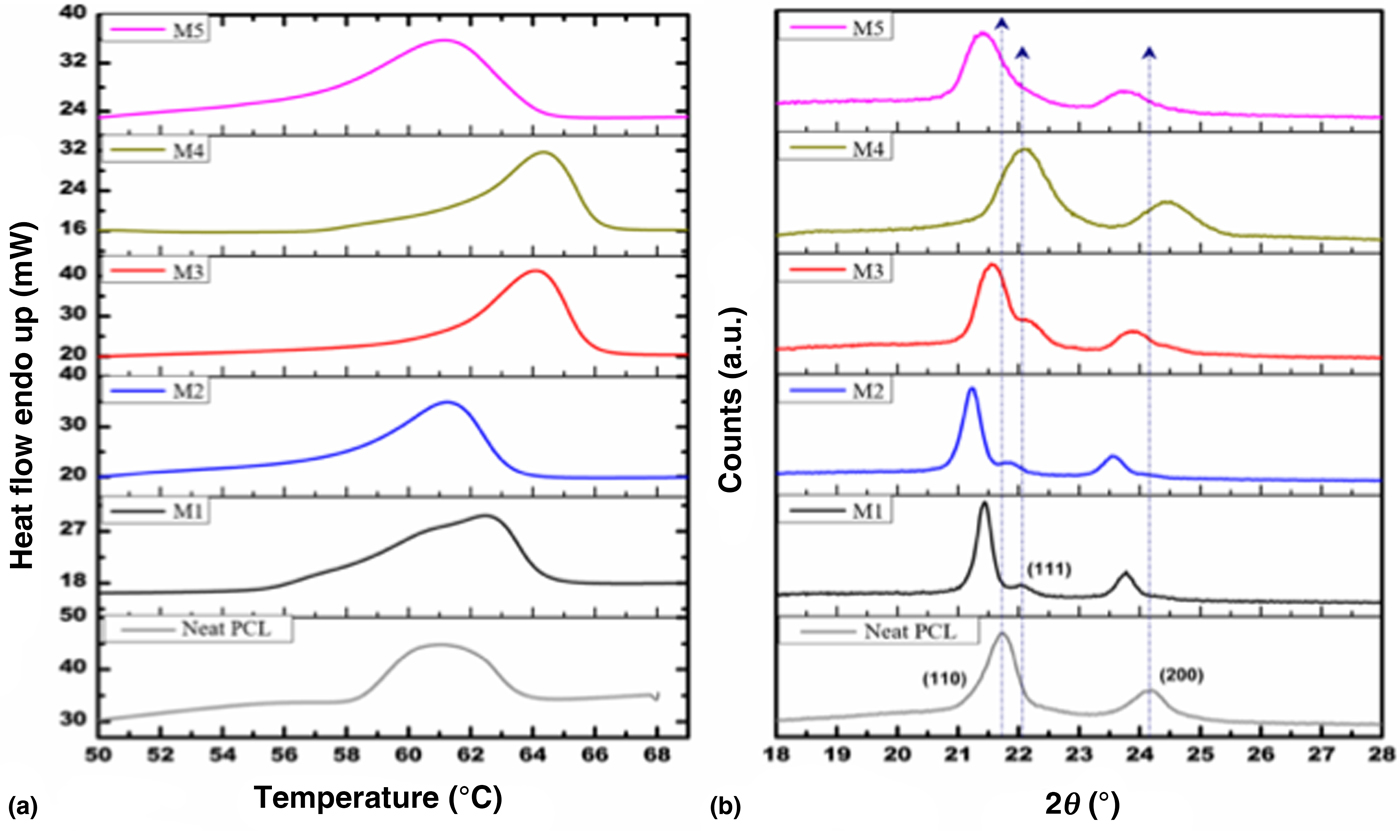
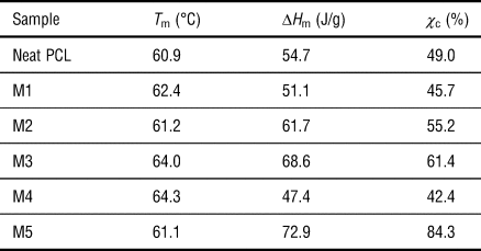
The degree of crystallinity and structural information of obtained mesh scaffolds were investigated by DSC and XRD, the results are shown in Fig. 3. The values of T m, ΔH m, and χ c of neat PCL and the obtained mesh scaffolds (M1–M5) were estimated by DSC and are listed in Table II. The thermogram of neat PCL (dotted line) shows a melting temperature of 60.9 °C, and the melting line shape exhibits a broader peak between 58 °C and 64 °C indicating the presence of different crystal domains. The thermogram for PCL mesh scaffold M1 (black line) shows three main contributions for the melting peak which indicates that the crystal domains are modified when the NF is produced. In this case, the calculated melting temperature is about 62.4 °C. Such changes could be attributed to the fact that the PCL polymeric chains are confined by the reduction of dimensionality (1D), during the NF formation, favoring and increasing the size of the crystals domains. On the other hand, the results for M2 and M3 (blue and red lines) show ΔH m values of 61.2 °C and 64 °C, respectively. As illustrated in Fig. 3(a), there is an improvement in the broadening of the melting line shape due to the incorporation of TiO2 which also favors the crystal size formation. The difference in melting temperature values is associated with the purity of TiO2 NPs in each scaffold. Furthermore, the presence of traces in M3 (P25 Degussa) promotes the crystallization of a larger size domain in comparison with M2 (synthesized TiO2). The T m calculated for M4 (dark yellow line) and M5 (magenta line) are 64.3 °C and 61.1 °C, respectively [Fig. 3(a)].
(a) Thermograms of the scaffolds samples M1–M5 (b) XRD diffractograms of the scaffold samples M1–M5.

Melting temperature (T m), enthalpy for melting (ΔH m) and degree of crystallinity (χ c) values retrieved from DSC.

The diffractogram for neat PCL (dotted line) shows two peaks at 21.7° and 24.1° corresponding to (110) and (200) planes, as it is shown in Fig. 3(b). These peaks are characteristic of the PCL orthorhombic unit cell.[Reference Dziadek, Menaszek, Zagrajczuk, Pawlik and Cholewa-Kowalska23, Reference Zhang and Qiu24] The diffractogram for M1 (black line) shows a decrease in intensity of the diffracted peaks and a shift to lower angles (21.4° and 23.7°, respectively), that suggests a compressive stress during the NFs formation. The results for M2 and M3 (blue and red lines) show additionally a peak in 21.8° which corresponds to the (111) plane, this indicates that the crystallinity is modified by the incorporation of TiO2. The diffractogram for M4 (dark yellow line) shows a broader peak centered in 22° indicating the contribution of several crystal domains. Such response is associated with collagenase agglomeration. Also, the diffractogram for M5 (magenta line) exhibits the contributions of different crystal domains present in the PCL-TiO2-collagenase composite material.
FTIR and Raman analysis
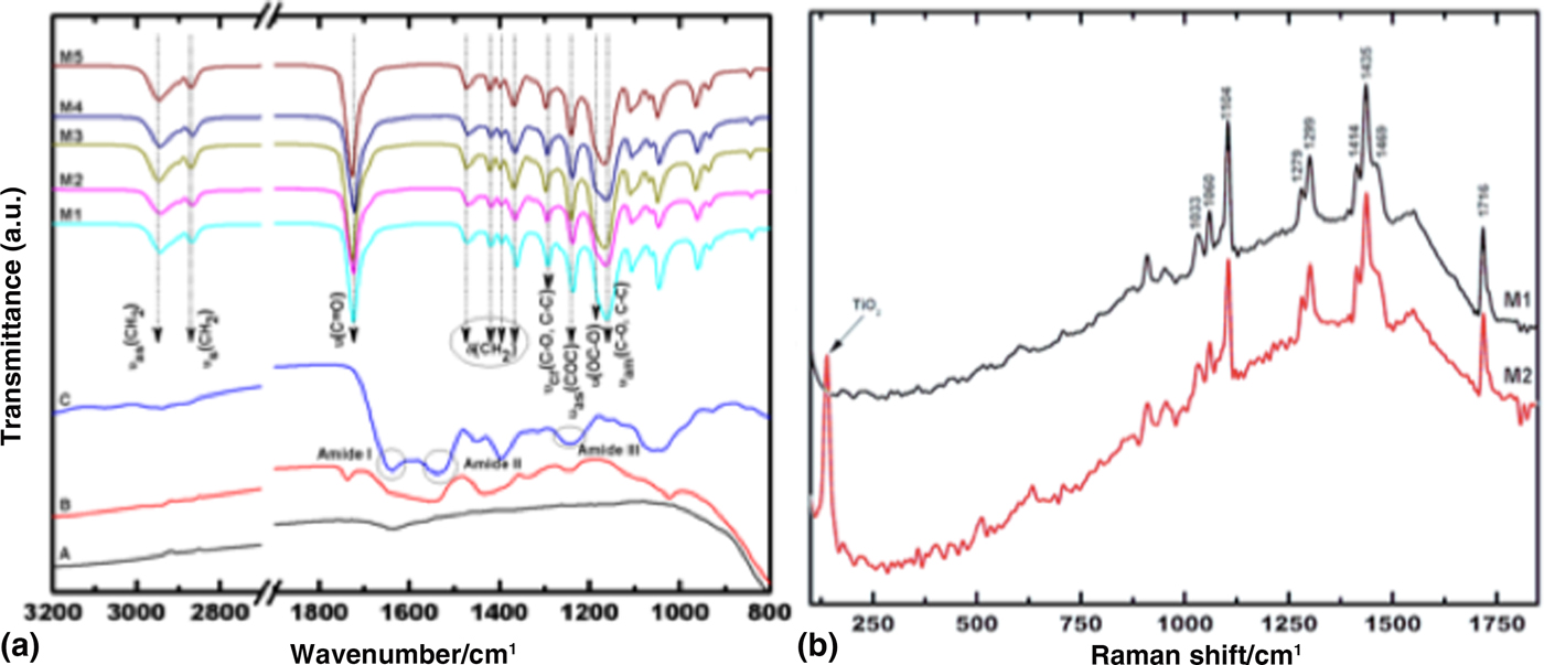
The spectra for all the mesh scaffolds show the same vibrational modes due to the low concentration of dopants within the NFs, exhibiting the response from the PCL [Fig. 4(a)]. The modes detected at 2949 and 2865 cm−1 correspond to an assymetric ( $ {\nu_{\rm as}} $) and symmetric (
 $ {\nu_{\rm s}} $) stretching modes originated by CH = O groups. In the region around 1473 to 1360 cm−1 the presence of CH2 and CH3 groups are detected and associated with bending (δ) modes. On the other hand, a strong band around 1726 cm−1 is detected and related to a C = O stretching (
 $ {\nu_{\rm s}} $) mode. The crystalline (
 $ {\nu_{\rm cr}} $) and amorphous (
 $ {\nu_{\rm am}} $) phases are assigned in 1294 and 1157 cm−1, respectively for C–O and C–C modes. The peaks detected at 240 and 1170 cm−1 correspond to an asymmetric (
 $ {\nu_{\rm as}} $) and symmetric (
 $ {\nu_{\rm s}} $) modes originated by COC groups.[Reference Del Ángel-Sánchez, Vázquez-Cuchillo, Salazar-Villanueva, Sánchez-Ramírez, Cruz-López and Aguilar-Elguezabal20] The spectrum for collagenase shows a band from 1600 to 1700 cm−1 and is associated with a C = O mode for amide I, the vibrational mode for N–H group is observed in a range from 1500 to 1550 cm−1 for amide II, and the band from 1200 cm−1 to 1300 cm−1 for amide III.[Reference Ghosal, Thomas, Kalarikkal and Gnanamanis25–Reference Abdelrazek, Hezma, El-khodary and Elzayat27] Since it was not possible to resolve the presence of TiO2 by FTIR, Raman spectroscopy was used to detect such NPs in the mesh scaffold M2 [Fig. 4(b)]. The Raman spectrum for M1 shows stretching modes
 $ {\nu_{\rm C=O}} $ at 1716 cm−1,
 $ {\nu_{\rm COC}} $ at 1104 cm−1, the mode
 $\delta _{{\rm C}{\rm H}_{\rm 2}}$ at 1435 cm−1 and the modes
 $\omega _{{\rm C}{\rm H}_{\rm 2}}$ at 1279 and 1299 cm−1. Such intense and narrow peaks indicate the presence of crystalline domains. The corresponding spectrum for the PCL-TiO2 system (M2) shows additionally a narrow mode around 140 cm−1indicating the presence of TiO2 NPs in the mesh scaffolds [Fig. 4(b)].
(a) FTIR spectra of the samples A, B, C and for the mesh scaffolds M1–M5. (b) Raman spectra of the scaffold samples M1 and M2.

Water CA and mechanical test
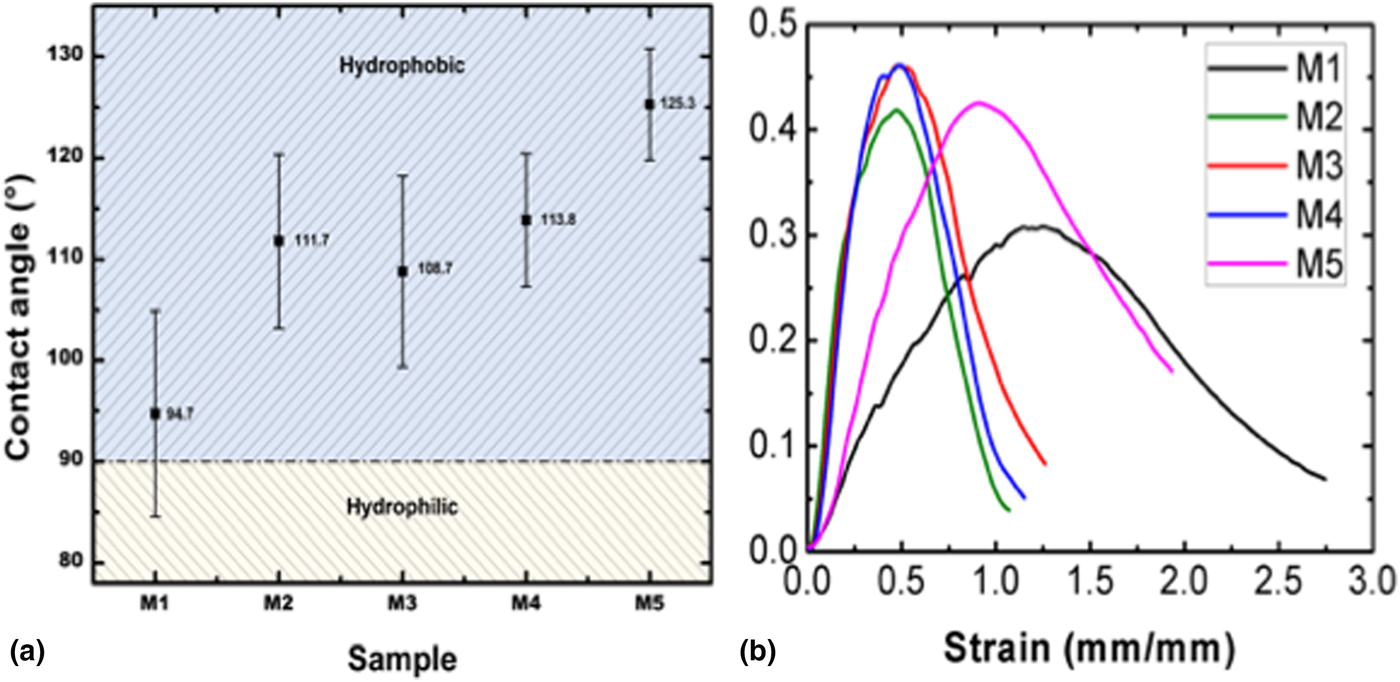
Water CA experimental measurements were performed in the scaffold samples to investigate their permeability [Fig. 5(a)]. The presence of CH2 groups in the backbone of PCL chains is the main reason for its hydrophobic property.[Reference Zhang and Qiu24] Such property can be modified by incorporating doping materials in the solutions for the NFs production shown in Fig. 5(a). The value obtained for the scaffold sample M1 is higher than the value reported in the Ref. Reference Dziadek, Menaszek, Zagrajczuk, Pawlik and Cholewa-Kowalska23 which is about 90°.
(a) Contact angle values of the developed mesh scaffolds M1–M5. (b) Experimental stress-strain curves of the scaffold samples M2–M5.

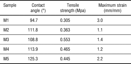
The incorporation of the dopants increases the hydrophobic performance in the scaffolds favoring cell growth. The chemical reactivity of the amphiphilic polymeric network creates a new network capable to bond covalently the hydrophobic and hydrophilic phases, obtaining a system with both permeability behavior (considering that the PCL segments form a hydrophobic core). During the tensile strength test, the mesh scaffolds (randomly oriented) were significantly deformed before they reached their yield point, the average values calculated are listed in Table III. The presence of the dopants in the PCL polymeric matrix increases its tensile strength performance and are similar to the results reported by Ghosal et al. in Ref. Reference Ghosal, Thomas, Kalarikkal and Gnanamanis25 for the PCL-collagen composite material, as it is shown in Fig. 5(b).
Contact angle and mechanical tests results.

Conclusions
This research paper describes the design, fabrication, and characterization of mesh scaffolds of PCL-TiO2-collagenase by using Forcespinning technique. The morphology and the diameters of the NFs were measured by SEM and it was found that the average diameter distribution was in the range of 100–300 nm. Also, for the scaffolds M4 and M5 it was observed some collagenase agglomerations in the NFs. The dependence of the crystallinity and structural formation were investigated by DSC and XRD, and it was found that a compressive stress component affects the crystalline and amorphous domains in the PCL polymeric matrix. The main functional groups in the system were detected by FTIR, while the presence of TiO2 NPs was observed by Raman spectroscopy. An improvement in the hydrophobicity property for the PCL-TiO2-collagenase scaffold was found during CA measurements. Finally, the mechanical resistance in the scaffolds (randomly oriented) was determined by uniaxial tensile tests and experimental data provided evidence that the incorporation of dopants in the polymeric matrix enhances the scaffolds mechanical performance. In conclusion, the results reported in this study meet the properties needed to build a scaffold able to promote cell culture for their application in tissue engineering.
Supplementary material
The supplementary material for this article can be found at https://doi.org/10.1557/mrc.2019.13
Acknowledgment
This work was supported by Consejo Nacional de Ciencia y Tecnología through the CONACyT-SENER Postdoctoral Grant CVU No. 256740 and 231013 (Grant No. 291205). National Lab in Additive Manufacturing, 3D Digitizing and Computed Tomography (MADiT) LN280867. Additionally, the authors want to thank the assistance of Engineer Cesar I. Borbolla Torres and M.Sc. Regina Lizbeth Vargas Mejia in the developing of this work.
Conflict of interest
None