Case report
A 43-year-old Caucasian man was admitted to our emergency department with recurrent symptoms of typical chest pain experienced after exercise. The physical examination was unremarkable. Electrocardiography was normal. Echocardiography showed normal biventricular function with competent atrioventricular and arterial valves. A coronary CT examination was performed with prospective ECG gating in a diastolic phase. CT findings reported anomalous origin of the left coronary artery from the opposite sinus with an interarterial course without coronary artery disease. As described in previous publications, Reference Harris, Whitehead, Shin, Keller, Weinberg and Fogel1,Reference Cheezum, Liberthson and Shah2 the coronary CT scan protocol allows us to assess: number of ostia and ostial morphology; presence of proximal vessel stenosis compared to normal distal reference and length of the stenosis; asymmetry index, defined as the ratio between the maximal and minimal diameter at the level of commissure between the right and left coronary leaflets; take-off angle; and take-off level of the anomalous coronary artery. Take-off angle is defined as the angle between the plane formed by the ostium centre to a point 5 mm along the coronary artery centreline, and a plane tangent to the aorta at the level of the coronary ostium. Reference Cheezum, Ghoshhajra and Bittencourt3 Acute angle is conventionally considered as the presence of an angle take-off below 45°. Reference Nasis, Machado, Cameron, Troupis, Meredith and Seneviratne4,Reference Virmani, Chun, Goldstein, Robinowitz and Mcallister5
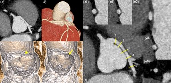
The radiological findings of the patient are shown in Figure 1. The anomalous left coronary artery presented a shared coronary ostium. The ostium of the left coronary artery appeared round and had a ratio of the vertical-to-horizontal measurements of 1. Proximal vessel stenosis of the anomalous coronary artery was not found. The cross-sectional area of the anomalous coronary artery obtained in double oblique view at the level of the anterior aortic commissure had a maximal/minimal diameter ratio of 1.87. The expected vertical-to-horizontal ratio value in the normal coronary artery is about 1. The take-off angle of the anomalous coronary artery was 65° with a take-off level below the aortic valve commissure.
Coronary CT identified features of the anomalous coronary artery in the presented case. ( a ): Multiplanar axial CT reconstruction at the level of the coronary artery origin demonstrates the left coronary artery coursing between the aorta and the pulmonary artery. ( b ): The vessel’s take-off level (below the commissure) is shown in three-dimensional rendered images. ( c, d): Virtual angioscopy post-processing technique is used to visualise the coronary ostia (yellow arrowhead). The yellow arrow points to the origin of the right coronary artery. ( e.1, e.2, e.4, e.5 ): Lumen cross-sectional area of the anomalous coronary artery obtained in double oblique view at different levels of the coronary artery. A normal vertical-to-horizontal ratio between the maximal and minimal diameter was found. ( e.3 ): Lumen cross-sectional area of the anomalous coronary artery obtained in double oblique view at the level of the anterior aortic commissure. This view is known as the “Angelini/Cheong cut”. The presence of an elliptical cross-sectional shape with an elevated ratio between the maximal and minimal diameter at the level of commissure between the right and left coronary leaflets was found.
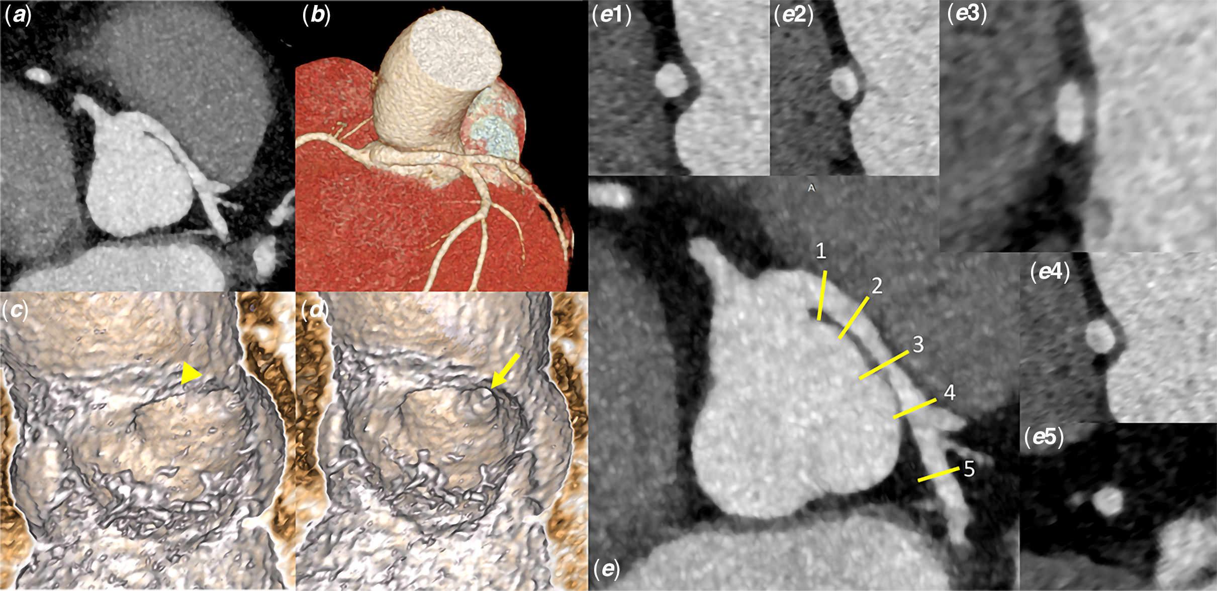
The anomalous left coronary artery had a round ostium with a non-acute take-off angle and no evidence of proximal vessel stenosis. The presence of an elliptical cross-sectional shape with a high ratio between the maximal and minimal diameter at the level of commissure between the right and left coronary leaflets was consistent with an intramural segment of the anomalous coronary artery.
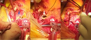
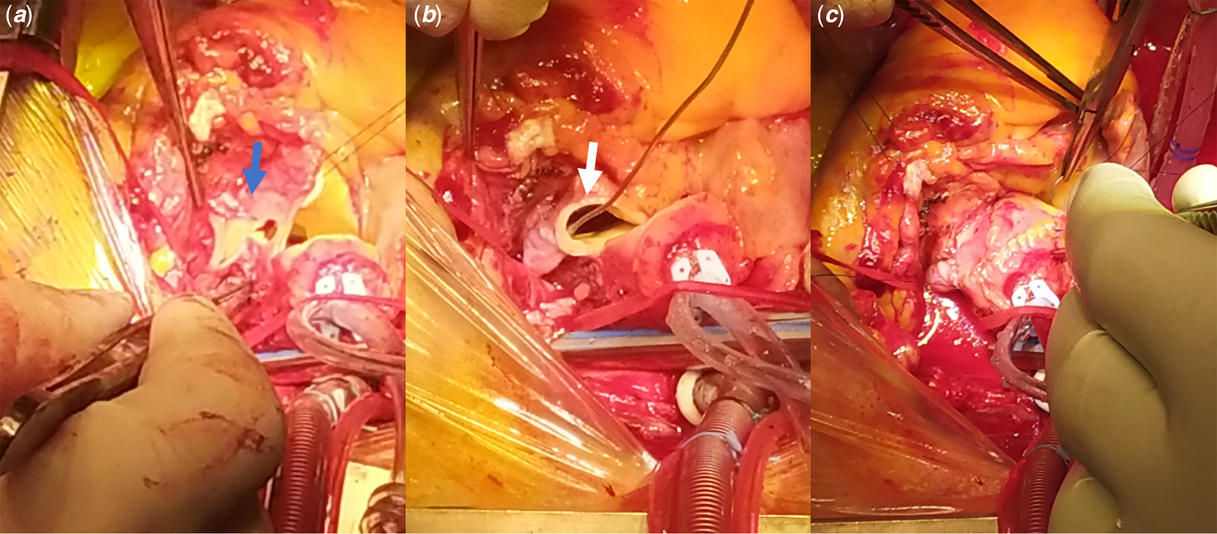
The patient underwent surgery in agreement with the AHA/ACC Guideline for the management of adult CHD. Reference Stout, Daniels and Aboulhosn6 Surgical inspection reported the following anatomical features: absent slit-like deformation of the coronary ostium with potential flap closure; absence of proximal intramural course or stenosis of the anomalous coronary artery; and; evidence of the anomalous left coronary artery within the aortic wall at the level of commissure between the right and left coronary cusp. The course of the anomalous left coronary within the aortic wall was consistent with an intramural segment of the anomalous coronary artery at the level of the septal commissure. “Anatomical” repair of the anomalous left coronary artery was performed (Fig 2). A new coronary ostium in the appropriate left coronary sinus was created using a patch of fresh autologous pericardium after aortocoronary incision. The postoperative period was uneventful and the patient was discharged from the hospital 7 days after surgery.
Intraoperative picture during surgical correction of the left anomalous coronary artery from the opposite sinus. ( a ): The aorta was transected at the level of the sino-tubular junction. The initial epicardial course of the left coronary artery was incised longitudinally. A vertical incision, expanded with a 5 mm tissue punch, was made in the left sinus towards the incised portion of the left coronary artery (blue arrow). ( b ): A patch of autologous pericardium was sutured into the aortocoronary incision to create a new left coronary ostium in the appropriate coronary sinus. A probe has been used to rule out obstruction of the new coronary ostium intraoperatively (white arrow). ( c ): Relationship of the new left coronary ostium and the aortic suture line.
Discussion
This report illustrates the case of a patient with an anomalous left coronary artery from the opposite coronary sinus with evidence of intramural course at the level of the septal commissure and no slit-like deformation of the coronary ostium or proximal intramural segment. The presence of an isolated intramural course at the level of the septal commissure without slit-like ostium or proximal intramural segment is an unusual finding if compared to previous publications. Miller et al state that all patients with intramural segments identified at surgery had coronary CT findings of acute take-off angle; slit-like orifice; and elliptical cross-sectional shape. Reference Miller, Anavekar, El Yaman, Burkhart, Miller and Julsrud7 Angelini et al described the universal presence of proximal intramural course in patients with the origin of the right coronary artery from the opposite sinus of Valsalva at the intravascular ultrasonography evaluation. Reference Angelini, Uribe, Monge, Tobis, Elayda and Willerson8 Cheezum et al define the presence of intramural course at the coronary CT scan in anomalous coronary arteries in cases with proximal vessel narrowing; acute take-off (define <45°); and separate ostium of the vessel from the aorta. Reference Cheezum, Ghoshhajra and Bittencourt3 This case suggests that the intramural course may not be ruled out based on the absence of slit-like ostium, acute take-off, or proximal vessel narrowing. Anomalous coronary arteries that have an interarterial and intramural course are the subgroup believed to be associated with an episode of sudden cardiac death. Reference Mosca and Phoon9 Several factors have been proposed to explain the mechanism of sudden cardiac death. The importance of the rigid peri-commissural area has been postulated as a potential mechanism able to restrict flow in the coronary artery. Reference Peñalver, Mosca, Weitz and Phoon10 This case report describes that anomalous coronary artery with an isolated intramural segment in the peri-commissural area with no evidence of slit-like ostium, acute take-off angle, and the proximal vessel narrowing is associated with typical chest pain and therefore reduced myocardial perfusion.
Moreover, knowledge of the location of the intramural course impacts surgical planning. Various surgical techniques have been used for surgical treatment in the anomalous aortic origin of a coronary artery. The unroofing procedure of the anomalous vessel along its proximal intramural segment has become the preferred management option. However, the absence of slit-like orifice or proximal intramural course of the anomalous coronary artery made unroofing procedure considered inappropriate.
Conclusion
We report on a patient with an anomalous left coronary artery from the opposite coronary sinus with evidence of intramural course at the level of the septal commissure and no slit-like deformation of the coronary ostium, acute take-off, or proximal vessel narrowing. Experts support those anomalous coronary arteries with intramural course are constantly associated with slit-like ostium, acute take-off, or proximal vessel narrowing. Although further investigation is required, we suggest that intramural course may not be ruled out based on the absence of slit-like ostium, acute take-off, or proximal vessel narrowing.
Acknowledgements
None.
Financial support
This research received no specific grant from any funding agency, commercial, or not-for-profit sectors.
Conflicts of interest
None.