Introduction
Obstructive sleep apnoea (OSA) is a common condition characterized by episodes of apnea or hypopnea during sleep, followed by an adverse cardiovascular effect.Reference Peppard, Young, Palta and Skatrud1–Reference Kessler, Chaouat and Weitzenblum4 Patients with univentricular physiology and Fontan-type palliation are at higher risk of developing cardiovascular complications than the general population,Reference Fishberger, Wernovsky and Gentles5 and their management with continuous positive airway pressure ventilation is clearly challenging. As a result, OSA may have greater cardiovascular consequences in adults with CHD and Fontan repairs than the general population.Reference Watson, Bushnell, Jones and Stout6
Case
A 29-year-old non-smoker male patient with a background of pulmonary atresia and intact ventricular septum unsuitable for biventricular repair was referred to our centre. The patient complained of shortness of breath, benign palpitations, atypical non-exertional chest pain, snoring, fatigue, and sleepiness; all these symptoms were attributed to his cardiac condition. His past medical history included a neonatal atrial septostomy, several palliative procedures including modified Blalock–Taussig shunt, a bidirectional Glenn anastomosis, and finally a total cavopulmonary connection completion. His subsequent medical history included a pacemaker implantation for sick sinus node syndrome.
On clinical examination, the patient was pink and well with an oxygen saturation of 94% in air. He was slightly overweight (a body mass index of 26.6 Kg/m2). Cardiac auscultation revealed a single second heart sound with a soft systolic heart murmur at the left sternal edge. The rest of the clinical examination was unremarkable. Transthoracic echocardiography showed an unobstructed total cavopulmonary connection and dilated left ventricle with moderately impaired systolic dysfunction. He also underwent a CT coronary angiogram, which showed the signs of an inferior myocardial infarction, which, in the presence of normal coronary arteries, could be related to one of his previous operations. The latter as well as a right hemidiaphragm dysfunction present on chest X-Ray could be responsible for the LV systolic function impairment and the patient’s symptoms.Reference Eisenberg and Eisenberg7
In addition, pulmonary function tests showed FVC 61% of predicted and FEV1 58% of predicted. There is a moderate gas transfer impairment (TLco: 41% predicted) and mixed ventilatory defect with concomitant restriction (TLC: 67% predicted) and obstruction (FEV1/FVC ratio: 62%), resulting in a degree of hyperinflation (RV/TLC: 135% predicted). (TLC: 67% predicted, RV: 93% predicted, RV/TLC: 135% predicted, TLco: 41% predicted, and Kco: 70% predicted).
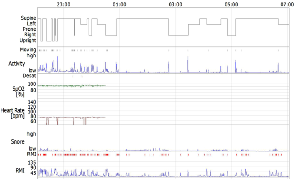
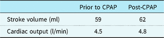
Sleep-disordered breathing suspected was mainly based on clinical symptoms, such as daytime sleepiness and fatigue. A higher suspicion of the condition was also raised based on the fact that there is an increased risk of sleep apnoea in the group of patients. Thus, the patient underwent a sleep study, which showed evidence of airway resistance with periods of nocturnal hypoventilation. Average oxygen saturations overnight were 94% but there were dips to a nadir of 70%. Mean transcutaneous carbon dioxide was satisfactory at 5.87 kPa; but, this rose during periods of probable rapid eye movement sleep to a maximum value of 8.4 kPa. These were associated with dips in oximetry. Sniff nasal inspiratory pressure was marginally low at 66 cmH2O (Figure 2). These results are in keeping with a degree of diaphragm dysfunction, probably related to his previous cardiac surgery and recent weight gain (Table 1). Nocturnal ventilation with the addition of continuous positive airway pressure ventilation was suggested. The effect of the ventilation therapy on the patient’s circulation was monitored by echocardiography. The continuous positive airway pressure was adjusted by cardiac output and oxygen saturation. Cardiac output was improved from 4.5 L/min to 4.7 L/min (Table 2, Figure 1a and b). The repeated lung function tests following the initiation of therapy showed no change in gas transfer, static lung volumes, and spirometric values. This would indicate a degree of airflow obstruction complicating this patient’s sleep study, which has had no effect in his circulation. On the other hand, his circulation did improve with therapy. In his latest follow up, the patient was clinically improved and was scheduled for further assessment.
2D TTE: Parasternal view of the LVOT. Spectral PW Doppler used for the measurement of the cardiac output (CO). (a) Pre-continuous positive airway pressure (CPAP) treatment. (b) Increase of CO post-treatment.

Sleep study: In the first half of the night, the patient had upper airway resistance elevated and snored with flattened airflow signals. Later in the night, he experienced three distinct periods of hypoventilation that were presumed to coincide with REM periods.

Diagnostic polysomnography data

AHI=Apnea/hypopnea index; RDI=Respiratory disturbance index; RMI=Respiratory mechanics instability.
Circulation parameters before and after the CPAP therapy

CPAP = Continuous positive airway pressure.
Discussion
Pulmonary vasoconstriction and reduction of systemic venous return resulting in the reduction of cardiac output form the pathophysiologic pathway of cardiac involvement in patients with obstructive sleep apnoea. Cardiac complications may have detrimental effect on patients with certain types of CHD.
Although there are many modifications of the Fontan operation, the end result includes redirection of the systemic venous blood to the pulmonary arteries, in the absence of subpulmonary ventricle. The systemic venous blood flow to the pulmonary arteries is dependent on the central venous pressure, which should be higher than the pulmonary artery pressure and exceed pulmonary vascular resistance. Factors causing an increase in the systemic or pulmonary artery pressure adversely affect the cardiac output. Furthermore, nocturnal hypoxia may cause vasoconstriction, reducing pulmonary blood flow and contributing to the failure of the Fontan circulation. On the other hand, positive airway pressure may also cause the failure of the Fontan circulation by increasing intrathoracic pressure and pulmonary arterial pressure, thus decreasing systemic venous return and cardiac output.Reference Young, Palta, Dempsey, Skatrud, Weber and Badr8
Close monitoring of Fontan patients after the initiation of continuous positive airway pressure ventilation therapy is paramount to assess the impact of administered therapy and to avoid adverse cardiovascular complications. Watson et al. described a method to titrate continuous positive airway pressure and identify invasively safe thresholds in the sleep laboratory in patients with Fontan circulation.Reference Lofland9 All four patients participated in the study benefited from therapy with clinical improvement and no short term complications.Reference Lofland9 It was suggested that the initial continuous positive airway pressure ventilation trial should be performed in a well-controlled environment at the cardiac catheterization laboratory. In our case, non-invasive echocardiographic assessment was implemented to choose the exact pressure used for continuous positive airway pressure ventilation. An increase in the cardiac output has been used as a marker for effective therapy. Obstructive sleep apnoea is relatively common in patients with a Fontan circulation. Treatment management of these patients is very challenging as meticulous monitoring of hemodynamics is essential. We report, herewith, a case of a Fontan patient presenting with obstructive sleep apnoea who was managed well non-invasively employing echocardiography.
Author ORCIDs
Maria Boutsikou 0000-0002-5457-2472
Financial Support
The research received no specific grant from any funding agency or commercial or non-for-profit sectors.
Acknowledgments
None.
Conflicts of Interest
None.