A sinus venous defect has been considered to be a disease entity different from an atrial septal defect because of a different development in utero, despite the similarities in the location of the defects, haemodynamics at the atrial level, and clinical manifestations.Reference Butts, Crean and Hlavacek 1 – Reference Sizarov, Anderson, Christoffels and Moorman 3 In this study, we report on an unusual case supporting this view.
Case report
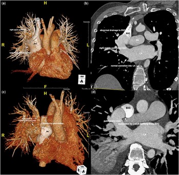
A 45-year-old man visited our hospital with wax-and-wane features of chest discomfort, dyspnoea, and palpitation. Atrial fibrillation was confirmed on electrocardiography. Echocardiography showed a large 31.5-mm diameter abnormal connection between the right upper pulmonary vein and the superior caval vein, a moderate degree of tricuspid regurgitation with an enlarged annulus, and an intact atrial septum. CT revealed an additional abnormal right pulmonary vein connected to the superior caval vein via a 9-mm-diameter orifice located 5 mm distal from the echocardiographically confirmed abnormal connection described above. Uniquely, the right middle pulmonary vein was also normally connected to the left atrium via a large opening (30 mm in diameter, Fig 1a and b). A catheterisation study detected mild pulmonary hypertension (mean pressure: 22–24 mmHg), elevated central venous pressure (17 mmHg), and increased pulmonary blood flow (Qp:Qs=4).
( a ) Preoperative three-dimensional (3D) CT image in the anteroposterior view. The two white arrows indicate the anomalous connections between the pulmonary veins (right upper and middle) and the superior vena cava (SVC). ( b ) A 3D-CT coronal view shows two connections to the right middle pulmonary vein: one with the SVC and the other with the left atrium (LA). ( c ) A right anterior (RA) oblique cranial view -CT image shows separated SVC blood flow and pulmonary venous flow after the patch baffling procedure. ( d ) An axial CT view shows the separate SVC and right pulmonary venous blood flow.

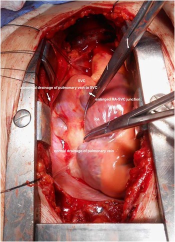
We redirected the blood flow from both right upper and middle pulmonary veins to the left atrium via the normal right middle pulmonary vein–left atrium pathway by constructing an intra-atrial baffle that redundantly covered both the anomalous windows between the right pulmonary veins and the superior caval vein (Fig 1c and d). On post-operative echocardiography, blood flow from the superior caval vein was not interrupted by this intra-atrial baffle; laminar flow was maintained because the superior caval vein and the right atrium were severely dilated (Fig 2). The modified Cox maze III procedure and tricuspid ring annuloplasty were concomitantly performed. Normal sinus rhythm was restored, and the degree of tricuspid regurgitation improved to ⩽grade 1 after the procedures. At discharge, the patient’s functional class had improved.
Operative view of the anomalous pulmonary venous connection to the superior vena cava (SVC) with an additional normal connection to the left atrium (LA). Despite the large exit connection to the LA, the SVC was severely dilated because of the anomalous connection with the right pulmonary veins. RA=right anterior.

Discussion
Butts et alReference Butts, Crean and Hlavacek 1 reported a series of cases supporting the theory that a sinus venosus defect is the consequence of fetal systemic-to-pulmonary veno-venous bridge persistence rather than any deficiency in the atrial septum. Persistent veno-venous connections present a spectrum of malformations, ranging from simple veno-venous malformations to the sinus venosus defect with a partially anomalous pulmonary venous connection. Of their three veno-venous connection cases included either extra-cardiac inter-pulmonary venous channels ran between an anomalously connected pulmonary vein and a normal pulmonary vein, or the anomalous pulmonary vein was normally connected to the left atrium. Crystal et alReference Crystal, Najashi, Williams, Redington and Anderson 4 also reported several cases exhibiting communication between the pulmonary vein and the right atrium, near the inferior caval vein, with retention of normal connections to the left atrium.
We report an unusual congenital anomalous feature that seems to lie somewhere on the sinus venosus defect developmental spectrum of Butts et al, probably between the partial anomalous pulmonary venous connection and the veno-venous connection. This is because we observed both an isolated partial anomalous pulmonary venous connection – between the right upper pulmonary vein and the superior caval vein – and a systemic–pulmonary veno-venous connection – between the right middle pulmonary vein and the superior caval vein – without any direct communication between the anomalously connected pulmonary veins – that is, the right upper and middle pulmonary veins. As these two pulmonary veins were clearly connected to the superior caval vein with an intact atrial septum, we cannot term this channel between the right middle pulmonary vein and the superior caval vein an atrial septal defect or a simple sinus venous defect. The features suggest that the patient’s right-sided pulmonary venous system developed unusually in utero. It seemed that the right middle pulmonary vein retained its initial connection with the primary pulmonary vein, which canalises from the mid-pharyngeal strand, finally resulting in retaining the normal connection with the left atrium,Reference Sizarov, Anderson, Christoffels and Moorman 3 whereas the primitive connection between the pulmonary upper pulmonary vein and the superior caval vein did not regress at all.
As the window between the right middle pulmonary vein and the superior caval vein was as large as that of the normal connection between the right middle pulmonary vein and the left atrium, a large amount of blood from the right middle pulmonary vein probably drained initially to the right atrium via the abnormal connection, and induced severe enlargement of the right atrium. In terms of treatment, intervention – that is, closing the 30-mm-diameter window between the right middle pulmonary vein and the superior caval vein with a stent graft of a device – seemed inappropriate, because another right upper pulmonary vein was connected to the superior caval vein without any direct communication with the left atrium at only 5 mm distal from the window between the right middle pulmonary vein and the superior caval vein. In addition, the window between the right upper pulmonary vein and the superior caval vein was rather large (9 mm in diameter) to be closed without making communication to the left atrium. Therefore, we placed a baffling patch to cover both orifices of the right upper and middle pulmonary veins. We incorporated some redundancy at the orifice of the right upper pulmonary vein to prevent interruption of blood flow from the right upper pulmonary vein; therefore, blood flow was re-directed by the baffling patch to the left atrium via the channel between the right middle pulmonary vein and the superior caval vein. Although the surgery was not so complicated, we nonetheless report this rare form of a systemic-to-pulmonary veno-venous connection with our distinctive CT and operative images.
Acknowledgements
None.
Financial Support
No external financial support was received for this study.
Conflicts of Interest
None.
Ethical Standards
This study was approved by the authors’ institutional review board.