Introduction
Carvajal syndrome, a variant of Naxos disease, is an autosomal recessive disorder caused by mutations in the DSP gene. It is clinically characterised by the triad of woolly hair, palmoplantar keratoderma, and dilated cardiomyopathy, primarily involving the left ventricle. Reference Protonotarios and Tsatsopoulou1 Patients often develop progressive heart failure in childhood or adolescence, with a high risk of sudden cardiac death. Reference Uzumcu, Norgett and Dindar2 Noncompaction cardiomyopathy, though less frequently described, can co-occur and contribute to the clinical complexity. Reference Grebur, Mester and Fekete3 Here, we present a case of Carvajal syndrome in a paediatric patient with features of dilated cardiomyopathy and left ventricular noncompaction, underscoring the importance of comprehensive clinical and genetic evaluation.
Case report
A 13-year-old male patient with autism, presented with a 1.5-month history of persistent cough, worsening over the past week. Two days prior to admission, he was evaluated at another facility for suspected viral upper respiratory infection and was prescribed symptomatic treatment. Due to elevated liver enzymes and clinical deterioration, he was referred to our centre for further evaluation.
On physical examination, the patient exhibited thin, fragile hair with patchy alopecia, nail anomalies, and ectodermal dysplasia in the lower extremities (Figure 1). Cardiovascular examination revealed a third heart sound (S3 gallop rhythm) and hepatomegaly extending 2 cm below the costal margin. Other findings included jugular venous distension and a mildly distended abdomen with no palpable masses.
In the left image, thin and fragile hair is observed, while the right image demonstrates nail anomalies and ectodermal dysplasia of the knees.

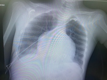
Initial investigations revealed elevated AST and ALT levels, cardiomegaly on chest X-ray (Figure 2), and free fluid in the abdomen on CT. Echocardiography demonstrated dilated ventricles, left ventricular noncompaction, and a reduced ejection fraction of 23%, raising suspicion for myocarditis or dilated cardiomyopathy (Video 1–2).
Chest X-ray of the patient.

The patient was admitted to the paediatric ICU and treated with intravenous furosemide (0.4 mg/kg/hour), milrinone (0.5 mcg/kg/min), and cefotaxime (3 × 50 mg/kg). Viral respiratory panel testing identified Haemophilus influenzae and Streptococcus pneumoniae. Electrocardiography showed normal QTc interval (380 ms) without ST-T changes or pre-excitation.
On the second day of admission, follow-up echocardiography showed left ventricular dilation (50 mm) with spherical trabeculation, ejection fraction of 31%, and severe left ventricular dysfunction. Tricuspid regurgitation was noted with an estimated right ventricular systolic pressure of 33 mmHg. The patient’s treatment was adjusted to include oral diuretics (furosemide 3 × 1 mg/kg), spironolactone (2 × 0,3 mg/kg), digoxin (2 × 5 mg), aspirin (1 × 100 mg), and bisoprolol (1 × 0,1 mg/kg). Magnesium sulphate infusion (10 mg/kg/hour) was administered for acute management.
On the 13th day of paediatric ICU stay, the patient was transferred to the ward with clinical stability. Genetic analysis confirmed a homozygous c.7912G > T nonsense variant in the DSP gene, consistent with Carvajal syndrome. The patient’s clinical presentation, characterised by woolly hair, nail anomalies, ectodermal dysplasia, and severe cardiomyopathy with noncompaction, was in line with this diagnosis.
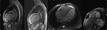
Cardiac MRI revealed severe global hypokinesia was observed in both the left and right ventricles on cine imaging. In late contrast-enhanced images, a non-ischaemic pattern of delayed enhancement was detected in the subepicardial and mid-myocardial regions of the anterior, lateral, and inferior walls at the basal and midventricular levels, forming a ring-like distribution. Minimal pericardial effusion was observed. Left ventricular non-ischaemic myocardial fibrosis and increased trabeculation were noted (NC/C: 15.8/4.4 = 3.59). Systolic dysfunction was present in both the left and right ventricles, with ejection fractions measured at 19 and 20%, respectively (Figure 3). The patient was referred to an advanced cardiac centre.
Cardiac MRI of the patient.

Discussion
Carvajal syndrome is a rare disorder with multisystem involvement, predominantly affecting the skin and cardiovascular system. Similar to prior reports, our patient presented with the hallmark features of woolly hair, palmoplantar keratoderma, and dilated cardiomyopathy. The co-occurrence of left ventricular noncompaction, as observed in this case, has been described but remains underreported in the literature. Reference Grebur, Mester and Fekete3
Previous studies highlight the high risk of heart failure and arrhythmias in Carvajal syndrome, often necessitating early recognition and aggressive management to prevent sudden cardiac death. Reference Uzumcu, Norgett and Dindar2,Reference Syrris, Ward and Evans4 Our patient’s reduced ejection fraction and severe left ventricular dysfunction required intensive care management with inotropic support and a combination of diuretics, beta-blockers, and aldosterone antagonists. While improvement in ejection fraction was observed, the prognosis remains guarded due to the progressive nature of the cardiomyopathy.
Genetic testing confirming the DSP mutation further supports the diagnosis and aligns with previous reports emphasising the utility of molecular diagnostics in distinguishing Carvajal syndrome from other cardiocutaneous syndromes. Reference Garcia-Gras, Lombardi and Giocondo5 Unlike Naxos disease, which primarily involves the right ventricle, Carvajal syndrome typically affects the left ventricle, as seen in this case. Reference Protonotarios and Tsatsopoulou1
Cardiac MRI findings provided additional insights into the disease pathology. The presence of severe global hypokinesia, non-ischaemic myocardial fibrosis, and increased trabeculation reinforced the diagnosis of left ventricular noncompaction cardiomyopathy. These findings are crucial, as noncompaction is associated with a higher risk of thromboembolism, arrhythmias, and progressive heart failure. Advanced imaging modalities such as cardiac MRI should be routinely incorporated into the diagnostic workflow to assess myocardial structure and function comprehensively Reference Jacquier, Thuny and Jop6
This case underscores the importance of a multidisciplinary approach involving dermatologists, cardiologists, and geneticists. The integration of clinical findings with advanced imaging and genetic testing is crucial for early diagnosis and tailored management.
Our report is limited by the absence of long-term follow-up data to assess the patient’s clinical progression and response to treatment. Future studies should aim to address these gaps and explore genotype–phenotype correlations in larger cohorts.
Conclusion
This case highlights the clinical and genetic complexity of Carvajal syndrome, emphasising the need for heightened awareness and a multidisciplinary approach. Early recognition and management are critical to mitigating the risk of heart failure and sudden cardiac death in affected individuals.
Supplementary material
The supplementary material for this article can be found at https://doi.org/10.1017/S1047951125001532.
Acknowledgements
None.
Financial support
None.