Introduction
The creation of a bidirectional cavopulmonary shunt for the patients with a functional single ventricle has become a routine intermediate procedure before total cavopulmonary connection. Reference Alsoufi, Manlhiot and Awan1–Reference Tan, Iyengar and Donath3 The bidirectional cavopulmonary shunt provides effective low-pressure pulmonary blood flow and ventricular volume unloading and contributes to a higher probability of achieving total cavopulmonary connection. Reference Scheurer, Hill and Vasuki4–Reference Ono, Burri and Mayr6 Despite these haemodynamic advantages in systemic circulation, there is a fundamental concern for the pulmonary circulation of bidirectional cavopulmonary shunt physiology, which is entirely dependent on the single passive venous flow through the superior caval vein. Low pulmonary vascular resistance is essential to establish bidirectional cavopulmonary shunt circulation. Elevated superior caval vein pressure following bidirectional cavopulmonary shunt could lead to the development of veno-venous collaterals from the upper systemic venous system to the lower systemic or pulmonary venous system, as a vascular shunt. Reference Van De Bruaene and Budts7 Compared to the aortopulmonary collaterals, the veno-venous collaterals develop under different circumstances. As aortopulmonary collaterals mostly grow to equalise deficit pulmonary blood flow, veno-venous collaterals on the other hand revolve around elevated upper systemic venous pressure and elevated trans-pulmonary gradient. The resulting reduced pulmonary artery blood flow can lead to insufficient development of the central pulmonary arteries. Desaturation can promote the development of aortopulmonary collaterals and can lead to a reduction in systemic ventricular function. With time, these collaterals might increase in size, behaving proportionally to the central venous pressure, and might eventually need catheter interventions. Reference McElhinney, Reddy, Hanley and Moore8–Reference Mahle, Wernovsky, Bridges, Linton and Paridon11 However, few studies addressed the prevalence of veno-venous collaterals, their therapy, and their impact on clinical outcomes.
In this study, we sought to determine the prevalence of veno-venous collaterals and of the coil or device embolisation of veno-venous collaterals in patients who underwent staged total cavopulmonary connection, to identify the risks for the development of veno-venous collaterals, and to determine the impact of veno-venous collaterals on mortality following total cavopulmonary connection.
Methods
Ethical statement
This study was approved by the Institutional Review Board of the Technical University of Munich (approved number 2023-422-S-KR on the 14th of August 2023). Because of the retrospective nature of the study, the need for individual patient consent was waived.
Patients and data collection
We retrospectively evaluated all patients who underwent staged Fontan palliations (bidirectional cavopulmonary shunt and total cavopulmonary connection) at the German Heart Center Munich from June 1995 to December 2022. Patients with conversion from the classic Fontan procedure to total cavopulmonary connection were excluded from this study. Medical records included baseline morphology and demographics as well as pre-, intra-, and postoperative data, using digital and paper chart reviews of each patient. The patients obtained outpatient follow-up with paediatric cardiologists. The institutional single ventricle database, which is regularly monitored, yielded the most follow-up data.
Surgical strategy and operative techniques
Bidirectional cavopulmonary shunt was performed using standard cardiopulmonary bypass, and ante-grade pulmonary blood flow was eliminated in most of the cases. Reference Ono, Burri and Mayr6,Reference Schreiber, Cleuziou, Cornelsen, Hörer, Eicken and Lange12 The azygos vein was routinely transected. The operative techniques for total cavopulmonary connection were described in previous reports, fenestration was used in high-risk patients only. Reference Schreiber, Hörer, Vogt, Cleuziou, Prodan and Lange13,Reference Ono, Kasnar-Samprec and Hager14
Haemodynamic evaluation by catheterisation and angiographic detection of veno-venous collaterals
Cardiac catheterisation was performed in all patients before bidirectional cavopulmonary shunt. Following bidirectional cavopulmonary shunt, cardiac catheterisation was performed when patients demonstrated symptoms, such as progressive cyanosis (under 75% of percutaneous oxygen saturation, SpO2) or superior caval vein syndrome. Cardiac catheterisation was performed in all patients before total cavopulmonary connection. Haemodynamic measurements included pulmonary artery pressure, left atrial pressure, systemic ventricular end-diastolic pressure, arterial pressure, and arterial oxygen saturation (SaO2). Trans-pulmonary gradient was calculated. Conventional angiography was obtained to identify evidence of veno-venous collaterals and any other lesions. The presence of veno-venous collaterals was determined by the angiogram of upper systemic veins, thoracic inferior caval vein, and pulmonary arteries. Reference Poterucha, Johnson and Taggart15 We defined veno-venous collaterals as originating from the superior systemic venous circulation if they had a clearly identifiable angiographic origin, supplying the other systemic or pulmonary parenchyma, and opacifying the pulmonary veins, inferior caval vein, atria, or a combination of them. Interventional coil embolisation of veno-venous collaterals was performed when it was technically feasible and clinically indicated (persistent deep cyanosis; SpO2 < 75% or veno-venous collaterals drain to the pulmonary veins). Patients who underwent embolisation had haemodynamic and clinical data collected at baseline and at routine ambulatory clinic follow-up and/or subsequent cardiac catheterisation. Angiograms in which collateral vessels were visualised but with ambiguous origin or distribution of blood flow were classified as veno-venous collateral negative.
Statistical analysis
Clinical characteristics and outcomes were compared between patients with and without veno-venous collaterals. Categorical variables are presented as absolute numbers and percentages. A chi-square test was used for comparison of categorical data. Continuous variables are expressed as medians with interquartile ranges or as means with standard deviations. An independent sample t-test was used to compare normally distributed variables. The Mann–Whitney U test was used for variables that were not normally distributed. Factors associated with the development of veno-venous collaterals were identified using a logistic regression analysis. Transplant-free survival after total cavopulmonary connection was calculated using the Kaplan–Meier method and a comparison with and without veno-venous collaterals was performed using log-rank test. The impact of veno-venous collaterals on clinical outcomes after total cavopulmonary connection was analysed using Cox regression models. Data analysis and graphing were performed with the Statistical Package for the Social Sciences (SPSS) version 28.0 for Windows (IBM, Ehningen, Germany) and R-statistical software (R foundation for statistical computing, Vienna, Austria).
Results
Patient characteristics and perioperative data
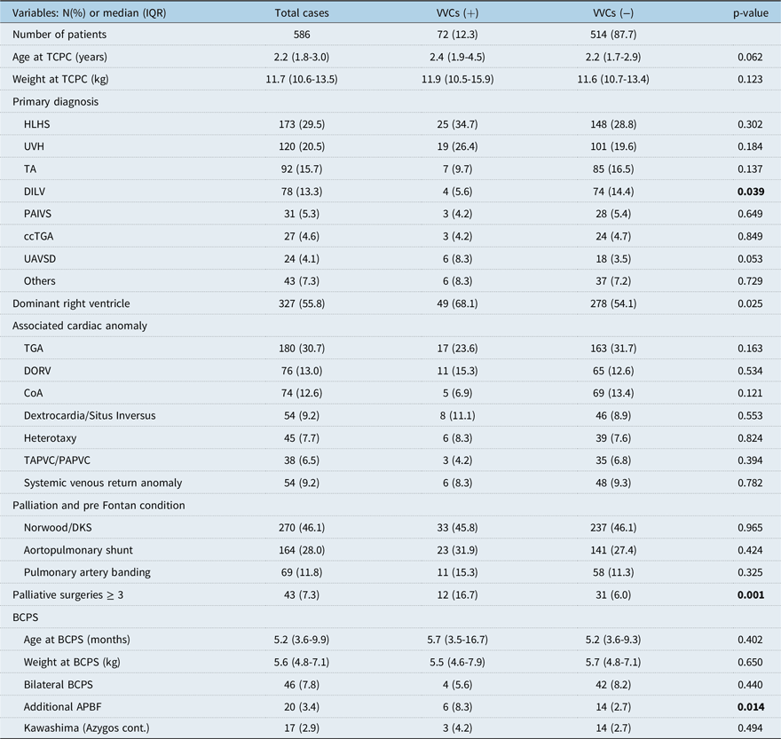
A total of 586 patients underwent staged Fontan palliation at our center during the study period. Veno-venous collaterals between bidirectional cavopulmonary shunt and total cavopulmonary connection were observed in 72 (12.3%) patients at a median period of 178 (interquartile range: 24–586) days after bidirectional cavopulmonary shunt and at a median period of 443 (interquartile range: 39–706) days before total cavopulmonary connection. Patients’ characteristics with and without veno-venous collaterals are presented in Table 1. Median age (2.4 vs. 2.2 years, p = 0.062) and weight (11.9 vs. 11.6 kg, p = 0.123) at total cavopulmonary connection were similar between the groups. The most frequent diagnosis was hypoplastic left heart syndrome in both groups (34.7 and 28.8%), respectively. The prevalence of veno-venous collaterals was lower in patients with double- inlet left ventricle (5.6 vs. 14.4%, p = 0.039) and was higher in patients with dominant right ventricle (68.1 vs. 54.1%, p = 0.025). In patients with veno-venous collaterals, the prevalence of patients who had palliative surgeries that were more than stage one and stage two, was higher compared to those without veno-venous collaterals (16.7 vs. 6.0%, p = 0.001). The prevalence of veno-venous collaterals was higher in patients who underwent additional pulmonary blood flow at the time of bidirectional cavopulmonary shunt (8.3 vs. 2.7%, p = 0.014). During the study period, 94 patients underwent bidirectional cavopulmonary shunt but did not reach total cavopulmonary connection. The veno-venous collaterals were found in 11 of 94 patients (11.7%), and the incidence of veno-venous collaterals was similar between the patients who reached total cavopulmonary connection and those who did not (12.3 vs. 11.7%, p = 0.872).
Baseline cohort characteristics in patients with and without VVCs before TCPC

TCPC = total cavopulmonary connection, HLHS: hypoplastic left heart syndrome; UVH = univentricular heart; TA = tricuspid atresia; DILV = double-inlet left ventricle; ccTGA = congenitaly corrected TGA, PAIVS = pulmonary atresia and intact ventricular seputum; UAVSD = unbalanced atrioventricular septal defect; TGA = transposition of the great arteries; DORV = double-outlet right ventricle; CoA = coarctation of the aorta; T(P)APVC = Total (partial)anomalous pulmonary venous connection; BCPS = bidirectional cavopulmonary shunt; APBF = ante-grade pulmonary blood flow.
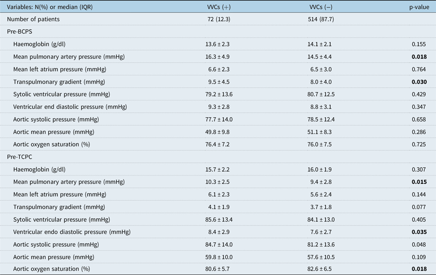
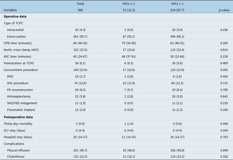
As for the origin of veno-venous collaterals, identified inflow origins are shown in Figure 1. The most frequent origin was the superior caval vein in 35 patients, followed by the brachiocephalic (innominate) vein in 33, the azygos/hemi-azygos vein in 16, and the venous angle (junction where the internal jugular vein and subclavian vein unite to brachiocephalic vein) in 7 cases (Figure 1A). As for the identified outflow (entry) origin, 48 were into the inferior caval vein, 19 into the pulmonary veins, 9 into the left atrium, and 4 into the coronary sinus (Figure 1B). A comparison of pre-bidirectional cavopulmonary shunt and pre-total cavopulmonary connection cardiac catheterisation data with and without veno-venous collaterals is shown in Table 2. In patients with veno-venous collaterals, mean pulmonary artery pressure (16.3 vs. 14.5 mmHg, p = 0.018), and trans-pulmonary gradient (9.5 vs. 8.0 mmHg, p = 0.030) were significantly higher compared to those without veno-venous collaterals before bidirectional cavopulmonary shunt. At the time of total cavopulmonary connection, mean pulmonary artery pressure (10.3 vs. 9.45 mmHg, p = 0.015) and end-diastolic pressure (8.4 vs. 7.6 mmHg, p = 0.035) were significantly higher compared to those without veno-venous collaterals. Aortic oxygen saturation (SaO2) was significantly lower compared to those without veno-venous collaterals (80.6 vs. 82.6 %, p = 0.018). Perioperative data are shown in Table 3. There was no significant difference in operative data and postoperative data between patients with and without veno-venous collaterals.
Location of the inflow origin of the VVCs between BCPS and TCPC.

Pre-BCPS and pre-TCPC cardiac catheterisazion data

BCPS = bidirectional cavopulmonary shunt; TCPC = total cavopulmonary connection.
Perioperative variables

Variables were presented in N(%) or median (IQR). RV = right ventricle; LV = left ventricle; TCPC = total cavopulmonary connection; CPB = cardiopulmonary bypass; AXC = aortic cross clamp; DKS = Dames–Kaye–Stansel anastomosis; AVV = atrioventricular valve; PA = pulmonary artery; SAS = subaortic stenosis; VSD = ventricular septal defect; ICU = intensive care unit.
Follow-up and impact of veno-venous collaterals on late outcome after total cavopulmonary connection
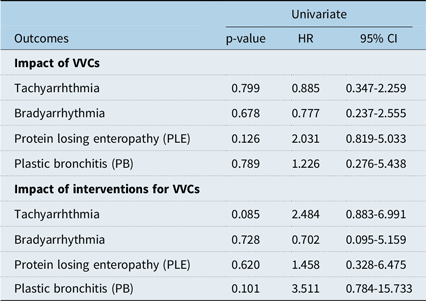
Median follow-up was 5.5 (interquartile range: 1.0–13.0, max 27.1) years. Transplant-free survival in patients with and without veno-venous collaterals is shown in Figure 2. There was no difference in transplant-free survival between the groups (p = 0.600). Impact of veno-venous collaterals between bidirectional cavopulmonary shunt and total cavopulmonary connection on morbidities after total cavopulmonary connection was analysed using the Cox model. The results are shown in Table 4. We could not find any significant impact of veno-venous collaterals on tachy-arrhythmia (p = 0.799, hazard ratio: 0.885), brady-arrhythmia (p = 0.678, hazard ratio: 0.777), protein losing enteropathy (p = 0.126, hazard ratio: 2.031), nor plastic bronchitis (p = 0.789, hazard ratio: 1.226).
Transplant-free survival after TCPC comparing the patients who demostrated VVCs before TCPC and those who did not. There was no impact of VVCs on transplant-free survival after TCPC (92.8 vs. 92.7% at 10 years, p = 0.563). TCPC: total cavopulmonary connection, VVCs: veno-venous collaterals.

Impact of VVCs on morbidities after TCPC using cox regression models

Intervention of veno-venous collaterals
Interventional closure of veno-venous collaterals was performed in 32 (5.5%) patients at a median period of 29 (interquartile range: 16–152) days after bidirectional cavopulmonary shunt. In these patients, median arterial oxygen saturation increased from 79 (70–82) to 81 (77–85) % after the procedure (p < 0.001). In 16 of these 32 (50.0%) patients, the increase in arterial oxygen saturation was 5% or more. The impact of interventional closure of veno-venous collaterals between bidirectional cavopulmonary shunt and total cavopulmonary connection on morbidities after total cavopulmonary connection was analysed using the Cox model. The results are shown in Table 4. We could not find any significant impact of veno-venous collaterals on tachy-arrhythmia (p = 0.085, hazard ratio: 2.484), brady-arrhythmia (p = 0.728, hazard ratio: 0.702), protein-losing enteropathy (p = 0.620, hazard ratio: 1.458), nor plastic bronchitis (p = 0.101, hazard ratio: 3.511).
Risk factor analysis for the development of veno-venous collaterals
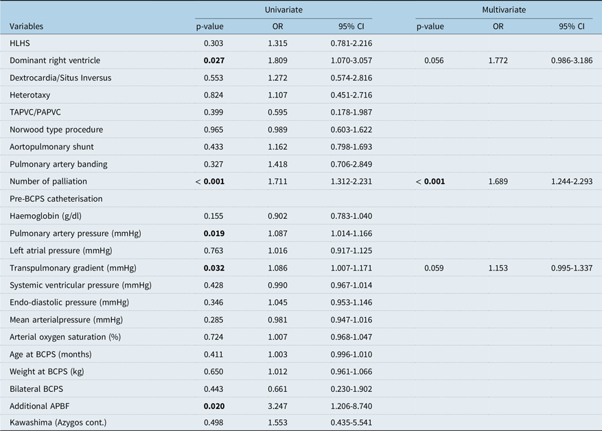
Results are shown in Table 5. Dominant right ventricle (p = 0.027, odds ratio: 1.809), the number of palliative procedures (p < 0.001, odds ratio: 1.711), pre-bidirectional cavopulmonary shunt pulmonary artery pressure (p = 0.019, odds ratio: 1.087), and pre-bidirectional cavopulmonary shunt trans-pulmonary gradient (p = 0.032, odds ratio: 1.086), and additional ante-grade pulmonary blood flow at bidirectional cavopulmonary shunt (p = 0.020, odds ratio: 3.247) showed a p < 0.10 in the univariate analysis. The multivariate analysis identified the number of palliative procedures (p < 0.001, odds ratio: 1.689) as an independent risk factor for the development of veno-venous collaterals.
Risk factor for the development of VVCs after BCPS

The bold values denotes significant in statistical analysis < 0.05. T(P)APVC = total (partial)anomalous pulmonary venous connection; BCPS = bidirectional cavopulmonary shunt; APBF = ante-grade pulmonary blood flow.
The association of bilateral bidirectional cavopulmonary shunt (p = 0.443, odds ratio: 0.661) or azygos/hemi-azygos continuation (Kawashima procedure, p = 0.498, odds ratio: 1.553) was not identified as a risk for the development of veno-venous collaterals between Glenn and Fontan.
Discussion
In this study, we report a prevalence of 12 % for veno-venous collaterals between bidirectional cavopulmonary shunt and total cavopulmonary connection. The most frequent inflow origins were the superior caval vein followed by the brachiocephalic (innominate) vein, while the most frequent outflow origin was the inferior caval vein. The development of veno-venous collaterals between bidirectional cavopulmonary shunt and total cavopulmonary connection did not influence clinical outcomes including mortality after total cavopulmonary connection. The need for further palliative procedures before total cavopulmonary connection, that is stage one and stage two palliation, and the pre-bidirectional cavopulmonary shunt trans-pulmonary gradient were independent risk factors for the development of veno-venous collaterals between bidirectional cavopulmonary shunt and total cavopulmonary connection.
Development of veno-venous collaterals after bidirectional cavopulmonary shunt
The establishment of passive pulmonary flow physiology following bidirectional cavopulmonary shunt produces upper systemic venous hypertension that might result in the formation of veno-venous collaterals, from the upper systemic veins to the lower systemic venous or pulmonary venous system (pulmonary veins or left atrium). Compared to aortopulmonary collaterals, fewer studies describe the prevalence of veno-venous collaterals during or after the staged Fontan palliation. McElhinney, et al. demonstrated that veno-venous collaterals were angiographically detectable in 18 of 54 (33%) patients after bidirectional cavopulmonary shunt, which was performed at a median age of 1.4 years, and postoperative angiography was performed at median of 15.4 months. Reference McElhinney, Reddy, Hanley and Moore8 They demonstrated that trans-pulmonary gradient early after bidirectional cavopulmonary shunt was higher, and mean SaO2 at follow-up was lower in patients who developed veno-venous collaterals. Magee, et al. identified that veno-venous collaterals in 31% of the patients at a median age of 1.3 years after bidirectional cavopulmonary shunt; increased trans-pulmonary gradient was an independent risk factor for the development of veno-venous collaterals. Reference Magee, McCrindle and Mawson9 The median time from bidirectional cavopulmonary shunt to postoperative catheterisation was 1.3 years. Heinemann, et al. showed that veno-venous collaterals were identified in 7 out of 14 (50%) patients after bidirectional cavopulmonary shunt, which was performed at a median age of 1.3 years, with a median follow-up of 5.8 years. Reference Heinemann, Breuer, Steger, Steil, Sieverding and Ziemer10 Our results demonstrated a prevalence of 12%, which was lower than in previous reports. The age at the time of bidirectional cavopulmonary shunt in these studies was significantly higher than in our cohort (median 1.4, 1.3, and 1.3 years in the previous studies and median 5.2 months in this study). We assume that performing bidirectional cavopulmonary shunt at an older age was the reason why the prevalence of veno-venous collaterals was that high in the previous studies. Nowadays, early bidirectional cavopulmonary shunt between 3 and 6 months of age has become a standard strategy. The benefit of an early bidirectional cavopulmonary shunt is to reduce the volume load on a systemic ventricle by conversation from a systemic to pulmonary artery shunt to Glenn circulation. Bidirectional cavopulmonary shunt provides stable haemodynamics, accelerates somatic development, and prevents the development of pulmonary hypertension. These beneficial effects of bidirectional cavopulmonary shunt are age-dependent and less likely to occur in older infants and children. Reference Mahle, Wernovsky, Bridges, Linton and Paridon11 Therefore, our patients had better haemodynamics after bidirectional cavopulmonary shunt and demonstrated a lower prevalence of veno-venous collaterals. However, risk factors for the development of veno-venous collaterals identified in the previous studies were quite consistent with our results. They are pre-bidirectional cavopulmonary shunt haemodynamic variables including pulmonary artery pressure and trans-pulmonary gradient. In our contemporary cohort of staged total cavopulmonary connection patients, the number of palliations was identified as a risk for the development of veno-venous collaterals. Repeated surgical explorations might trigger the development of veno-venous collaterals.
Intervention for veno-venous collaterals after bidirectional cavopulmonary shunt
Interventional coil embolisation of veno-venous collaterals has been reported in previous studies. McElhinney, et al. reported that 10 of 31 veno-venous collaterals were coil embolised with a median increase in SaO2 of 16%. Reference McElhinney, Reddy, Hanley and Moore8 Magee, et al. reported that coil occlusion was performed in 4 of 32 patients who developed veno-venous collaterals. Reference Magee, McCrindle and Mawson9 They also demonstrated that veno-venous collaterals flow was reduced after surgical revision of the bidirectional cavopulmonary shunt pathway. Heineman, et al. showed that catheter intervention was performed in 5 of the 16 patients (31.2%), and SaO2 significantly improved after coil embolisation of veno-venous collaterals in all patients. Reference Heinemann, Breuer, Steger, Steil, Sieverding and Ziemer10 Several case reports demonstrated the successful interventional closure of veno-venous collaterals by a vascular plug or duct occluder after bidirectional cavopulmonary shunt. Reference Karur, Mahima and Nanjappa16–Reference Abdel-Aziz, Tanase and Ewert19 Our results also showed the improvement of SaO2 after veno-venous collaterals closure. It is to note that interventional embolisation was performed relatively early after bidirectional cavopulmonary shunt (median period of 29 days), due to the low arterial oxygen saturation. We did not close the veno-venous collaterals found at the cardiac catheterisation before total cavopulmonary connection, because veno-venous collaterals from the superior caval vein to the inferior caval vein disappear after the Fontan completion.
Impact of veno-venous collaterals on outcomes after total cavopulmonary connection
Regarding the impact of veno-venous collaterals on outcomes after total cavopulmonary connection, no significant difference in transplant-free survival was observed in patients with and without veno-venous collaterals. We also found no difference in morbidities after total cavopulmonary connection in patients with and without veno-venous collaterals. Veno-venous collaterals after bidirectional cavopulmonary shunt might act as a natural shunt to prevent the elevation of the upper systemic venous pressure in the Glenn circulation, and they might disappear or reduce after the Fontan completion. Veno-venous collaterals developing after the Fontan procedure have other mechanisms. Veno-venous collaterals after the Fontan procedure were observed from the systemic venous to the pulmonary venous systems. The prevalence of veno-venous collaterals after the Fontan procedure was documented in our previous study. Reference Nguyen Cong, Schaeffer and Palm20
Study limitations
This study was limited by its retrospective and single-center study design. Surgical and medical management may have changed during the study period, probably influencing the long-term outcomes. Veno-venous collaterals were detected by cardiac catheterisation, and the flow of veno-venous collaterals could not be quantified.
Conclusions
The prevalence of veno-venous collaterals between bidirectional cavopulmonary shunt and total cavopulmonary connection was 12%. The most frequent location of inflow of veno-venous collaterals was the superior caval vein. The interventional closure of veno-venous collaterals improved the arterial oxygen saturation in half of the patients. Veno-venous collaterals might be triggered by the elevating pressure of upper systemic veins and also by more surgical palliations. Veno-venous collaterals that developed between bidirectional cavopulmonary shunt and total cavopulmonary connection had no impact on transplant-free survival or late complications following total cavopulmonary connection.
Financial support
This study had no financial support.
Competing interests
The authors declare no potential conflicts of interest concerning the research, authorship, or publication of this article.