Introduction
Sialodochitis fibrinosa is a rare salivary gland disease that was first described by Kussmaul in 1879.Reference Kussmaul1 We report a case of a 61-year-old man who presented with a 4-year history of bilateral painless parotid swellings associated with eating and drinking. Differential diagnoses were explored, but magnetic resonance imaging and histopathological studies confirmed the diagnosis of sialodochitis fibrinosa.
The conservative measures trialled included reviewing atopic and dietary factors. Salivary gland massage to empty excess saliva was also unsuccessful. Staged bilateral total parotidectomy provided a curative outcome for this patient. Dissection of the nerve fibres was challenging, as they were splayed and stretched across multiple mucous-filled cysts.
Although bilateral total parotidectomy can provide a curative outcome, a staged approach is necessary to reduce the risk of bilateral facial nerve injury and allow time for nerve rehabilitation.
Case report
A 61-year-old man presented to the oral and maxillofacial surgery department in 2012 with a 4-year history of bilateral painless parotid swelling, occurring most mealtimes, requiring massage of the glands to empty excess saliva.
At this time, he was offered three options: continuation of conservative management, or consideration of transoral bilateral duct ligation or bilateral parotidectomy. He decided to continue with conservative management.
The patient had a past medical history of hypertension, hypercholesterolaemia, anxiety and obesity. Medication included lisinopril 10 mg once daily, amlodipine 5 mg once daily and ezetimibe 10 mg once daily. His only reported allergy was to simvastatin (myalgia).
In September 2018, he presented to the ENT emergency clinic with bilateral parotid swelling and a four-week history of progressive enlargement of the left parotid gland following a viral upper respiratory tract infection.
On physical examination, he had a left-sided fluctuant 12.5 × 5 cm swelling and a right-sided fluctuant 3 × 2 cm swelling. There was no erythema or tenderness. Facial nerve function was intact and intra-oral examination findings were unremarkable, except for the lack of manual saliva expression from the left Stensen's duct. There were no palpable cervical lymph nodes.
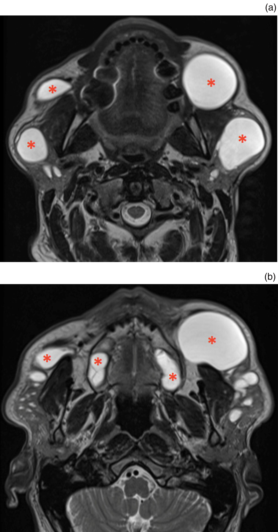
Magnetic resonance imaging revealed a beaded appearance of the parotid ducts bilaterally, with massively dilated segments alternating with ductal strictures (Figure 1), in keeping with an extreme case of chronic sialodochitis. There were no filling defects. The submandibular glands were unremarkable. Human immunodeficiency virus screening results were negative. Microbiological examination demonstrated candida only. Blood tests did not demonstrate eosinophilia.
Axial T2-weighted magnetic resonance imaging of neck soft tissue with contrast. (a & b) Both images depict multiple cystic dilations (asterisks) in the intraglandular ducts of the parotid glands bilaterally.

Other differential diagnoses had been considered, including Sjögren's syndrome, sarcoidosis, lymphoma, immunoglobulin G4 related disease, and bacterial or viral infectious sialadenitis and sialolithiasis. However, based on the clinical findings and investigations, the diagnosis of bilateral sialodochitis was considered most likely. This was later confirmed following histopathological assessment of the tissue sample.
The patient underwent a left total parotidectomy in December 2018. He sustained left facial palsy House–Brackmann grade V, affecting the left temporal and zygomatic division and the marginal mandibular nerve. This had spontaneously improved to grade II at the six-month follow-up review in May 2019. The patient was delighted with the outcome of the initial surgery and opted to have surgical treatment for the right parotid gland.
He underwent a right total parotidectomy in August 2019. His immediate post-operative recovery was complicated by a neck haematoma, requiring surgical evacuation and wash-out of the right parotid bed under general anaesthesia. Bipolar haemostasis was used and the wound was packed with bismuth iodoform paraffin paste gauze. Two days later, the dressing was removed and the wound was closed formally in the operating theatre.
Intra-operatively, the appearance of each gland was grossly abnormal. Multiple thick-walled mucous-filled cysts measuring approximately 1–2 cm in diameter were spread throughout the gland, embedded in firm fibrous tissue. The appearance was similar to that of a cystic hygroma. Branches of the facial nerve were splayed and stretched between and around the cysts. Fastidious sharp dissection of nerve fibres from the fibrous tissue under microscopic control was the only way to preserve the bulk of nerve function. Antegrade and retrograde dissection were used with continuous nerve monitoring.
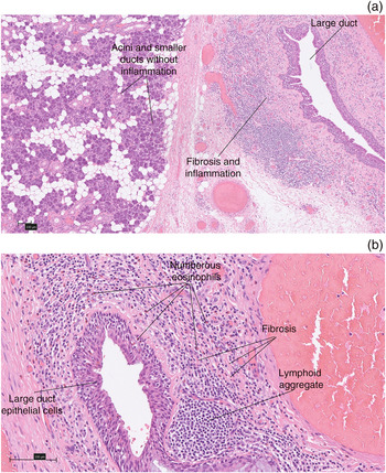
Histological assessment of the tissue demonstrated periductal fibrosis, with infiltration of lymphocytes, plasma cells, and abundant eosinophils, consistent with the condition (Figure 2).
Parotid gland biopsy haematoxylin and eosin stains ((a) ×6 magnification and (b) ×30 magnification), showing eosinophilic inflammation, fibrosis and lymphoid aggregates focused around the dilated intralobular ducts.

The patient was reviewed in the out-patient clinic in September 2019, where he was reported to be recovering well; however, he had a persistent left-sided marginal mandibular nerve weakness. He was discharged from ENT with full remission of his facial swelling and pain. Apart from some altered sensation in the neck and ears, which would be expected after a parotidectomy, the patient feels that both his preceding symptoms (pain and swelling after eating) and subsequent facial nerve weakness are no longer a problem. He feels the treatments have largely improved his quality of life.
Discussion
Sialodochitis fibrinosa is a rare salivary gland disease that was first reported by Kussmaul in 1879. It is characterised by recurrent salivary gland swelling and mucofibrinous plugs causing pain.Reference Kussmaul1 Diagnosis can be challenging because of its rarity, and it presents similarly to acute inflammatory and chronic conditions causing salivary gland swelling. Patients may present with one or more symptoms, including multiple recurrent salivary gland swelling, dehydration, reduced salivary flow, thick secretions from the salivary ducts, a history of allergic diseases and association with food.Reference Chikamatsu, Shino, Fukuda, Sakakura and Furuya2 The presence of mucofibrinous plugs may lead to acute parotitis and chronic sialadenitis.
The diagnosis of sialodochitis fibrinosa is one of exclusion, with no widely adopted case definition. One reviewReference Baer, Okuhama, Eisele, Tversky and Gniadek3 offers the definition of recurrent painful major salivary gland swelling associated with eosinophil-rich mucus plugs, or the histopathological finding of sialodochitis with a periductal eosinophilic infiltrate.
Treatment is based on symptoms and is largely supportive. Depending on the patient's symptoms, sialodochitis fibrinosa can be managed conservatively in the first instance. This can involve compressive massages of the salivary glands, and the administration of antihistamines with or without steroids. There is a fine balance between allowing conservative management for as long as possible and preventing continued fibrosis and parotid enlargement to complicate future surgery. A few recent papers have described successful outcomes following interventional sialendoscopy.Reference Gallo, Capaccio, Benazzo, De Campora, De Vincentiis and Farneti4,Reference Schwarz, Stuermer and Luers5 For those with massive parotid enlargement or where conservative measures fail, such as in our patient, parotidectomy can be offered to provide a curative outcome.Reference Hayashi, Onda, Ohata, Takano and Shibahara6,Reference Flores, Alvarez, Sanchinel, Andrus, Malpartida and Giraldez7
• Sialodochitis fibrinosa should be considered a potential cause of recurrent parotid or submandibular swelling after systemic and infectious causes have been excluded
• Sialodochitis fibrinosa should initially be managed conservatively, with operative management reserved for cases refractory to treatment
• Investigation into sialodochitis fibrinosa should include a detailed history of the patient's diet, and indicators of allergy or atopy
• Staged approach surgery may be a better way of managing the condition, to avoid simultaneous facial nerve injury and allow time for facial nerve rehabilitation
Competing interests
None declared