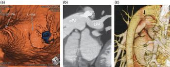
A12-year-old boy visited our outpatient clinic for regular follow up during 3 years following occlusion of a patent arterial duct using a coil. At the last visit, he was noted to have an ejection systolic murmur graded at 3 out of 6. During previous successful occlusion of his arterial duct using a 0.052″ Gianturco and 0.038 Flipper coils, the minimum diameter of the arterial duct was measured at 3.1 millimetres, with the ratio of pulmonary to systemic flow calculated at 2.2 to 1, and with a gradient of 4 mmHg between the pulmonary trunk and the left pulmonary artery. Doppler echocardiography on this occasion showed accelerated blood flow of 1.8 m/s in the left pulmonary artery, compared to 1.1 m/s in the right pulmonary artery. Perfusion lung scanning using radionuclide showed significantly decreased flow to the left lung, with only 30% of the total flow going to the left lung. Multi-detector computed tomography revealed 1.5 loops of the coils protruding into the left pulmonary artery (Fig. 1a, the blue structure indicating the 0.052″ Gianturco coil).Reference Hayabuchi, Mori and Kagami1 The longitudinal view of the left pulmonary artery, along with a view of the aorta, showed this coil not only protruding into, but also retracting the left pulmonary artery. Figure 1b shows the longitudinal view, and 1c shows the close relationship of the aorta and the left pulmonary artery. The arrow indicates the coil. We plan to offer further intervention should the stenosis progress. Our experience shows that multi-detector computed tomography is an easy and clinically useful tool with which to evaluate the complicated nature of pulmonary arterial stenosis that can occur after occlusion of the arterial duct with coils.
Figure 1