Introduction
Vestibular schwannomas are benign, slow-growing tumours that arise from the abnormal proliferation of perineural Schwann cells of the vestibular nerve.Reference Hougaard, Norgaard, Pedersen, Bibby and Ovesen1,Reference Springborg, Fugleholm, Poulsgaard, Cayé-Thomasen, Thomsen and Stangerup2 As the natural history of vestibular schwannoma is nowadays better understood, we are able to individualise the treatment. The available options are dependent mainly upon tumour size, growth rate, symptomatology, patient age and the degree of co-morbidity severity, and include watchful waiting, stereotactic radiosurgery and microsurgery.
For many patients and particularly for those with larger tumours, microsurgery remains the treatment of choice.Reference Gal, Shinn and Huang3 Microsurgery outcomes have improved significantly over the years. The translabyrinthine approach for vestibular schwannoma resection is thought to be a very reliable technique for tumour removal, and has the benefit of providing excellent access regardless of tumour size.Reference Wu and Sterker4 However, in contrast to the middle cranial fossa and retrosigmoid approaches, which can in certain cases be considered hearing-preserving, in the translabyrinthine approach, hearing on the ipsilateral side is sacrificed.Reference Springborg, Fugleholm, Poulsgaard, Cayé-Thomasen, Thomsen and Stangerup2 Hearing rehabilitation can be a challenge in such cases. When it comes to patients with neurofibromatosis type 2 (NF2) suffering from bilateral vestibular schwannoma, global deafness might be inevitable,Reference Lustig, Yeagle, Driscoll, Blevins, Francis and Niparko5,Reference Roehm, Mallen-St Clair, Jethanamest, Golfinos, Shapiro and Waltzman6 with very limited hearing rehabilitation options available.
Recently, the possibility of a translabyrinthine approach for vestibular schwannoma resection, with cochlear nerve preservation and cochlear implantation, has been raised.Reference Belal7–Reference Kim, Han, Kim and Moon9 However, because of surgical trauma, vascular compromise and subsequent inflammation, the residual cochlea can develop fibrosis and neo-ossification. Labyrinthine ossification similar to this can be demonstrated in middle-ear infections, meningitis, labyrinthitis and sepsis. The degree and time period in which this occurs is variable, with some patients showing evidence as early as 2 weeks post-surgery, and others showing evidence between 4 months and 11 years post-surgery, or not at all.Reference Belal7,Reference Hassepass, Arndt, Aschendorff, Laszig and Wesarg8 Such processes (fibrosis and neo-ossification) are extremely significant in cochlear implantation cases, as extensive cochlear fibrosis can even prevent implantation.Reference Kameswaran, Kumar, Murali, Natarajan and Krishnan10
Reports on the time frame of cochlear fibrosis and ossification are lacking. Our study therefore aimed to radiologically assess the time period in which cochlear obliteration develops and hence the period when patency for cochlear implant insertion is compromised. This will help determine the ideal time period for staged cochlear implant insertion for auditory rehabilitation and possible restoration of binaural hearing.
Materials and methods
Study settings
A retrospective cohort study was carried out in a tertiary, referral, academic centre, with a catchment area of approximately 2.2 million. The study was approved by the local audit board and no further ethical approval was required.
Patient selection
We reviewed the vestibular schwannoma database for cases presenting over a 15-year period. Only patients who had undergone a translabyrinthine approach for vestibular schwannoma resection as the sole treatment, with available post-operative magnetic resonance imaging (MRI) scans, were enrolled. Patients with vestibular schwannoma resected by other approaches (e.g. retrosigmoid or middle cranial fossa approaches), or those who had undergone prior or subsequent stereotactic radiosurgery within two years of vestibular schwannoma resection, were excluded. Only patients with available scans for at least one year post-operatively were included.
Imaging aspects
The post-operative MRI scans were reviewed for fluid signal within the cochlea. All MRI scans analysed had slice thicknesses of between 0.8 mm and 3 mm. Assessments were conducted of fluid-enhanced MRI scans, including: 17 fast imaging employing steady-state acquisition (‘FIESTA’) scans, 10 T2-weighted scans, 30 T1-weighted pre-fluid-attenuated inversion recovery (‘pre-FLAIR’) scans, 20 T1-weighted post-fast spin echo scans, 2 T1-weighted three-dimensional scans and 1 T1-weighted turbo spin echo scan.
All scans were assessed independently by two reviewers; in cases of discrepancy, the final decision was reached with consensus. The scans were examined in both the axial and coronal planes.
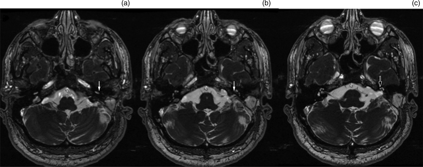
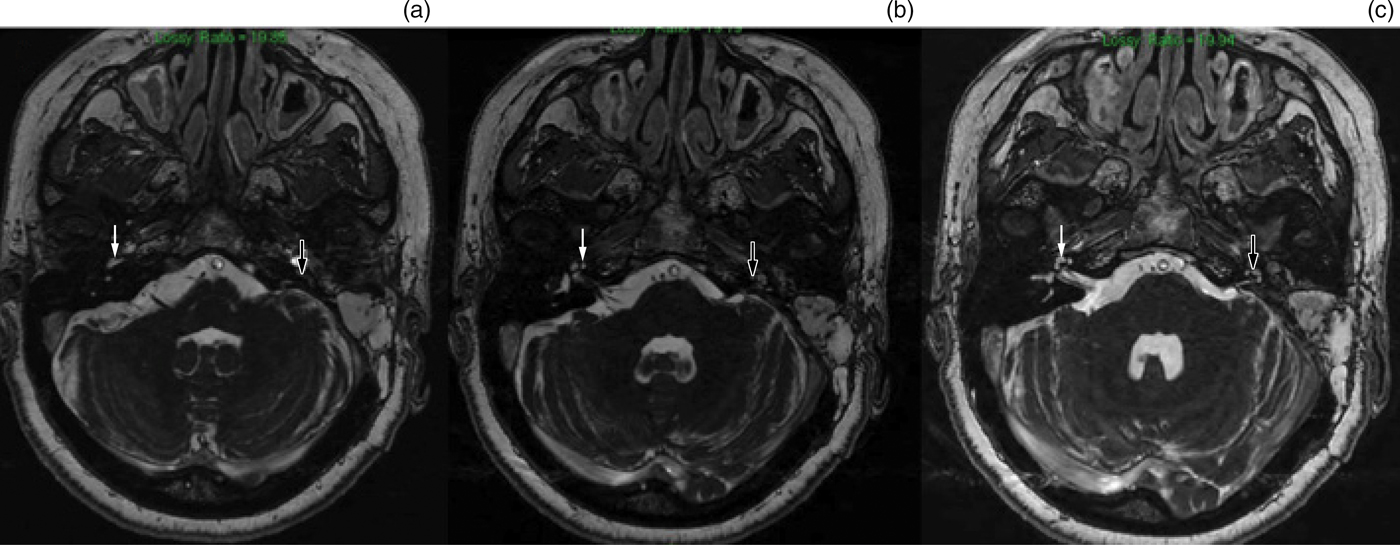
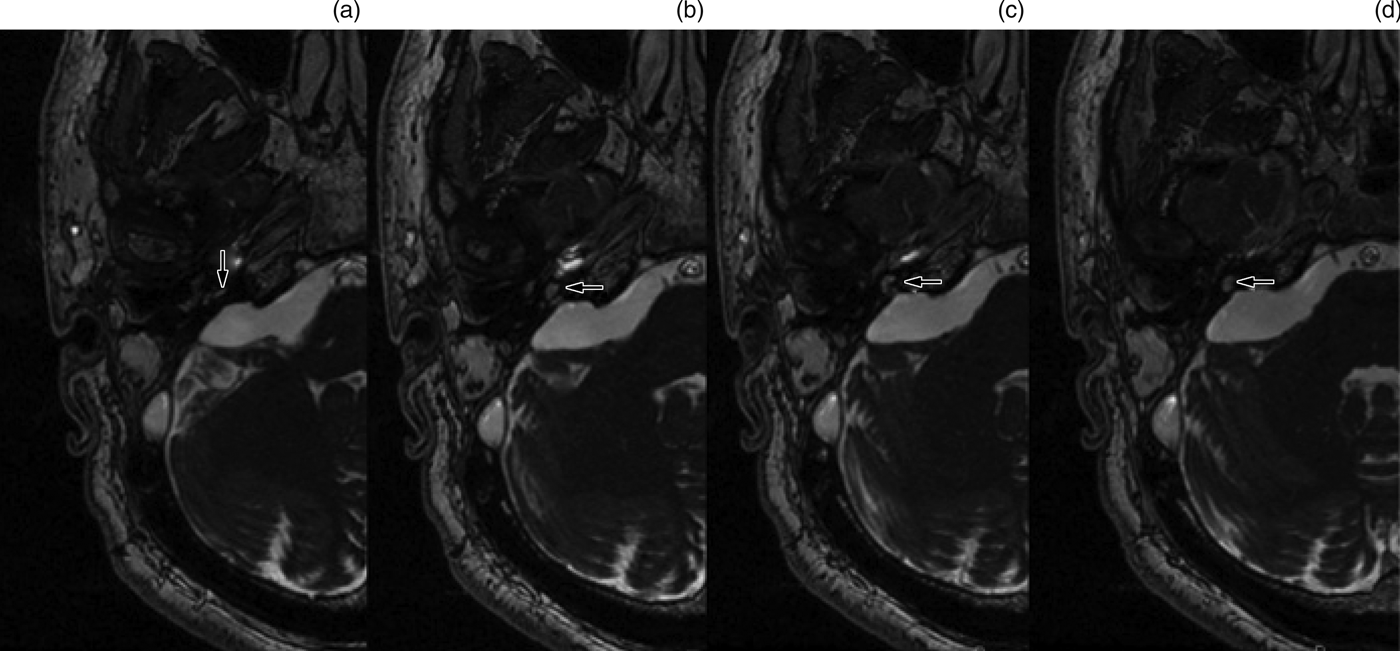
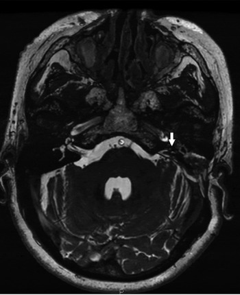
The MRI scans were assessed based on the ossification grading system developed by Isaacson et al.Reference Isaacson, Booth, Kutz, Lee and Roland11 The cochleae were classified according to four grades: grade 1 – no cochlear obstruction (Figure 1); grade 2 – obstruction of the scala tympani of the proximal inferior basal turn up to 6 mm from the round window membrane (Figure 2); grade 3 – obstruction of scala tympani of the entire basal turn with possible involvement of the middle turn (Figure 3); and grade 4 – minimal to no fluid signal in the scala tympani and vestibuli (Figure 4).
Axial, three-dimensional, fast imaging employing steady-state acquisition (‘FIESTA’) magnetic resonance imaging scans of patient 6, inferior to superior, bilaterally through the cochlea, demonstrating grade 1 cochlear obstruction (no obstruction). (a) Patent right inferior basal turn, with hypointense linear, osseous spiral lamina (solid arrow). This separates the posterior-medial scala tympani from the anterior-lateral scala vestibule (vestibular schwannoma). (b) Patent ascending basal turn of the cochlea (solid arrow). (c) Patent middle and apical turns of the cochlea (hollow arrow). P = posterior

Axial, three-dimensional, fast imaging employing steady-state acquisition (‘FIESTA’) magnetic resonance imaging scans of patient 14, inferior to superior, through both cochleae, corresponding to grade 2 (partial) cochlear obstruction. (a) Normal, non-obstructed right inferior basal turn (solid arrow), with almost complete obstruction of the scala vestibule and proximal obstruction of the scala tympani of the left inferior basal turn (hollow arrow). (b) Intact ascending basal turn (hollow arrow). (c) Intact middle and apical turns of the left cochlea (hollow arrow).
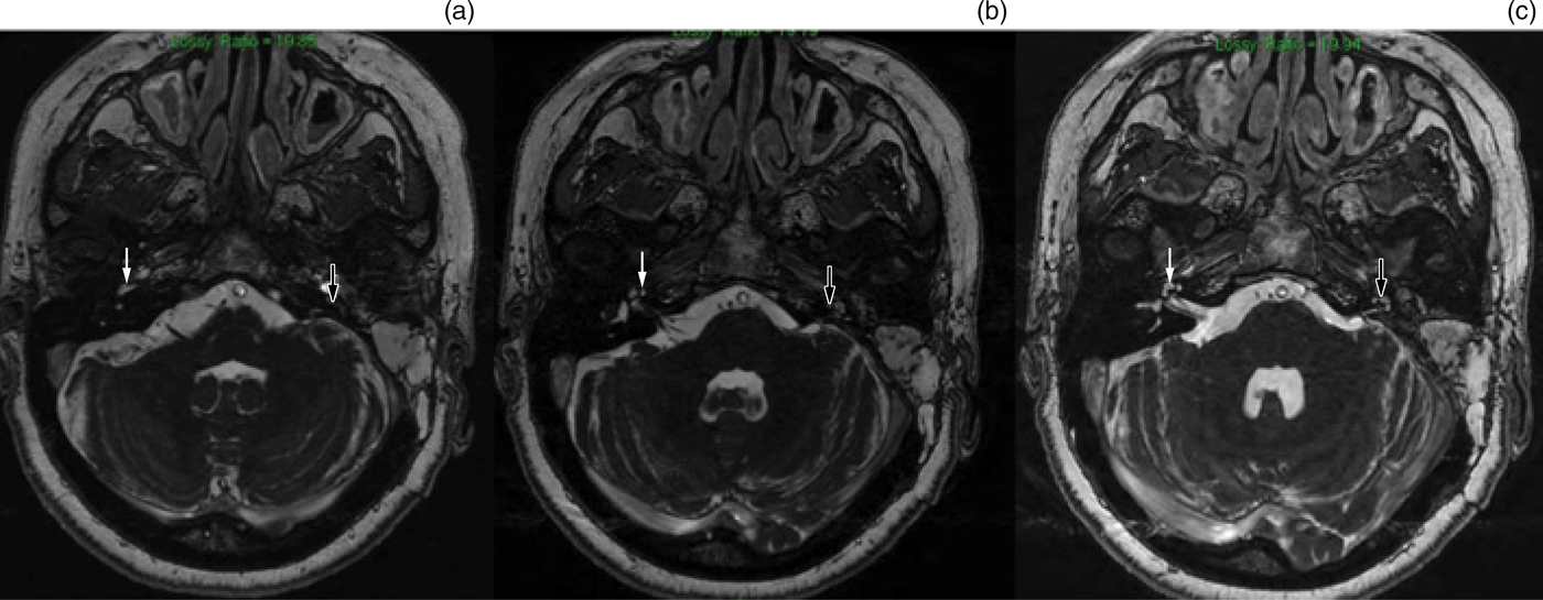
Axial, three-dimensional, fast imaging employing steady-state acquisition (‘FIESTA’) magnetic resonance imaging scan of patient 5, demonstrating grade 3 (partial) cochlear obstruction, inferior to superior sequential slices. (a) Obstruction of the inferior basal turn showing slight enhancement of the scala tympani (arrow). (b) Obstruction of the ascending basal turn (arrow). (c & d) Obstruction extending to the middle turn (arrow).

Axial, three-dimensional, fast imaging employing steady-state acquisition (‘FIESTA’) magnetic resonance imaging follow-up scan of patient 14, demonstrating grade 4 (complete) cochlear obstruction, with no fluid signal from the left cochlea (arrow) compared to the right. P = posterior

The follow-up time ranged from 4 months to 151 months. We also included 10 patients whose follow-up scans for the first four years post-operatively were missing but who had long follow-up scans available, in order to obtain information on long-term cochlear patency.
Data collection and analysis
We collected demographic data, namely patient age at operation, gender and follow-up duration. Data were recorded and organised on Excel® software spreadsheets.
Initially, we examined cochlear patency at 6 months, at more than 6 months but less than 12 months, and at 12 months following translabyrinthine approach resection, in a cross-sectional way, to determine in what percentage of cases and when the radiologically indicated cochlear obliteration would take place.
We also looked longitudinally at the MRI scans conducted after one year and four years’ follow up, where available, to determine the long-term behaviour of the cochlea. The data were entered into an Excel spreadsheet and a descriptive statistical summary was compiled of the cross-sectional analysis of the time series.
The analysis of variance test was employed (using SPSS 23 software; IBM, Armonk, New York, USA) to assess the statistical significance between the cochlear obliteration at different time-points: six months, six months to one year, more than one year but less than four years, and more than four years since the surgery. The level of significance was set at 0.05.
Results
A total of 39 patients out of 120 who underwent a translabyrinthine approach met the inclusion criteria, 17 of whom were female and 22 male. Inadequate imaging scan quality and incomplete data were the main reasons for the large number of exclusions. Mean patient age was 58.02 ± 11.17 years (range, 39–78 years).
Twelve patients underwent post-operative scanning at six months: 75 per cent (n = 9) showed no evidence of cochlear obstruction and 25 per cent (n = 3) showed partial cochlear obstruction (one grade 2 and two grade 3 partial obstruction cases). Interestingly, no patients demonstrated complete cochlear obstruction.
Thirteen patients underwent MRI scanning between 6 months and 12 months post-operatively: 38.5 per cent (n = 5) continued to demonstrate radiologically unobstructed (grade 1) cochleae, 38.5 per cent (n = 5) demonstrated partial (grade 2 or 3) obstruction and 23.01 per cent (n = 3) showed complete (grade 4) cochlear obstruction.
The sequential analysis of the time series suggested a distinct increase in morphological changes in the cochlea between six months and one year following a translabyrinthine approach for vestibular schwannoma resection, with cases of unobstructed cochleae decreasing dramatically from 75 per cent to 38.5 per cent within this time period. However, subsequent analysis suggested that after one year there appears to be a relative plateau in the rate of obstruction.
Comparison of the cochlear ossification at each time point (using analysis of variance) revealed a statistically significant progression of the cochlear obliteration with time (p = 0.0098).
After one year, 33.3 per cent of patients scanned, continued to show no evidence of cochlear obstruction, while 18 out of 27 patients demonstrated partial (25.9 per cent) or complete obstruction (40.7 per cent).
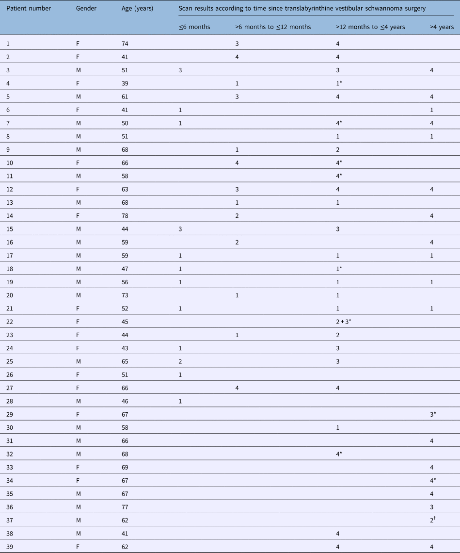
Surprisingly, of the patients who were followed up and scanned beyond four years, 26.3 per cent continued to demonstrate no obstruction. Table 1 shows a detailed breakdown of the cases, while Table 2 summarises the findings.
Detailed demographic data and scan results

Data on right side of table represent grades of cochlear obstruction. *Indicates two scans in the same time period. †Indicates three scans in the same time period. F = female; M = male
Cochlear obliteration summary results

Data represent percentages (and numbers) of patients with each grade of cochlear obstruction. In patients who underwent multiple scans, the highest grade was accepted.
Discussion
Herein, we show that ipsilateral cochlear implantation is still a possibility following translabyrinthine vestibular schwannoma resection, provided that the cochlear nerve has been preserved. The lack of definite radiological evidence of cochlear obliteration in the first six months suggests that sequential implantation is safe during this time frame. However, between six months and one year post-operatively, the chance of cochlear fibrosis increases. Still, even at this stage, cochlear implantation remains possible in selected cases.
• Cochlear fibrosis and obliteration following labyrinthectomy can make cochlear implantation challenging or even impossible
• The time frame for a patent cochlea and safe cochlear implantation following labyrinthectomy is unclear
• Our study demonstrates that advanced cochlear fibrosis often takes more than six months after translabyrinthine resection
• This indicates there is a six-month time window for safer cochlear implantation
• This is important for patients with bilateral vestibular schwannoma (neurofibromatosis 2) or disease in their only hearing ear, who might benefit from resection and implantation
• Although around 40 per cent of cases have a patent cochlea one year following labyrinthectomy, cochlear fibrosis progresses significantly between six months and one year
Hearing loss following vestibular schwannoma resection can be detrimental to patients’ quality of life. Currently used methods of auditory rehabilitation are: hearing aids, with contralateral routing of the sound; and bone conducting or bone-anchored hearing aids, which utilise the functional auditory apparatus of the contralateral ear. However, such options have certain limitations in patients with only one hearing ear. In addition, they are not feasible in patients with NF2 and bilateral vestibular schwannoma. The lack of hearing rehabilitation options has led to trials of cochlear implantation following vestibular schwannoma resection, with positive results in terms of improved binaural hearing.Reference Lustig, Yeagle, Driscoll, Blevins, Francis and Niparko5–Reference Kameswaran, Kumar, Murali, Natarajan and Krishnan10 Detrimental in this strategy are the continuity and functionality of the ipsilateral cochlear nerve, and the patency of the cochlear turns.
Previous studies have shown that despite progressive ossification, spiral ganglion cells can survive and result in auditory improvement following electrical stimulation.Reference Wanna, Carlson, Blachon, Noble, Dawant and Labadie12 Furthermore, drill-out techniques now allow cochlear implantation to be performed on progressively ossified cochleae.Reference Hassepass, Arndt, Aschendorff, Laszig and Wesarg8,Reference Wanna, Carlson, Blachon, Noble, Dawant and Labadie12,Reference Nichani, Green, Hans, Bruce, Henderson and Ramsden13 Clinically, however, the outcomes can be negatively impacted by ossified cochleae.Reference Hassepass, Arndt, Aschendorff, Laszig and Wesarg8,Reference Nichani, Green, Hans, Bruce, Henderson and Ramsden13 We show, to our knowledge for the first time, that the cochlea can stay radiologically patent for at least six months post-operatively (75 per cent of cochleae were completely patent and 25 per cent were partially patent). Thus, cochlear implantation is a possibility for hearing rehabilitation as a second-stage procedure, and following a post-operative MRI scan that rules out residual tumour. This is an option provided that the cochlear nerve has not been damaged during the operation.
Cochlear obliteration and magnetic resonance imaging
Cochlear ossification is a progressive process that evolves from initial acute inflammation, to fibrosis, and later to neo-ossification and obstruction of the cochlear turns. Changes are seen histologically as early as 17 hours following a translabyrinthine approach, and can span 4–11 years.Reference Belal7 These changes have been attributed to compromised arterial and venous drainage, particularly within the internal auditory meatus.Reference Isaacson, Booth, Kutz, Lee and Roland11 This is supported by radiologically patent cochleae, demonstrated after labyrinthectomy performed for Ménière's disease, where the internal auditory meatus remains intact.Reference Sargent, Liao and Gonda14 However, all the patients in our study had undergone vestibular schwannoma resection from the internal auditory meatus and the cerebellopontine angle, in addition to labyrinthectomy; this can explain the gradual fibrosis in most cases.
The capacity of T2-weighted MRI scans to establish patency of the cochlear turns is well established, and such scans can demonstrate the progressive degree of ossification.Reference Isaacson, Booth, Kutz, Lee and Roland11,Reference Booth, Roland, Kutz, Lee and Isaacson15,Reference Beutner, Mathys, Turowski, Schipper and Klenzner16 The ossification grading system developed by Isaacson et al.Reference Isaacson, Booth, Kutz, Lee and Roland11 was utilised within this study. Fast imaging employing steady-state acquisition scanning with a slice thickness of 1 mm or less is suitable for this grading. However, in fluid-enhanced T1-weighted imaging scans with more than 1 mm slice thickness, the differentiation between grades of partial ossification is difficult.
There is an important gap in the literature regarding comparable research. One similar study assessed the degree of cochlear ossification during the follow up of 14 patients.Reference Booth, Roland, Kutz, Lee and Isaacson15 At one year post-operatively, complete cochlear obstruction was demonstrated in 33.3 per cent of cases, while 11.1 per cent of cochleae remained completely unobstructed. Our results are more promising, showing fibrosis and obliteration in a smaller number of patients. This may be attributed to differences in: the numbers of patients included (we present the largest series), surgical techniques, surgical experience, scanning techniques and interpretation of findings. Our accuracy and experience in interpreting the scans will continue to improve as MRI techniques evolve.
To implant or not to implant?
This study covers a follow-up period of up to 151 months (over 12 years), demonstrating an unobstructed cochlea in 26.3 per cent of cases, partial obstruction in 26.1 per cent and complete obstruction in 52 per cent beyond 4 years. The most significant change in cochlear ossification seems to occur within the first year, with an unobstructed cochlea in 75 per cent of cases within six months post-surgery, decreasing to 38.5 per cent between six months and one year. This gives us a relatively safe window for cochlear implantation of six months. Thereafter, the percentage of approximately 40 per cent makes this window a grey zone, with cochlear implantation still being an option for a significant number of patients.
Following a long period (four years), the cochlea can still be patent in about 20 per cent of cases, which is promising. However, given the long time frame in those cases, detailed analysis of imaging is needed, and the possibility of atrophy or dysfunction of the unstimulated cochlear nerve should be seriously considered. Cochlear nerve anatomical integrity and function are vital factors when determining patient candidacy for cochlear implantation; hence, advanced assessment such as promontory testing is crucial.
In order to avoid progressive neural dysfunction and neo-ossification, simultaneous cochlear implantation can be performed, and this has been demonstrated successfully in a number of cases.Reference Kim, Han, Kim and Moon9,Reference Arriaga and Marks17 However, as this study has revealed that the cochlea remains patent and potentially implantable for a reasonable time period post-operatively, it is arguable that a staged approach to cochlear implantation should be advocated. This would reduce potential operating theatre time, enable better control of post-operative complications, and allow early follow up to assess for residual disease or recurrence. The importance of discrete, early post-operative scanning is emphasised with such a strategy, preferentially using heavily T2-weighted images. We found that such techniques made fluid signal in the cochlea easier to detect.
Our study suggests that cochlear implantation for hearing rehabilitation should be increasingly considered in the management of vestibular schwannoma, and not only in patients with NF2 or with vestibular schwannoma in the only hearing ear, provided that the cochlear nerve can be preserved.
Strengths and weaknesses
As our study involves a long time period, the heterogeneity of the MRI techniques and the variation in scan slice thicknesses are the main weak points of this work. Of note, the scans were primarily performed to assess any residual vestibular schwannoma, and potential growth following near total or subtotal resection; therefore, post-contrast images were the standard, while techniques that allow us to better assess fluid signal were not always included. Additionally, there were also variations regarding the time-points when the patients underwent MRI; such weaknesses can be attributed to the retrospective nature of the study.
In order to overcome the above points, we performed a detailed radiological study, enrolling a relatively large number of patients and scans compared to previous studies, but also excluding a large number of patients with data of inadequate quality. Additionally, we included cases with an extensive follow up, which gave us some indication of what actually happened to the cochlea following labyrinthectomy many years down the line.
Our data provide additional information about a relatively unexplored field of hearing rehabilitation, which could be applicable not only in patients undergoing a translabyrinthine approach for vestibular schwannoma resection, but also in patients undergoing labyrinthectomy for other reasons such as refractory Ménière's disease.
Competing interests
None declared